|
Articular Tubercle
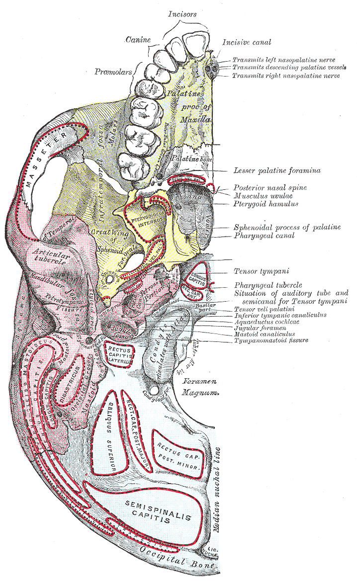
The articular tubercle (eminentia articularis) is a bony eminence on the temporal bone in the skull. It is a rounded eminence of the anterior root of the posterior end of the outer surface of the squama temporalis. This tubercle forms the front boundary of the mandibular fossa, and in the fresh state is covered with cartilage. The mandibular condyle normally moves over the articular tubercle during physiologic maximal opening of the jaw. The articular tubercle is the site of attachment of the lateral ligament of the temporomandibular joint In anatomy, the temporomandibular joints (TMJ) are the two joints connecting the jawbone to the skull. It is a bilateral Synovial joint, synovial articulation between the temporal bone of the skull above and the condylar process of mandible be .... Earth's Lab- Retrieved 2018-08-30< ... [...More Info...] [...Related Items...] OR: [Wikipedia] [Google] [Baidu] |
Temporal Bone
The temporal bone is a paired bone situated at the sides and base of the skull, lateral to the temporal lobe of the cerebral cortex. The temporal bones are overlaid by the sides of the head known as the temples where four of the cranial bones fuse. Each temple is covered by a temporal muscle. The temporal bones house the structures of the ears. The lower seven cranial nerves and the major vessels to and from the brain traverse the temporal bone. Structure The temporal bone consists of four parts—the squamous, mastoid, petrous and tympanic parts. The squamous part is the largest and most superiorly positioned relative to the rest of the bone. The zygomatic process is a long, arched process projecting from the lower region of the squamous part and it articulates with the zygomatic bone. Posteroinferior to the squamous is the mastoid part. Fused with the squamous and mastoid parts and between the sphenoid and occipital bones lies the petrous part, which is shaped li ... [...More Info...] [...Related Items...] OR: [Wikipedia] [Google] [Baidu] |
Base Of Skull
The base of skull, also known as the cranial base or the cranial floor, is the most Anatomical terms of location#Superior and inferior, inferior area of the human skull, skull. It is composed of the endocranium and the lower parts of the Calvaria (skull), calvaria. Structure Structures found at the base of the skull are for example: Bones There are five bones that make up the base of the skull: *Ethmoid bone *Sphenoid bone *Occipital bone *Frontal bone *Temporal bone Sinuses *Occipital sinus *Superior sagittal sinus *Superior petrosal sinus Foramina of the skull *Foramen cecum (frontal bone), Foramen cecum *Optic foramen *Foramen lacerum *Foramen rotundum *Foramen magnum *Foramen ovale (skull), Foramen ovale *Jugular foramen *Internal auditory meatus *Mastoid foramen *Sphenoidal emissary foramen *Foramen spinosum Sutures *Frontoethmoidal suture *Sphenofrontal suture *Sphenopetrosal suture *Sphenoethmoidal suture *Petrosquamous suture *Sphenosquamosal suture Other *Sph ... [...More Info...] [...Related Items...] OR: [Wikipedia] [Google] [Baidu] |
Skull
The skull, or cranium, is typically a bony enclosure around the brain of a vertebrate. In some fish, and amphibians, the skull is of cartilage. The skull is at the head end of the vertebrate. In the human, the skull comprises two prominent parts: the neurocranium and the facial skeleton, which evolved from the first pharyngeal arch. The skull forms the frontmost portion of the axial skeleton and is a product of cephalization and vesicular enlargement of the brain, with several special senses structures such as the eyes, ears, nose, tongue and, in fish, specialized tactile organs such as barbels near the mouth. The skull is composed of three types of bone: cranial bones, facial bones and ossicles, which is made up of a number of fused flat and irregular bones. The cranial bones are joined at firm fibrous junctions called sutures and contains many foramina, fossae, processes, and sinuses. In zoology, the openings in the skull are called fenestrae, the most ... [...More Info...] [...Related Items...] OR: [Wikipedia] [Google] [Baidu] |
Squama Temporalis
The squamous part of temporal bone, or temporal squama, forms the front and upper part of the temporal bone, and is scale-like, thin, and translucent. Surfaces Its outer surface is smooth and convex; it affords attachment to the temporal muscle, and forms part of the temporal fossa; on its hinder part is a vertical groove for the middle temporal artery. A curved line, the ''temporal line'', or ''supramastoid crest'', runs backward and upward across its posterior part; it serves for the attachment of the temporal fascia, and limits the origin of the temporalis muscle. The boundary between the squamous part and the mastoid portion of the bone, as indicated by traces of the original suture, lies about 1 cm. below this line. Projecting from the lower part of the squamous part is a long, arched process, the '' zygomatic process''. This process is at first directed lateralward, its two surfaces looking upward and downward; it then appears as if twisted inward upon itself, a ... [...More Info...] [...Related Items...] OR: [Wikipedia] [Google] [Baidu] |
Mandibular Fossa
The mandibular fossa, also known as the glenoid fossa in some dental literature, is the depression in the temporal bone that articulates with the mandible. Structure In the temporal bone, the mandibular fossa is bounded anteriorly by the articular tubercle and posteriorly by the tympanic portion of the temporal bone, which separates it from the external acoustic meatus. The fossa is divided into two parts by a narrow slit, the petrotympanic fissure (Glaserian fissure). It is concave in shape to receive the condyloid process of the mandible. Development The mandibular fossa develops from condylar cartilage. This may be stimulated by SOX9 or ALK2, as has been seen in mouse models. Function The condyloid process of the mandible articulates with the temporal bone of the skull at the mandibular fossa. Clinical significance Problems with morphogenesis during embryonic development can lead to the mandibular fossa not forming. This may be caused by mutations to SOX9 or ALK ... [...More Info...] [...Related Items...] OR: [Wikipedia] [Google] [Baidu] |
Mandibular Condyle
The condyloid process or condylar process is the process on the human and other mammalian species' mandibles that ends in a condyle, the mandibular condyle. It is thicker than the coronoid process of the mandible and consists of two portions: the condyle and the constricted portion which supports it, the neck. Condyle The most superior part of the mandible, the condyle presents an articular surface for articulation with the articular disk of the temporomandibular joint; it is convex from before backward and from side to side, and extends further on the posterior than on the anterior surface. Its long axis is directed medialward and slightly backward, and if prolonged to the middle line will meet that of the opposite condyle near the anterior margin of the foramen magnum. At the lateral extremity of the condyle is a small tubercle for the attachment of the temporomandibular ligament. The articular surface of the condyle is covered by fibrous tissue, and interfaces with an articu ... [...More Info...] [...Related Items...] OR: [Wikipedia] [Google] [Baidu] |
Temporomandibular Joint
In anatomy, the temporomandibular joints (TMJ) are the two joints connecting the jawbone to the skull. It is a bilateral Synovial joint, synovial articulation between the temporal bone of the skull above and the condylar process of mandible below; it is from these bones that its name is derived. The joints are unique in their bilateral function, being connected via the mandible. Structure The main components are the joint capsule, articular disc, mandibular condyles, articular surface of the temporal bone, temporomandibular ligament, stylomandibular ligament, sphenomandibular ligament, and lateral pterygoid muscle. Capsule The articular capsule (capsular ligament) is a thin, loose envelope, attached above to the circumference of the mandibular fossa and the articular tubercle immediately in front; below, to the neck of the condyle of the mandible. Its loose attachment to the neck of the mandible allows for free movement. Articular disc The unique feature of the temporomand ... [...More Info...] [...Related Items...] OR: [Wikipedia] [Google] [Baidu] |


