|
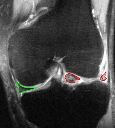
Tear Of Meniscus
A tear of a meniscus is a rupturing of one or more of the fibrocartilage strips in the knee called meniscus (anatomy), menisci. When doctors and patients refer to "torn cartilage" in the knee, they actually may be referring to an injury to a meniscus at the top of one of the Tibia, tibiae. Menisci can be torn during innocuous activities such as walking or squatting position, squatting. They can also be torn by Physical trauma, traumatic force encountered in sports or other forms of physical exertion. The traumatic action is most often a twisting movement at the knee while the leg is bent. In older adults, the meniscus can be damaged following prolonged 'wear and tear'. Especially acute injuries (typically in younger, more active patients) can lead to displaced tears which can cause mechanical symptoms such as clicking, catching, or locking during motion of the joint. The joint will be in pain when in use, but when there is no load, the pain goes away. A tear of the medial meniscus ... [...More Info...] [...Related Items...] OR: [Wikipedia] [Google] [Baidu] |
Orthopedics
Orthopedic surgery or orthopedics (American and British English spelling differences, alternative spelling orthopaedics) is the branch of surgery concerned with conditions involving the musculoskeletal system. Orthopedic surgeons use both surgical and nonsurgical means to treat musculoskeletal Physical trauma, trauma, Spinal disease, spine diseases, Sports injury, sports injuries, degenerative diseases, infections, tumors and congenital disorders. Etymology Nicholas Andry coined the word in French as ', derived from the Ancient Greek words ("correct", "straight") and ("child"), and published ''Orthopedie'' (translated as ''Orthopædia: Or the Art of Correcting and Preventing Deformities in Children'') in 1741. The word was Assimilation (linguistics), assimilated into English as ''orthopædics''; the Typographic ligature, ligature ''æ'' was common in that era for ''ae'' in Greek- and Latin-based words. As the name implies, the discipline was initially developed with atte ... [...More Info...] [...Related Items...] OR: [Wikipedia] [Google] [Baidu] |
Viscoelastic
In materials science and continuum mechanics, viscoelasticity is the property of materials that exhibit both Viscosity, viscous and Elasticity (physics), elastic characteristics when undergoing deformation (engineering), deformation. Viscous materials, like water, resist both shear flow and Strain (materials science), strain linearly with time when a Stress (physics), stress is applied. Elastic materials strain when stretched and immediately return to their original state once the stress is removed. Viscoelastic materials have elements of both of these properties and, as such, exhibit time-dependent strain. Whereas elasticity is usually the result of chemical bond, bond stretching along crystallographic planes in an ordered solid, viscosity is the result of the diffusion of atoms or molecules inside an amorphous material.Meyers and Chawla (1999): "Mechanical Behavior of Materials", 98-103. Background In the nineteenth century, physicists such as James Clerk Maxwell, Ludwig Boltzm ... [...More Info...] [...Related Items...] OR: [Wikipedia] [Google] [Baidu] |
Valgus Stress
The valgus stress test or medial stress test is a test for damage to the medial collateral ligament of the knee. It involves placing the leg into extension, with one hand placed as a pivot Pivot may refer to: *Pivot, the point of rotation in a lever system *More generally, the center point of any rotational system *Pivot joint, a kind of joint between bones in the body *Pivot turn, a dance move Companies *Incitec Pivot, an Austra ... on the knee. With the other hand placed upon the foot applying an abducting force, an attempt is then made to force the leg at the knee into valgus. If the knee is seen to open up on the medial side, this is indicative of medial collateral ligament damage and may also indicate capsular or cruciate ligament laxity. There are two versions of this test: valgus at 0 degrees and valgus at 30 degrees. When performing the test at 30 degrees, the MCL is the primary stabilizer; the joint capsule is also tested. When tested at 0 degrees, the MCL, medial jo ... [...More Info...] [...Related Items...] OR: [Wikipedia] [Google] [Baidu] |
Varus Deformity
A varus deformity is an excessive inward angulation ( medial angulation, that is, towards the body's midline) of the distal segment of a bone or joint. The opposite of varus is called valgus. The terms varus and valgus always refer to the direction that the distal segment of the joint points. For example, in a valgus deformity of the knee, the distal part of the leg below the knee is deviated ''outward, in relation to the femur,'' resulting in a '' knock-kneed'' appearance. Conversely, a ''varus'' deformity at the knee results in a '' bowlegged'' with the distal part of the leg deviated ''inward, in relation to the femur''. However, in relation to the mid-line of the body, the knee joint is deviated towards the mid-line. Terminology The terminology is made confusing by the etymology of these words. * The terms ''varus'' and ''valgus'' are both Latin, but confusingly, their Latin meanings conflict with their current usage. In current usage, as noted above, a varus deformity ... [...More Info...] [...Related Items...] OR: [Wikipedia] [Google] [Baidu] |
Extension (kinesiology)
Motion, the process of movement, is described using specific anatomical terms. Motion includes movement of organs, joints, limbs, and specific sections of the body. The terminology used describes this motion according to its direction relative to the anatomical position of the body parts involved. Anatomists and others use a unified set of terms to describe most of the movements, although other, more specialized terms are necessary for describing unique movements such as those of the hands, feet, and eyes. In general, motion is classified according to the anatomical plane it occurs in. ''Flexion'' and ''extension'' are examples of ''angular'' motions, in which two axes of a joint are brought closer together or moved further apart. ''Rotational'' motion may occur at other joints, for example the shoulder, and are described as ''internal'' or ''external''. Other terms, such as ''elevation'' and ''depression'', describe movement above or below the horizontal plane. Many anatomic ... [...More Info...] [...Related Items...] OR: [Wikipedia] [Google] [Baidu] |
Flexion
Motion, the process of movement, is described using specific anatomical terminology, anatomical terms. Motion includes movement of Organ (anatomy), organs, joints, Limb (anatomy), limbs, and specific sections of the body. The terminology used describes this motion according to its direction relative to the anatomical position of the body parts involved. Anatomy, Anatomists and others use a unified set of terms to describe most of the movements, although other, more specialized terms are necessary for describing unique movements such as those of the hands, feet, and eyes. In general, motion is classified according to the anatomical plane it occurs in. ''Flexion'' and ''extension'' are examples of ''angular'' motions, in which two axes of a joint are brought closer together or moved further apart. ''Rotational'' motion may occur at other joints, for example the shoulder, and are described as ''internal'' or ''external''. Other terms, such as ''elevation'' and ''depression'', descri ... [...More Info...] [...Related Items...] OR: [Wikipedia] [Google] [Baidu] |
McMurray Test
The McMurray test, also known as the McMurray circumduction test is used to evaluate individuals for tears in the meniscus of the knee. A tear in the meniscus may cause a pedunculated tag of the meniscus which may become jammed between the joint surfaces. To perform the test, the knee is held by one hand, which is placed along the joint line, and flexed to complete flexion while the foot is held by the sole (of the foot) with the other hand. The examiner then rotates the leg internally while extending the knee to 90 degrees of flexion. If a "thud" or "click" is felt along with pain, this constitutes a "positive McMurray test" for a tear in the posterior portion of the lateral meniscus. Likewise, external rotation of the leg can be applied to test the posterior portion of the medial meniscus. The McMurray test is named after Thomas Porter McMurray, a British orthopedic surgeon Orthopedic surgery or orthopedics (American and British English spelling differences, alternative ... [...More Info...] [...Related Items...] OR: [Wikipedia] [Google] [Baidu] |
Physician
A physician, medical practitioner (British English), medical doctor, or simply doctor is a health professional who practices medicine, which is concerned with promoting, maintaining or restoring health through the Medical education, study, Medical diagnosis, diagnosis, prognosis and therapy, treatment of disease, injury, and other physical and mental impairments. Physicians may focus their practice on certain disease categories, types of patients, and methods of treatment—known as Specialty (medicine), specialities—or they may assume responsibility for the provision of continuing and comprehensive medical care to individuals, families, and communities—known as general practitioner, general practice. Medical practice properly requires both a detailed knowledge of the Discipline (academia), academic disciplines, such as anatomy and physiology, pathophysiology, underlying diseases, and their treatment, which is the science of medicine, and a decent Competence (human resources ... [...More Info...] [...Related Items...] OR: [Wikipedia] [Google] [Baidu] |
Fibular Collateral Ligament
The lateral collateral ligament (LCL, long external lateral ligament or fibular collateral ligament) is an extrinsic ligament of the knee located on the lateral side of the knee. Its superior attachment is at the lateral epicondyle of the femur (superoposterior to the popliteal groove); its inferior attachment is at the lateral aspect of the head of fibula (anterior to the apex). The LCL is not fused with the joint capsule. Inferiorly, the LCL splits the tendon of insertion of the biceps femoris muscle. Structure The LCL measures some 5 cm in length. It is rounded, and is more narrow and less broad compared to the medial collateral ligament. It extends obliquely inferoposteriorly from its superior attachment to its inferior attachment. In contrast to the medial collateral ligament, it is not fused with either the capsular ligament nor the lateral meniscus. Because of this, the LCL is more flexible than its medial counterpart, and is therefore less susceptible to injury. ... [...More Info...] [...Related Items...] OR: [Wikipedia] [Google] [Baidu] |
Ligament
A ligament is a type of fibrous connective tissue in the body that connects bones to other bones. It also connects flight feathers to bones, in dinosaurs and birds. All 30,000 species of amniotes (land animals with internal bones) have ligaments. It is also known as ''articular ligament'', ''articular larua'', ''fibrous ligament'', or ''true ligament''. Comparative anatomy Ligaments are similar to tendons and fasciae as they are all made of connective tissue. The differences among them are in the connections that they make: ligaments connect one bone to another bone, tendons connect muscle to bone, and fasciae connect muscles to other muscles. These are all found in the skeletal system of the human body. Ligaments cannot usually be regenerated naturally; however, there are periodontal ligament stem cells located near the periodontal ligament which are involved in the adult regeneration of periodontist ligament. The study of ligaments is known as . Humans Other ligame ... [...More Info...] [...Related Items...] OR: [Wikipedia] [Google] [Baidu] |
Femoral Condyle
The lower extremity of femur (or distal extremity) is the lower end of the femur (thigh bone) in human and other animals, closer to the knee. It is larger than the upper extremity of femur, is somewhat cuboid in form, but its transverse diameter is greater than its antero-posterior; it consists of two oblong eminences known as the lateral condyle and medial condyle. Condyles Anteriorly, the condyles are slightly prominent and are separated by a smooth shallow articular depression called the patella surface. Posteriorly, they project considerably and a deep notch, the intercondylar fossa of femur, is present between them. The lateral condyle is the more prominent and is the broader both in its antero-posterior and transverse diameters, the medial condyle is the longer and, when the femur is held with its body perpendicular, projects to a lower level. When, however, the femur is in its natural oblique position the lower surfaces of the two condyles lie practically in the sam ... [...More Info...] [...Related Items...] OR: [Wikipedia] [Google] [Baidu] |
Tibial Plateau
Tibial may refer to: * Tibia bone * Tibial nerve * Anterior tibial artery * Posterior tibial artery * Anterior tibial vein * Posterior tibial vein The posterior tibial veins are veins of the leg in humans. They drain the posterior compartment of the leg and the plantar surface of the foot to the popliteal vein. Structure The posterior tibial veins receive blood from the medial and lat ... * Insect tibia {{Disambiguation ... [...More Info...] [...Related Items...] OR: [Wikipedia] [Google] [Baidu] |


