|
Pterygoid Plexus
The pterygoid plexus (; in Merriam-Webster Online Dictionary '. from ''pteryx'', "wing" and ''eidos'', "shape") is a of considerable size, and is situated between the and |
Maxillary Vein
The maxillary vein, or internal maxillary vein, is a vein of the head. It is a short trunk which accompanies the first part of the maxillary artery. It is formed by a confluence of the veins of the pterygoid plexus and the interpterygoid emissary vein, and passes posteriorly between the sphenomandibular ligament and the neck of the mandible. It unites with the superficial temporal vein to form the retromandibular vein. Structure The maxillary vein is a short trunk which accompanies the first part of the maxillary artery. It is formed from the merging of the veins of the pterygoid plexus, and the interpterygoid emissary vein. It passes posteriorly between the sphenomandibular ligament and the neck of the mandible. It unites with the superficial temporal vein. It drains into the retromandibular vein (posterior facial vein). The maxillary vein anastomoses with the retroglenoid vein. Development The maxillary vein may be the embryological origin of the central retinal vein T ... [...More Info...] [...Related Items...] OR: [Wikipedia] [Google] [Baidu] |
Foramen Vesalii
In the base of the skull, in the great wings of the sphenoid bone, medial to the foramen ovale, a small aperture, the sphenoidal emissary foramen, may occasionally be seen (it is often absent) opposite the root of the pterygoid process. When present, it opens below near the scaphoid fossa. Vesalius was the first to describe and illustrate this foramen, and it thus sometimes bears the name of foramen Vesalii (meaning foramen of Vesalius). Other names include foramen venosum and canaliculus sphenoidalis. Importance If at all present, the sphenoidal emissary foramen gives passage to a small vein (vein of Vesalius) that connects the pterygoid plexus with the cavernous sinus. The importance of this passage lies in the fact that an infected thrombus from an extracranial source may reach the cavernous sinus. The mean area of the foramen is small, which may suggest that it plays a minor role in the dynamics of blood circulation in the venous system of the head. Structure The sphenoidal ... [...More Info...] [...Related Items...] OR: [Wikipedia] [Google] [Baidu] |
External Jugular Vein
The external jugular vein receives the greater part of the blood from the exterior of the cranium and the deep parts of the face, being formed by the junction of the posterior division of the retromandibular vein with the posterior auricular vein. Structure It commences in the substance of the parotid gland, on a level with the angle of the mandible, and runs perpendicularly down the neck, in the direction of a line drawn from the angle of the mandible to the middle of the clavicle superficial to the sternocleidomastoideus. In its course it crosses the sternocleidomastoideus obliquely, and in the subclavian triangle perforates the deep fascia, and ends in the subclavian vein lateral to or in front of the scalenus anterior, piercing the roof of the posterior triangle. It is separated from the sternocleidomastoideus by the investing layer of the deep cervical fascia, and is covered by the platysma, the superficial fascia, and the integument; it crosses the cutaneous cervical nerv ... [...More Info...] [...Related Items...] OR: [Wikipedia] [Google] [Baidu] |
Posterior Auricular Vein
The posterior auricular vein is a vein of the head. It begins from a plexus with the occipital vein and the superficial temporal vein, descends behind the auricle, and drains into the external jugular vein. Structure The posterior auricular vein begins upon the side of the head, in a plexus which communicates with the tributaries of the occipital vein and the superficial temporal vein. It descends behind the auricle. It joins the posterior division of the retromandibular vein. It drains into the external jugular vein. It receive the stylomastoid vein, and some tributaries from the cranial surface of the auricle. Variation The posterior auricular vein may drain into the internal jugular vein or a posterior jugular vein if there are variations in the external jugular vein The external jugular vein receives the greater part of the blood from the exterior of the cranium and the deep parts of the face, being formed by the junction of the posterior division of the retromandib ... [...More Info...] [...Related Items...] OR: [Wikipedia] [Google] [Baidu] |
Retromandibular Vein
The retromandibular vein (temporomaxillary vein, posterior facial vein) is a major vein of the face. Anatomy Origin The retromandibular vein is formed by the union of the superficial temporal and maxillary veins. Course It descends in the substance of the parotid gland, superficial to the external carotid artery (but beneath the facial nerve), between the ramus of the mandible and the sternocleidomastoideus muscle. It terminates by dividing into two branches: * an ''anterior'', which passes forward and joins anterior facial vein, to form the common facial vein, which then drains into the internal jugular vein. * a ''posterior'', which is joined by the posterior auricular vein and becomes the external jugular vein. Function The retromandibular vein provides venous drainage to the superior cranium, and significant drainage to the ear. Clinical significance Parrot's sign is a sensation of pain when pressure is applied to the retromandibular region. Additional images ... [...More Info...] [...Related Items...] OR: [Wikipedia] [Google] [Baidu] |
Superficial Temporal Vein
The superficial temporal vein is a vein of the side of the head. It begins on the side and vertex of the skull in a network of veins which communicates with the frontal vein and supraorbital vein, with the corresponding vein of the opposite side, and with the posterior auricular vein and occipital vein. It ultimately crosses the posterior root of the zygomatic arch, enters the parotid gland, and unites with the internal maxillary vein to form the posterior facial vein. Structure It begins on the side and vertex of the skull in a network () which communicates with the frontal vein and supraorbital vein, with the corresponding vein of the opposite side, and with the posterior auricular vein and occipital vein. From this network frontal and parietal branches arise, and join above the zygomatic arch to form the trunk of the vein, which is joined by the middle temporal vein emerging from the temporalis muscle. It then crosses the posterior root of the zygomatic arch, enters the sub ... [...More Info...] [...Related Items...] OR: [Wikipedia] [Google] [Baidu] |
Maxillary Vein
The maxillary vein, or internal maxillary vein, is a vein of the head. It is a short trunk which accompanies the first part of the maxillary artery. It is formed by a confluence of the veins of the pterygoid plexus and the interpterygoid emissary vein, and passes posteriorly between the sphenomandibular ligament and the neck of the mandible. It unites with the superficial temporal vein to form the retromandibular vein. Structure The maxillary vein is a short trunk which accompanies the first part of the maxillary artery. It is formed from the merging of the veins of the pterygoid plexus, and the interpterygoid emissary vein. It passes posteriorly between the sphenomandibular ligament and the neck of the mandible. It unites with the superficial temporal vein. It drains into the retromandibular vein (posterior facial vein). The maxillary vein anastomoses with the retroglenoid vein. Development The maxillary vein may be the embryological origin of the central retinal vein T ... [...More Info...] [...Related Items...] OR: [Wikipedia] [Google] [Baidu] |
Cavernous Sinus Thrombosis
The cavernous sinus within the human head is one of the dural venous sinuses creating a cavity called the lateral sellar compartment bordered by the temporal bone of the skull and the sphenoid bone, lateral to the sella turcica. Structure The cavernous sinus is one of the dural venous sinuses of the head. It is a network of veins that sit in a cavity. It sits on both sides of the sphenoidal bone and pituitary gland, approximately 1 × 2 cm in size in an adult. The carotid siphon of the internal carotid artery, and cranial nerves III, IV, V (branches V1 and V2) and VI all pass through this blood filled space. Both sides of cavernous sinus is connected to each other via intercavernous sinuses. The cavernous sinus lies in between the inner and outer layers of dura mater. Nearby structures * Above: optic tract, optic chiasma, internal carotid artery. * Inferiorly: foramen lacerum, and the junction of the body and greater wing of sphenoid bone. * Medially: pituitary gla ... [...More Info...] [...Related Items...] OR: [Wikipedia] [Google] [Baidu] |
Foramen Lacerum
The foramen lacerum ( la, lacerated piercing) is a triangular hole in the base of skull. It is located between the sphenoid bone, the apex of the petrous part of the temporal bone, and the basilar part of the occipital bone. Structure The foramen lacerum ( la, lacerated piercing) is a triangular hole in the base of skull. It is located between 3 bones: * the sphenoid bone, forming the anterior border. * the apex of petrous part of the temporal bone, forming the posterolateral border. * the basilar part of occipital bone, forming the posteromedial border. It is the junction point of 3 sutures of the skull: * the petroclival (petrooccipital) suture. * the sphenopetrosal suture. * the sphenooccipital suture. It is situated anteromedial to the carotid canal. Development The foramen lacerum fills with cartilage after birth. Function The foramen lacerum transmits many structures, including: * the artery of the pterygoid canal. * the recurrent artery of the foramen laceru ... [...More Info...] [...Related Items...] OR: [Wikipedia] [Google] [Baidu] |
Foramen Ovale (skull)
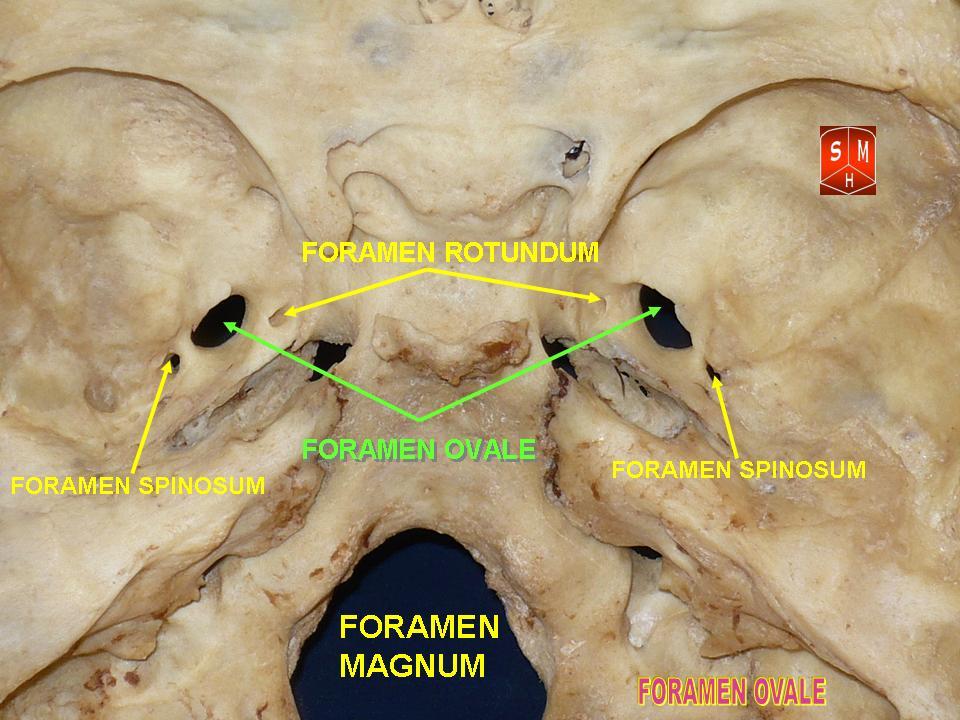
The foramen ovale (Latin: oval window) is a hole in the posterior part of the sphenoid bone, posterolateral to the foramen rotundum. It is one of the larger of the several holes (the foramina) in the skull. It transmits the mandibular nerve, a branch of the trigeminal nerve. Structure The foramen ovale is an opening in the greater wing of the sphenoid bone. The foramen ovale is one of two cranial foramina in the greater wing, the other being the foramen spinosum. The foramen ovale is posterolateral to the foramen rotundum and anteromedial to the foramen spinosum. Posterior and medial to the foramen is the opening for the carotid canal. Variation Similar to other foramina, the foramen ovale differs in shape and size throughout the natural life. The earliest perfect ring-shaped formation of the foramen ovale was observed in the 7th fetal month and the latest in 3 years after birth, in a study using over 350 skulls.In a study conducted on 100 skulls, the foramen ovale was d ... [...More Info...] [...Related Items...] OR: [Wikipedia] [Google] [Baidu] |
Cavernous Sinus
The cavernous sinus within the human head is one of the dural venous sinuses creating a cavity called the lateral sellar compartment bordered by the temporal bone of the skull and the sphenoid bone, lateral to the sella turcica. Structure The cavernous sinus is one of the dural venous sinuses of the head. It is a network of veins that sit in a cavity. It sits on both sides of the sphenoidal bone and pituitary gland, approximately 1 × 2 cm in size in an adult. The carotid siphon of the internal carotid artery, and cranial nerves III, IV, V (branches V1 and V2) and VI all pass through this blood filled space. Both sides of cavernous sinus is connected to each other via intercavernous sinuses. The cavernous sinus lies in between the inner and outer layers of dura mater. Nearby structures * Above: optic tract, optic chiasma, internal carotid artery. * Inferiorly: foramen lacerum, and the junction of the body and greater wing of sphenoid bone. * Medially: pituitary gla ... [...More Info...] [...Related Items...] OR: [Wikipedia] [Google] [Baidu] |
Maxillary Artery
The maxillary artery supplies deep structures of the face. It branches from the external carotid artery just deep to the neck of the mandible. Structure The maxillary artery, the larger of the two terminal branches of the external carotid artery, arises behind the neck of the mandible, and is at first imbedded in the substance of the parotid gland; it passes forward between the ramus of the mandible and the sphenomandibular ligament, and then runs, either superficial or deep to the lateral pterygoid muscle, to the pterygopalatine fossa. It supplies the deep structures of the face, and may be divided into mandibular, pterygoid, and pterygopalatine portions. First portion The ''first'' or ''mandibular '' or ''bony'' portion passes horizontally forward, between the neck of the mandible and the sphenomandibular ligament, where it lies parallel to and a little below the auriculotemporal nerve; it crosses the inferior alveolar nerve, and runs along the lower border of the lateral pte ... [...More Info...] [...Related Items...] OR: [Wikipedia] [Google] [Baidu] |
