|
Pupillary Reflex
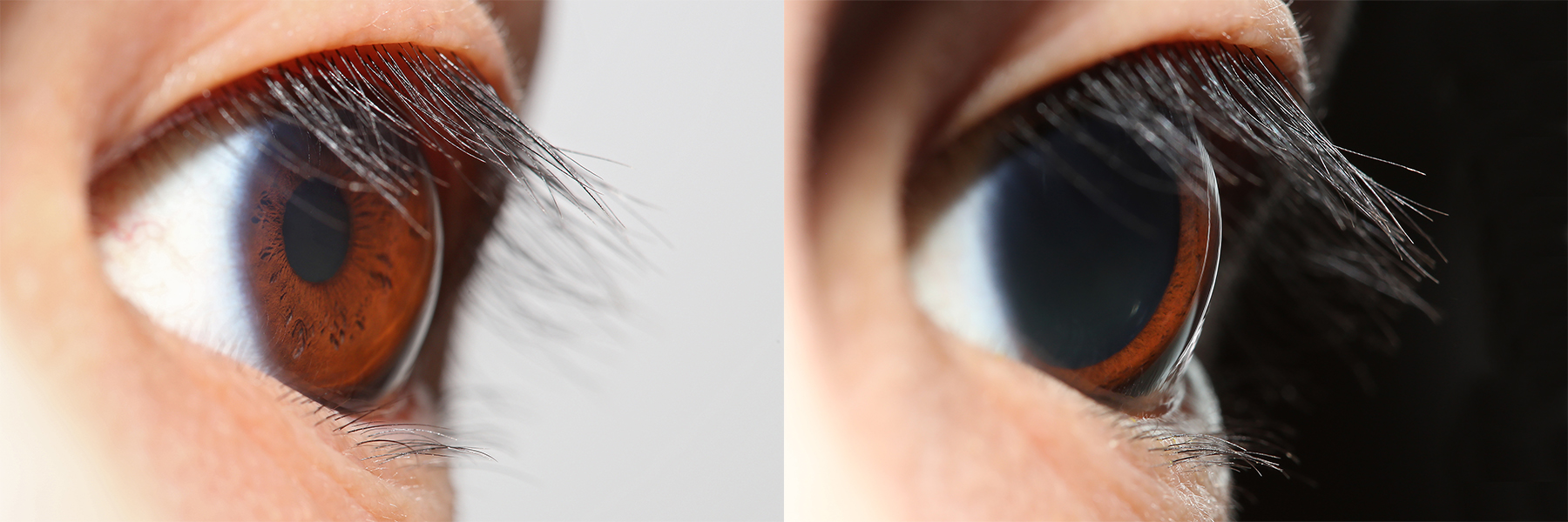
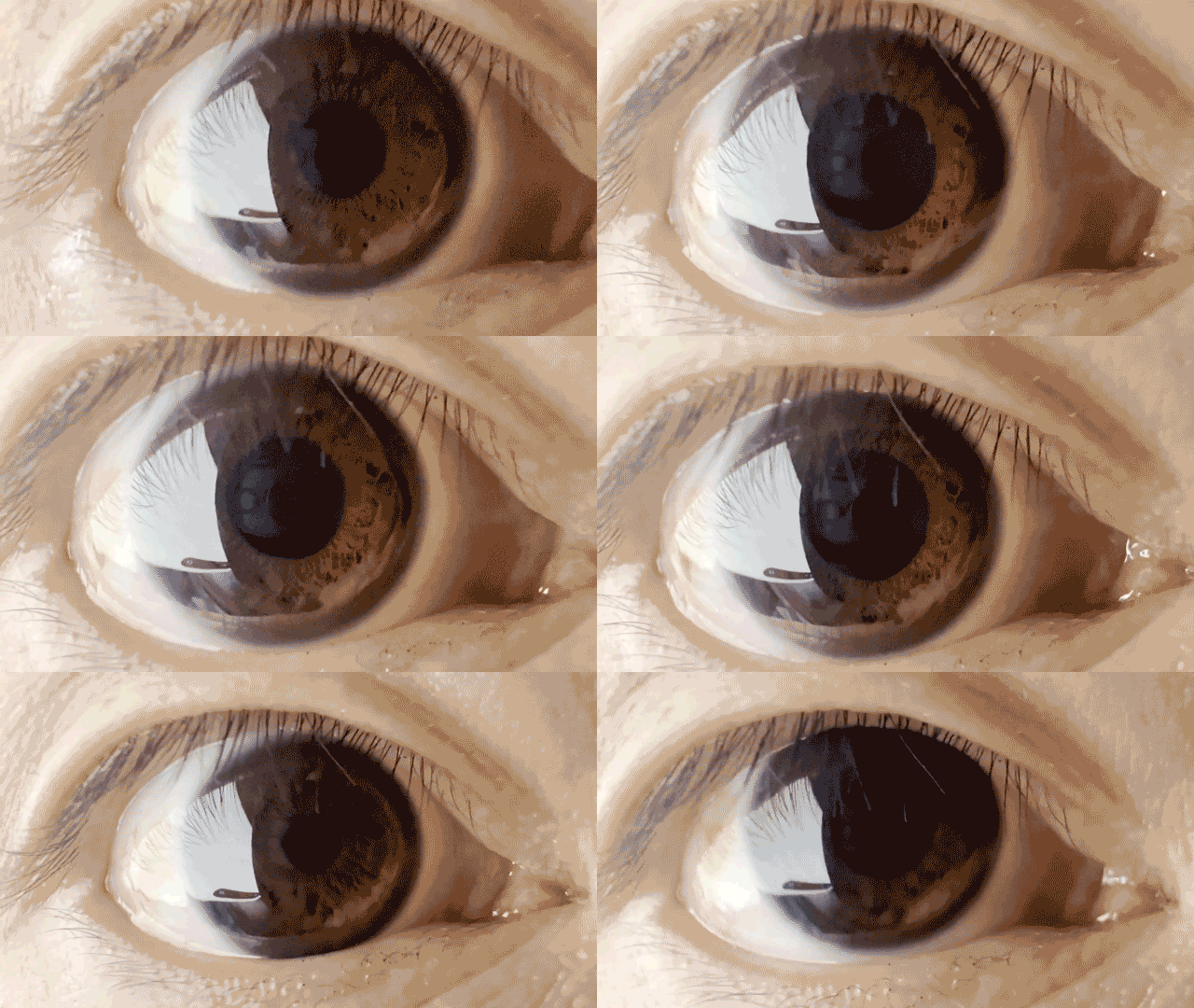
Pupillary reflex refers to one of the reflexes associated with pupillary function. These include the pupillary light reflex and accommodation reflex. Although the pupillary response, in which the pupil dilates or constricts due to light is not usually called a "reflex", it is still usually considered a part of this topic. Adjustment to close-range vision is known as "the near response", while relaxation of the ciliary muscle to view distant objects is known as the "far response". In "the near response" there are three processes that occur to focus an image on the retina. Convergence of the eyes, or the orientation of the visual axis of each eye towards an object in order to focus its image on each fovea, is the first of the three responses. This can be observed by the cross-eyed movement of the eyes when a finger is held up in front of a face and moved towards the face. Next, constriction of the pupil occurs. Because the lens cannot refract In physics, refraction is the r ... [...More Info...] [...Related Items...] OR: [Wikipedia] [Google] [Baidu] |
Reflex
In biology, a reflex, or reflex action, is an involuntary, unplanned sequence or action and nearly instantaneous response to a stimulus. Reflexes are found with varying levels of complexity in organisms with a nervous system. A reflex occurs via neural pathways in the nervous system called reflex arcs. A stimulus initiates a neural signal, which is carried to a synapse. The signal is then transferred across the synapse to a motor neuron, which evokes a target response. These neural signals do not always travel to the brain, so many reflexes are an automatic response to a stimulus that does not receive or need conscious thought. Many reflexes are fine-tuned to increase organism survival and self-defense. This is observed in reflexes such as the startle reflex, which provides an automatic response to an unexpected stimulus, and the feline righting reflex, which reorients a cat's body when falling to ensure safe landing. The simplest type of reflex, a short-latency reflex, has ... [...More Info...] [...Related Items...] OR: [Wikipedia] [Google] [Baidu] |
Pupil
The pupil is a hole located in the center of the iris of the eye that allows light to strike the retina.Cassin, B. and Solomon, S. (1990) ''Dictionary of Eye Terminology''. Gainesville, Florida: Triad Publishing Company. It appears black because light rays entering the pupil are either absorbed by the tissues inside the eye directly, or absorbed after diffuse reflections within the eye that mostly miss exiting the narrow pupil. The size of the pupil is controlled by the iris, and varies depending on many factors, the most significant being the amount of light in the environment. The term "pupil" was coined by Gerard of Cremona. In humans, the pupil is circular, but its shape varies between species; some cats, reptiles, and foxes have vertical slit pupils, goats and sheep have horizontally oriented pupils, and some catfish have annular types. In optical terms, the anatomical pupil is the eye's aperture and the iris is the aperture stop. The image of the pupil as seen from o ... [...More Info...] [...Related Items...] OR: [Wikipedia] [Google] [Baidu] |
Pupillary Light Reflex
The pupillary light reflex (PLR) or photopupillary reflex is a reflex that controls the diameter of the pupil, in response to the intensity ( luminance) of light that falls on the retinal ganglion cells of the retina in the back of the eye, thereby assisting in adaptation of vision to various levels of lightness/darkness. A greater intensity of light causes the pupil to constrict ( miosis/myosis; thereby allowing less light in), whereas a lower intensity of light causes the pupil to dilate ( mydriasis, expansion; thereby allowing more light in). Thus, the pupillary light reflex regulates the intensity of light entering the eye. Light shone into one eye will cause both pupils to constrict. Terminology The pupil is the dark circular opening in the center of the iris and is where light enters the eye. By analogy with a camera, the pupil is equivalent to aperture, whereas the iris is equivalent to the diaphragm. It may be helpful to consider the ''Pupillary reflex'' as an Iris ... [...More Info...] [...Related Items...] OR: [Wikipedia] [Google] [Baidu] |
Accommodation Reflex
The accommodation reflex (or ''accommodation-convergence reflex'') is a reflex action of the eye, in response to focusing on a near object, then looking at a distant object (and vice versa), comprising coordinated changes in vergence, lens shape ( accommodation) and pupil size. It is dependent on cranial nerve II (afferent limb of reflex), superior centers (interneuron) and cranial nerve III (efferent limb of reflex). The change in the shape of the lens is controlled by ciliary muscles inside the eye. Changes in contraction of the ciliary muscles alter the focal distance of the eye, causing nearer or farther images to come into focus on the retina; this process is known as accommodation. The reflex, controlled by the parasympathetic nervous system, involves three responses: pupil constriction, lens accommodation, and convergence. A near object (for example, a computer screen) subtends a large area in the visual field, i.e. the eyes receive light from wide angles. When moving fo ... [...More Info...] [...Related Items...] OR: [Wikipedia] [Google] [Baidu] |
Pupillary Response
Pupillary response is a physiological response that varies the size of the pupil between 1.5 mm and 8 mm, via the optic and oculomotor cranial nerve. A constriction response (miosis), is the narrowing of the pupil, which may be caused by scleral buckles or drugs such as opiates/ opioids or anti-hypertension medications. Constriction of the pupil occurs when the circular muscle, controlled by the parasympathetic nervous system (PSNS), contracts, and also to an extent when the radial muscle relaxes. A dilation response (mydriasis), is the widening of the pupil and may be caused by adrenaline; anticholinergic agents; stimulant drugs such as MDMA, cocaine, and amphetamines; and some hallucinogenics (e.g. LSD). Dilation of the pupil occurs when the smooth cells of the radial muscle, controlled by the sympathetic nervous system (SNS), contract, and also when the cells of the iris sphincter muscle relax. The responses can have a variety of causes, from an involuntary reflex reac ... [...More Info...] [...Related Items...] OR: [Wikipedia] [Google] [Baidu] |
Ciliary Muscle
The ciliary muscle is an intrinsic muscle of the eye formed as a ring of smooth muscleSchachar, Ronald A. (2012). "Anatomy and Physiology." (Chapter 4) . in the eye's middle layer, the uvea ( vascular layer). It controls accommodation for viewing objects at varying distances and regulates the flow of aqueous humor into Schlemm's canal. It also changes the shape of the lens within the eye but not the size of the pupil which is carried out by the sphincter pupillae muscle and dilator pupillae. The ciliary muscle, pupillary sphincter muscle and pupillary dilator muscle sometimes are called intrinsic ocular muscles or intraocular muscles. Structure Development The ciliary muscle develops from mesenchyme within the choroid and is considered a cranial neural crest derivative. Nerve supply The ciliary muscle receives parasympathetic fibers from the short ciliary nerves that arise from the ciliary ganglion. The parasympathetic postganglionic fibers are part of cranial n ... [...More Info...] [...Related Items...] OR: [Wikipedia] [Google] [Baidu] |
Retina
The retina (; or retinas) is the innermost, photosensitivity, light-sensitive layer of tissue (biology), tissue of the eye of most vertebrates and some Mollusca, molluscs. The optics of the eye create a focus (optics), focused two-dimensional image of the visual world on the retina, which then processes that image within the retina and sends nerve impulses along the optic nerve to the visual cortex to create visual perception. The retina serves a function which is in many ways analogous to that of the photographic film, film or image sensor in a camera. The neural retina consists of several layers of neurons interconnected by Chemical synapse, synapses and is supported by an outer layer of pigmented epithelial cells. The primary light-sensing cells in the retina are the photoreceptor cells, which are of two types: rod cell, rods and cone cell, cones. Rods function mainly in dim light and provide monochromatic vision. Cones function in well-lit conditions and are responsible fo ... [...More Info...] [...Related Items...] OR: [Wikipedia] [Google] [Baidu] |
Fovea Centralis
The ''fovea centralis'' is a small, central pit composed of closely packed Cone cell, cones in the eye. It is located in the center of the ''macula lutea'' of the retina. The ''fovea'' is responsible for sharp central visual perception, vision (also called foveal vision), which is necessary in humans for activities for which visual detail is of primary importance, such as reading (activity), reading and driving. The fovea is surrounded by the ''parafovea'' belt and the ''perifovea'' outer region. The ''parafovea'' is the intermediate belt, where the Retinal ganglion cell, ganglion cell layer is composed of more than five layers of cells, as well as the highest density of cones; the ''perifovea'' is the outermost region where the ganglion cell layer contains two to four layers of cells, and is where visual acuity is below the optimum. The ''perifovea'' contains an even more diminished density of cones, having 12 per 100 micrometres versus 50 per 100 micrometres in the most centra ... [...More Info...] [...Related Items...] OR: [Wikipedia] [Google] [Baidu] |
Eye Lens
The lens, or crystalline lens, is a transparent biconvex structure in most land vertebrate eyes. Relatively long, thin fiber cells make up the majority of the lens. These cells vary in architecture and are arranged in concentric layers. New layers of cells are recruited from a thin epithelium at the front of the lens, just below the basement membrane surrounding the lens. As a result the vertebrate lens grows throughout life. The surrounding lens membrane referred to as the lens capsule also grows in a systematic way, ensuring the lens maintains an optically suitable shape in concert with the underlying fiber cells. Thousands of suspensory ligaments are embedded into the capsule at its largest diameter which suspend the lens within the eye. Most of these lens structures are derived from the epithelium of the embryo before birth. Along with the cornea, aqueous, and vitreous humours, the lens refracts light, focusing it onto the retina. In many land animals the shape of the lens c ... [...More Info...] [...Related Items...] OR: [Wikipedia] [Google] [Baidu] |
Refraction
In physics, refraction is the redirection of a wave as it passes from one transmission medium, medium to another. The redirection can be caused by the wave's change in speed or by a change in the medium. Refraction of light is the most commonly observed phenomenon, but other waves such as sound waves and Wind wave, water waves also experience refraction. How much a wave is refracted is determined by the change in wave speed and the initial direction of wave propagation relative to the direction of change in speed. Optical Prism (optics), prisms and Lens (optics), lenses use refraction to redirect light, as does the human eye. The refractive index of materials varies with the wavelength of light,R. Paschotta, article ochromatic dispersion in th, accessed on 2014-09-08 and thus the angle of the refraction also varies correspondingly. This is called dispersion (optics), dispersion and causes prism (optics), prisms and rainbows to divide white light into its constituent spectral ... [...More Info...] [...Related Items...] OR: [Wikipedia] [Google] [Baidu] |


