|
Mandibular Canal
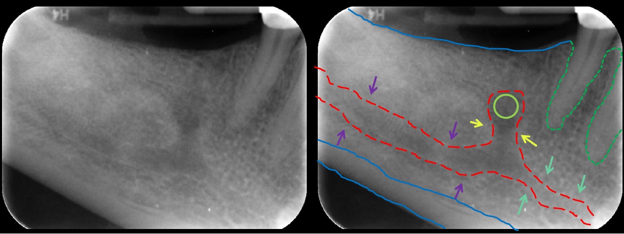
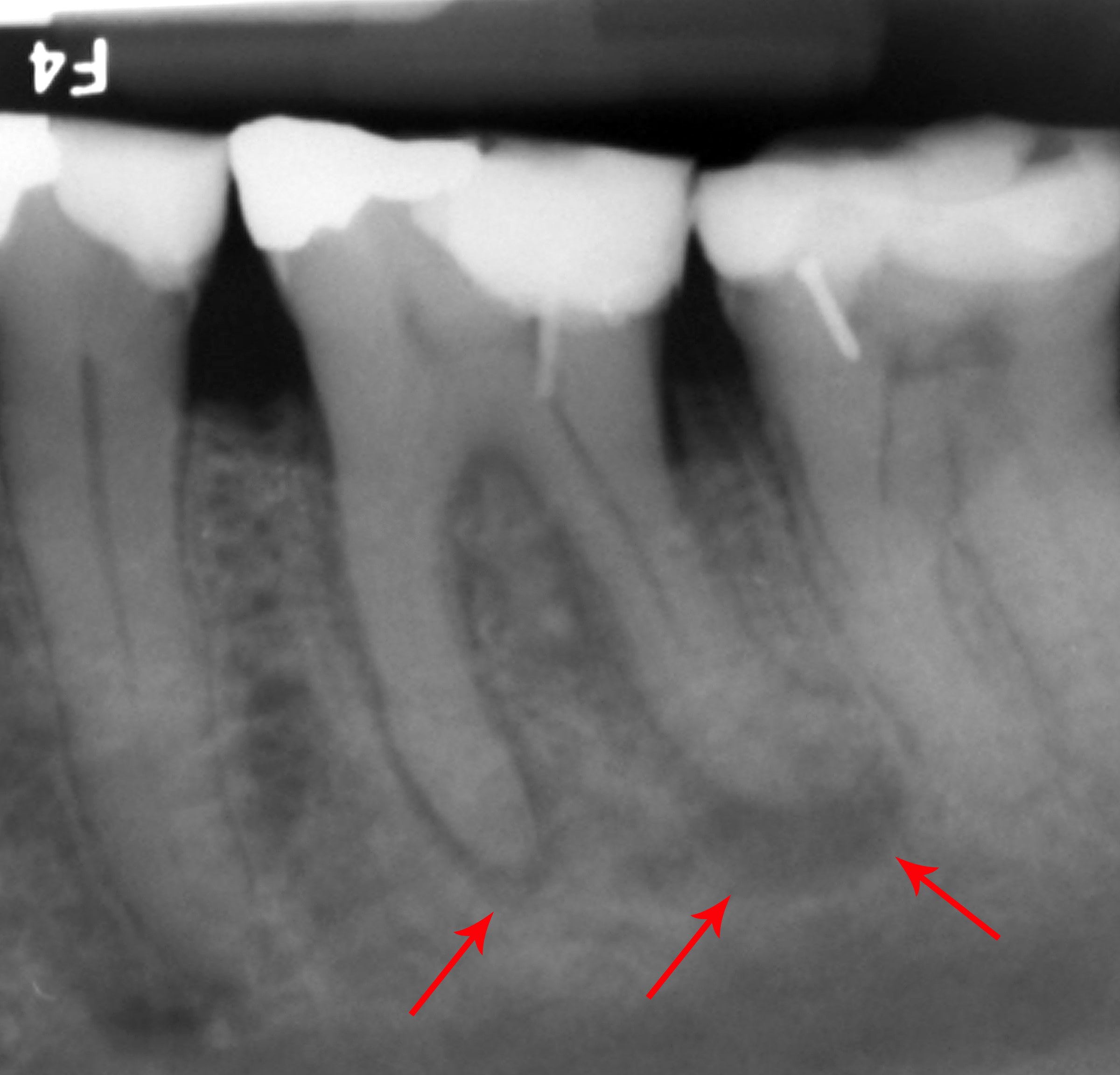
In human anatomy, the mandibular canal is a canal within the mandible that contains the inferior alveolar nerve, inferior alveolar artery, and inferior alveolar vein. It runs obliquely downward and forward in the ramus, and then horizontally forward in the body, where it is placed under the alveoli and communicates with them by small openings. On arriving at the incisor teeth, it turns back to communicate with the mental foramen, giving off a small canal known as the mandibular incisive canal, which run to the cavities containing the incisor teeth. It carries branches of the inferior alveolar nerve and artery. The mandibular canal is continuous with two foramina: the mental foramen which opens in the mental region of the mandible and carried the distal fibres of the inferior alveolar nerve as the mental nerve; and the mandibular foramen on medial aspect of ramus, into which the mandibular nerve enters to become the inferior alveolar nerve. The mandibular canal often runs cl ... [...More Info...] [...Related Items...] OR: [Wikipedia] [Google] [Baidu] |
Mandibular Incisive Canal Highlighted
In jawed vertebrates, the mandible (from the Latin ''mandibula'', 'for chewing'), lower jaw, or jawbone is a bone that makes up the lowerand typically more mobilecomponent of the mouth (the upper jaw being known as the maxilla). The jawbone is the skull's only movable, posable bone, sharing joints with the cranium's temporal bones. The mandible hosts the lower teeth (their depth delineated by the alveolar process). Many muscles attach to the bone, which also hosts nerves (some connecting to the teeth) and blood vessels. Amongst other functions, the jawbone is essential for chewing food. Owing to the Neolithic advent of agriculture (), human jaws evolved to be smaller. Although it is the strongest bone of the facial skeleton, the mandible tends to deform in old age; it is also subject to fracturing. Surgery allows for the removal of jawbone fragments (or its entirety) as well as regenerative methods. Additionally, the bone is of great forensic significance. Structure In h ... [...More Info...] [...Related Items...] OR: [Wikipedia] [Google] [Baidu] |
Foramen
In anatomy and osteology, a foramen (; : foramina, or foramens ; ) is an opening or enclosed gap within the dense connective tissue (bones and deep fasciae) of extant and extinct amniote animals, typically to allow passage of nerves, artery, arteries, veins or other soft tissue structures (e.g. muscle tendon) from one body compartment to another. Skull The skulls of vertebrates have foramina through which nerves, arteries, veins, and other structures pass. The human skull has many foramina, collectively referred to as the cranial foramina. Spine Within the vertebral column (spine) of vertebrates, including the Human vertebral column, human spine, each bone has an opening at both its top and bottom to allow nerves, arteries, veins, etc. to pass through. Other * Apical foramen, the hole at the tip of the root of a tooth * Foramen ovale (heart), a hole between the venous and arterial sides of the fetal heart * Vertebra#Cervical vertebrae, Transverse foramen, one of a pair ... [...More Info...] [...Related Items...] OR: [Wikipedia] [Google] [Baidu] |
Molar (tooth)
The molars or molar teeth are large, flat teeth at the back of the mouth. They are more developed in mammals. They are used primarily to grind food during chewing. The name ''molar'' derives from Latin, ''molaris dens'', meaning "millstone tooth", from ''mola'', millstone and ''dens'', tooth. Molars show a great deal of diversity in size and shape across the mammal groups. The third molar of humans is sometimes vestigial. Human anatomy In humans, the molar teeth have either four or five cusps. Adult humans have 12 molars, in four groups of three at the back of the mouth. The third, rearmost molar in each group is called a wisdom tooth. It is the last tooth to appear, breaking through the front of the gum at about the age of 20, although this varies among individuals and populations, and in many cases the tooth is missing. The human mouth contains upper (maxillary) and lower (mandibular) molars. They are: maxillary first molar, maxillary second molar, maxillary third mol ... [...More Info...] [...Related Items...] OR: [Wikipedia] [Google] [Baidu] |
Ramus Of The Mandible
In jawed vertebrates, the mandible (from the Latin ''mandibula'', 'for chewing'), lower jaw, or jawbone is a bone that makes up the lowerand typically more mobilecomponent of the mouth (the upper jaw being known as the maxilla). The jawbone is the skull's only movable, posable bone, sharing joints with the cranium's temporal bones. The mandible hosts the lower teeth (their depth delineated by the alveolar process). Many muscles attach to the bone, which also hosts nerves (some connecting to the teeth) and blood vessels. Amongst other functions, the jawbone is essential for chewing food. Owing to the Neolithic advent of agriculture (), human jaws evolved to be smaller. Although it is the strongest bone of the facial skeleton, the mandible tends to deform in old age; it is also subject to fracturing. Surgery allows for the removal of jawbone fragments (or its entirety) as well as regenerative methods. Additionally, the bone is of great forensic significance. Structure ... [...More Info...] [...Related Items...] OR: [Wikipedia] [Google] [Baidu] |
Root Canal Treatment
Root canal treatment (also known as endodontic therapy, endodontic treatment, or root canal therapy) is a treatment sequence for the infected pulp of a tooth that is intended to result in the elimination of infection and the protection of the decontaminated tooth from future microbial invasion. It is generally done when the cavity is too big for a normal filling. Root canals, and their associated pulp chamber, are the physical hollows within a tooth that are naturally inhabited by nerve tissue, blood vessels and other cellular entities. Endodontic therapy involves the removal of these structures, disinfection and the subsequent shaping, cleaning, and decontamination of the hollows with small files and irrigating solutions, and the obturation (filling) of the decontaminated canals. Filling of the cleaned and decontaminated canals is done with an inert filling such as gutta-percha and typically a zinc oxide eugenol-based cement. Epoxy resin is employed to bind gutta-percha i ... [...More Info...] [...Related Items...] OR: [Wikipedia] [Google] [Baidu] |
Dental Extraction
A dental extraction (also referred to as tooth extraction, exodontia, exodontics, or informally, tooth pulling) is the removal of teeth from the dental alveolus (socket) in the alveolar bone. Extractions are performed for a wide variety of reasons, but most commonly to remove teeth which have become unrestorable through tooth decay, periodontal disease, or dental trauma, especially when they are associated with toothache. Sometimes impacted wisdom teeth (wisdom teeth that are stuck and unable to grow normally into the mouth) cause recurrent infections of the gum ( pericoronitis), and may be removed when other conservative treatments have failed (cleaning, antibiotics and operculectomy). In orthodontics, if the teeth are crowded, healthy teeth may be extracted (often bicuspids) to create space so the rest of the teeth can be straightened. Procedure Extractions could be categorized into non-surgical (simple) and surgical, depending on the type of tooth to be removed and o ... [...More Info...] [...Related Items...] OR: [Wikipedia] [Google] [Baidu] |
Wisdom Tooth
The third molar, commonly called wisdom tooth, is the most posterior of the three molars in each quadrant of the human dentition. The age at which wisdom teeth come through ( erupt) is variable, but this generally occurs between late teens and early twenties. Most adults have four wisdom teeth, one in each of the four quadrants, but it is possible to have none, fewer, or more, in which case the extras are called supernumerary teeth. Wisdom teeth may become stuck ( impacted) and not erupt fully, if there is not enough space for them to come through normally. Impacted wisdom teeth are still sometimes removed for orthodontic treatment, believing that they move the other teeth and cause crowding, though this is disputed. Impacted wisdom teeth may suffer from tooth decay if oral hygiene becomes more difficult. Wisdom teeth which are partially erupted through the gum may also cause inflammation and infection in the surrounding gum tissues, termed pericoronitis. More conservativ ... [...More Info...] [...Related Items...] OR: [Wikipedia] [Google] [Baidu] |
Mandibular Nerve
In neuroanatomy, the mandibular nerve (V) is the largest of the three divisions of the trigeminal nerve, the fifth Cranial nerves, cranial nerve (CN V). Unlike the other divisions of the trigeminal nerve (ophthalmic nerve, maxillary nerve) which contain only Afferent nerve fiber, afferent fibers, the mandibular nerve contains both afferent and Efferent nerve fiber, efferent fibers. These nerve fibers innervate structures of the lower jaw and face, such as the tongue, lower lip, and chin. The mandibular nerve also innervates the muscles of mastication. Structure Course The large sensory root of mandibular nerve emerges from the lateral part of the trigeminal ganglion and exits the cranial cavity through the Foramen ovale (skull), foramen ovale. The motor root (Latin: ''radix motoria'' s. ''portio minor''), the small motor root of the trigeminal nerve, passes under the trigeminal ganglion and through the Foramen ovale (skull), foramen ovale to unite with the sensory root just out ... [...More Info...] [...Related Items...] OR: [Wikipedia] [Google] [Baidu] |
Mandibular Foramen
The mandibular foramen is an opening on the internal surface of the ramus of the mandible. It allows for divisions of the mandibular nerve and blood vessels to pass through. Structure The mandibular foramen is an opening on the internal surface of the ramus of the mandible. It allows for divisions of the mandibular nerve and blood vessels to pass through. Variation There are two distinct anatomies to its rim. * In the common form the rim is V-shaped, with a groove separating the anterior and posterior parts. * In the horizontal-oval form there is no groove, and the rim is horizontally oriented and oval in shape, the anterior and posterior parts connected. Rarely, a bifid inferior alveolar nerve may be present, in which case a second mandibular foramen, more inferiorly placed, exists and can be detected by noting a doubled mandibular canal on a radiograph. Function The mandibular nerve is one of three branches of the trigeminal nerve, and the only one having motor innervat ... [...More Info...] [...Related Items...] OR: [Wikipedia] [Google] [Baidu] |
Mental Nerve
The mental nerve is a sensory nerve of the face. It is a branch of the posterior trunk of the inferior alveolar nerve, itself a branch of the mandibular nerve (CN V3), itself a branch of the trigeminal nerve (CN V). It provides sensation to the front of the chin and the lower lip, as well as the gums of the anterior mandibular (lower) teeth. It can be blocked with local anaesthesia for procedures on the chin, lower lip, and mucous membrane of the inner cheek. Problems with the nerve cause chin numbness. Structure The mental nerve is a branch of the posterior trunk of the inferior alveolar nerve. This is a branch of the mandibular nerve (CN V3), itself a branch of the trigeminal nerve (CN V). It emerges from the mental foramen in the mandible. It divides into three branches beneath the depressor anguli oris muscle. One branch descends to the skin of the chin. Two branches ascend to the skin and mucous membrane of the lower lip. These branches communicate freely with the facial n ... [...More Info...] [...Related Items...] OR: [Wikipedia] [Google] [Baidu] |
Dennis Tarnow
Dennis Perry Tarnow (born May 28, 1946) is an American dentist specializing in periodontics, prosthodontics and implant dentistry and is known for his mark on dental implant research and education. He is currently director of implant dentistry at Columbia University College of Dental Medicine and former chairman of the department of periodontics and implant dentistry at New York University College of Dentistry. He is a sought after speaker on the subject of implant dentistry.Ohio Dental Association - Dr. Dennis Tarnow to present full-day program at the 142nd Annual Session April 1, 2008 Early years and education Tarnow was born and raised in |


