|
Dilaceration
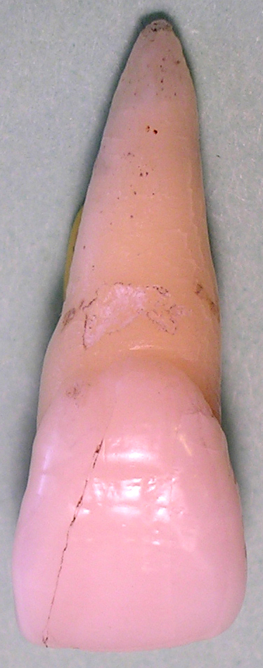
Dilaceration is a developmental disturbance in shape of teeth. It refers to an angulation, or a sharp bend or curve, in the root or crown of a formed tooth. This disturbance is more likely to affect the maxillary incisors and occurs in permanent dentition. Although this may seem more of an aesthetics issue, an impacted maxillary incisor will cause issues related to occlusion, phonetics, mastication, and psychology on young patients. Description The condition is thought to be due to trauma or possibly a delay in tooth eruption relative to bone remodeling gradients during the period in which tooth is forming. Standerwick RG. A possible etiology for the dilaceration and flexion of permanent tooth roots relative to bone remodeling gradients in alveolar bone. Dent Hypotheses [...More Info...] [...Related Items...] OR: [Wikipedia] [Google] [Baidu] |
Turner's Hypoplasia
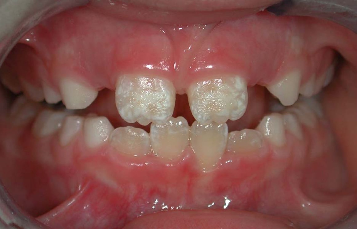
Enamel hypoplasia is a defect of the teeth in which the enamel is deficient in quantity, caused by defective enamel matrix formation during enamel development, as a result of inherited and acquired systemic condition(s). It can be identified as missing tooth structure and may manifest as pits or grooves in the crown of the affected teeth, and in extreme cases, some portions of the crown of the tooth may have no enamel, exposing the dentin. It may be generalized across the dentition or localized to a few teeth. Defects are categorized by shape or location. Common categories are pit-form, plane-form, linear-form, and localised enamel hypoplasia. Hypoplastic lesions are found in areas of the teeth where the enamel was being actively formed during a systemic or local disturbance. Since the formation of enamel extends over a long period of time, defects may be confined to one well-defined area of the affected teeth. Knowledge of chronological development of deciduous and permanen ... [...More Info...] [...Related Items...] OR: [Wikipedia] [Google] [Baidu] |
Maxillary Central Incisor
The maxillary central incisor is a human tooth in the front upper jaw, or maxilla, and is usually the most visible of all teeth in the mouth. It is located mesial (closer to the midline of the face) to the maxillary lateral incisor. As with all incisors, their function is for shearing or cutting food during mastication (chewing). There is typically a single cusp on each tooth, called an incisal ridge or incisal edge. Formation of these teeth begins at 14 weeks in utero for the deciduous (baby) set and 3–4 months of age for the permanent set. There are some minor differences between the deciduous maxillary central incisor and that of the permanent maxillary central incisor. The deciduous tooth appears in the mouth at 8–12 months of age and shed at 6–7 years, and is replaced by the permanent tooth around 7–8 years of age. The permanent tooth is larger and is longer than it is wide. The maxillary central incisors contact each other at the midline of the face. The mand ... [...More Info...] [...Related Items...] OR: [Wikipedia] [Google] [Baidu] |
Endodontics
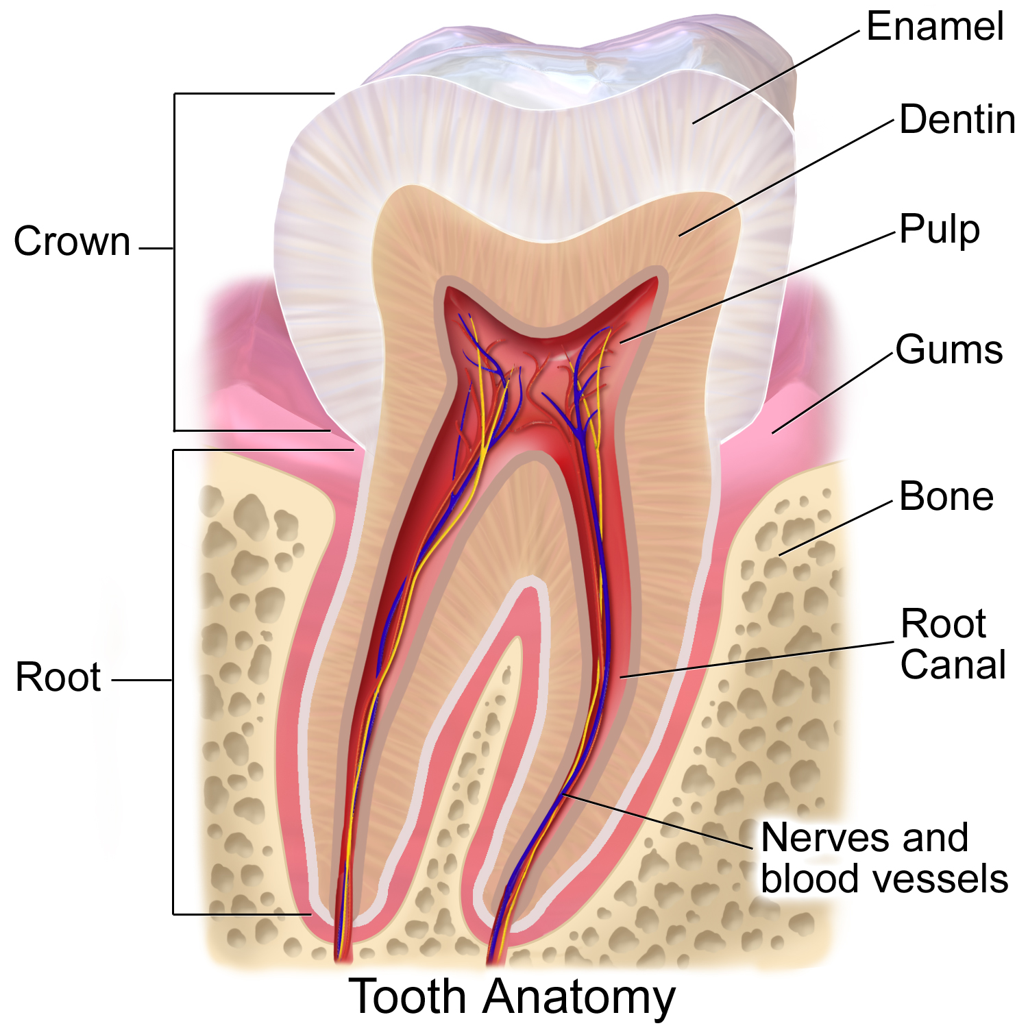
Endodontics (from the Greek roots ''endo-'' "inside" and ''odont-'' "tooth") is the dental specialty concerned with the study and treatment of the dental pulp. Overview Endodontics encompasses the study (practice) of the basic and clinical sciences of normal dental pulp, the etiology, diagnosis, prevention, and treatment of diseases and injuries of the dental pulp along with associated periradicular conditions. In clinical terms, endodontics involves either preserving part, or all of the dental pulp in health, or removing all of the pulp in irreversible disease. This includes teeth with irreversibly inflamed and infected pulpal tissue. Not only does endodontics involve treatment when a dental pulp is present, but also includes preserving teeth which have failed to respond to non-surgical endodontic treatment, or for teeth that have developed new lesions, e.g., when root canal re-treatment is required, or periradicular surgery. Endodontic treatment is one of the most comm ... [...More Info...] [...Related Items...] OR: [Wikipedia] [Google] [Baidu] |
Smith–Magenis Syndrome
Smith–Magenis Syndrome (SMS), also known as 17p- syndrome, is a microdeletion syndrome characterized by an abnormality in the short (p) arm of chromosome 17. It has features including intellectual disability, facial abnormalities, difficulty sleeping, and numerous behavioral problems such as self-harm. Smith–Magenis syndrome affects an estimated between 1 in 15,000 to 1 in 25,000 individuals. Signs and symptoms Facial features of children with Smith–Magenis syndrome include a broad and square face, deep-set eyes, large cheeks, and a prominent jaw, as well as a flat nose bridge (in the young child; as the child ages it becomes more ski-jump shaped). Eyes tend to be deep-set, close together and upwards-slanted. Eyebrows are heavy with lateral extension. The mouth is the most noticeable feature; both upper and lower lips are full, and the mouth is wide. The mouth curves downwards and the upper lip curves outwards, due to a fleshy philtrum. These facial features become more no ... [...More Info...] [...Related Items...] OR: [Wikipedia] [Google] [Baidu] |
Tooth Enamel
Tooth enamel is one of the four major Tissue (biology), tissues that make up the tooth in humans and many other animals, including some species of fish. It makes up the normally visible part of the tooth, covering the Crown (tooth), crown. The other major tissues are dentin, cementum, and Pulp (tooth), dental pulp. It is a very hard, white to off-white, highly mineralised substance that acts as a barrier to protect the tooth but can become susceptible to degradation, especially by acids from food and drink. Calcium hardens the tooth enamel. In rare circumstances enamel fails to form, leaving the underlying dentin exposed on the surface. Features Enamel is the hardest substance in the human body and contains the highest percentage of minerals (at 96%),Ross ''et al.'', p. 485 with water and organic material composing the rest.Ten Cate's Oral Histology, Nancy, Elsevier, pp. 70–94 The primary mineral is hydroxyapatite, which is a crystalline calcium phosphate. Enamel is formed o ... [...More Info...] [...Related Items...] OR: [Wikipedia] [Google] [Baidu] |
Dentition
Dentition pertains to the development of teeth and their arrangement in the mouth. In particular, it is the characteristic arrangement, kind, and number of teeth in a given species at a given age. That is, the number, type, and morpho-physiology (that is, the relationship between the shape and form of the tooth in question and its inferred function) of the teeth of an animal. Animals whose teeth are all of the same type, such as most non-mammalian vertebrates, are said to have '' homodont'' dentition, whereas those whose teeth differ morphologically are said to have '' heterodont'' dentition. The dentition of animals with two successions of teeth (deciduous, permanent) is referred to as ''diphyodont'', while the dentition of animals with only one set of teeth throughout life is ''monophyodont''. The dentition of animals in which the teeth are continuously discarded and replaced throughout life is termed ''polyphyodont''. The dentition of animals in which the teeth are set in so ... [...More Info...] [...Related Items...] OR: [Wikipedia] [Google] [Baidu] |
Root Canal Treatment
Root canal treatment (also known as endodontic therapy, endodontic treatment, or root canal therapy) is a treatment sequence for the infected pulp of a tooth which is intended to result in the elimination of infection and the protection of the decontaminated tooth from future microbial invasion. Root canals, and their associated pulp chamber, are the physical hollows within a tooth that are naturally inhabited by nerve tissue, blood vessels and other cellular entities. Together, these items constitute the dental pulp. Endodontic therapy involves the ''removal'' of these structures, disinfection and the subsequent shaping, cleaning, and decontamination of the hollows with small files and irrigating solutions, and the ''obturation'' (filling) of the decontaminated canals. Filling of the cleaned and decontaminated canals is done with an inert filling such as gutta-percha and typically a zinc oxide eugenol-based cement. Epoxy resin is employed to bind gutta-percha in some root ... [...More Info...] [...Related Items...] OR: [Wikipedia] [Google] [Baidu] |
Permanent Teeth
Permanent teeth or adult teeth are the second set of teeth formed in diphyodont mammals. In humans and old world simians, there are thirty-two permanent teeth, consisting of six maxillary and six mandibular molars, four maxillary and four mandibular premolars, two maxillary and two mandibular canines, four maxillary and four mandibular incisors. Timeline The first permanent tooth usually appears in the mouth at around six years of age, and the mouth will then be in a transition time with both primary (or deciduous dentition) teeth and permanent teeth during the mixed dentition period until the last primary tooth is lost or shed. The first of the permanent teeth to erupt are the permanent first molars, right behind the last 'milk' molars of the primary dentition. These first permanent molars are important for the correct development of a permanent dentition. Up to thirteen years of age, 28 of the 32 permanent teeth will appear. The full permanent dentition is completed much late ... [...More Info...] [...Related Items...] OR: [Wikipedia] [Google] [Baidu] |
Neoplasm
A neoplasm () is a type of abnormal and excessive growth of tissue. The process that occurs to form or produce a neoplasm is called neoplasia. The growth of a neoplasm is uncoordinated with that of the normal surrounding tissue, and persists in growing abnormally, even if the original trigger is removed. This abnormal growth usually forms a mass, when it may be called a tumor. ICD-10 classifies neoplasms into four main groups: benign neoplasms, in situ neoplasms, malignant neoplasms, and neoplasms of uncertain or unknown behavior. Malignant neoplasms are also simply known as cancers and are the focus of oncology. Prior to the abnormal growth of tissue, as neoplasia, cells often undergo an abnormal pattern of growth, such as metaplasia or dysplasia. However, metaplasia or dysplasia does not always progress to neoplasia and can occur in other conditions as well. The word is from Ancient Greek 'new' and 'formation, creation'. Types A neoplasm can be benign, potentially ma ... [...More Info...] [...Related Items...] OR: [Wikipedia] [Google] [Baidu] |
Cyst
A cyst is a closed sac, having a distinct envelope and cell division, division compared with the nearby Biological tissue, tissue. Hence, it is a cluster of Cell (biology), cells that have grouped together to form a sac (like the manner in which water molecules group together to form a bubble); however, the distinguishing aspect of a cyst is that the cells forming the "shell" of such a sac are distinctly abnormal (in both appearance and behaviour) when compared with all surrounding cells for that given location. A cyst may contain air, fluids, or semi-solid material. A collection of pus is called an abscess, not a cyst. Once formed, a cyst may resolve on its own. When a cyst fails to resolve, it may need to be removed surgically, but that would depend upon its type and location. Cancer-related cysts are formed as a defense mechanism for the body following the development of mutations that lead to an uncontrolled cellular division. Once that mutation has occurred, the affected cell ... [...More Info...] [...Related Items...] OR: [Wikipedia] [Google] [Baidu] |
Axenfeld–Rieger Syndrome
Axenfeld–Rieger syndrome is a rare autosomal dominance (genetics), dominant disorder, which affects the development of the teeth, eyes, and abdominal region. Axenfeld–Rieger syndrome is part of the so-called iridocorneal or anterior segment dysgenesis syndromes, which were formerly known as anterior segment cleavage syndromes, anterior chamber segmentation syndromes or mesodermal dysgenesis. Although the exact classification of this set of signs and symptoms is somewhat confusing in current scientific literature, most authors agree with the classification cited here. Axenfeld Anomaly is known as the development of a posterior embryotoxon, associated with strands of the iris adhered to a Schwalbe line that has been displaced anteriorly, which when added to glaucoma is called Axenfeld Syndrome. Rieger's Anomaly is defined by a universe of congenital anomalies of the iris, such as Iris hypoplasia with glaucoma, iris hypoplasia, corectopia or polycoria.Rieger H. Contributions to th ... [...More Info...] [...Related Items...] OR: [Wikipedia] [Google] [Baidu] |


