|
Cancer-associated Fibroblast
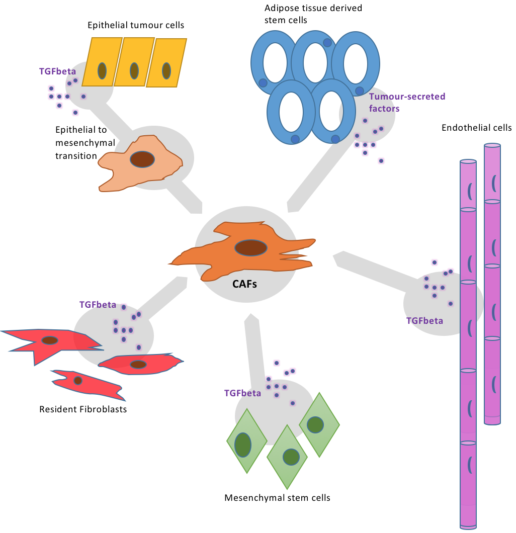
A cancer-associated fibroblast (CAF) (also known as tumour-associated fibroblast; carcinogenic- associated fibroblast; activated fibroblast) is a cell type within the tumor microenvironment that promotes tumorigenic features by initiating the remodelling of the extracellular matrix or by secreting cytokines. CAFs are a complex and abundant cell type within the tumour microenvironment; the number cannot decrease, as they are unable to undergo apoptosis. CAFs have been found to be abundant in a tumour stroma. Myofibroblasts and fibroblasts make up CAFs. The functions of these CAFs have been known to stimulate angiogenesis, supporting the formation of tumours and thus proliferation of cancer cell and metastasis. Cancer cells are usually also drug resistant, which is contributed by CAFs. As such, this interaction is being studied for potential anti-cancer therapy. Normal fibroblasts aid in the production of components of the extracellular matrix such as collagens, fibres, glycosaminogl ... [...More Info...] [...Related Items...] OR: [Wikipedia] [Google] [Baidu] |
Fibroblast
A fibroblast is a type of cell (biology), biological cell that synthesizes the extracellular matrix and collagen, produces the structural framework (Stroma (tissue), stroma) for animal Tissue (biology), tissues, and plays a critical role in wound healing. Fibroblasts are the most common cells of connective tissue in animals. Structure Fibroblasts have a branched cytoplasm surrounding an elliptical, speckled cell nucleus, nucleus having two or more nucleoli. Active fibroblasts can be recognized by their abundant Endoplasmic reticulum#Rough endoplasmic reticulum, rough endoplasmic reticulum. Inactive fibroblasts (called fibrocytes) are smaller, spindle-shaped, and have a reduced amount of rough endoplasmic reticulum. Although disjointed and scattered when they have to cover a large space, fibroblasts, when crowded, often locally align in parallel clusters. Unlike the epithelial cells lining the body structures, fibroblasts do not form flat monolayers and are not restricted by a ... [...More Info...] [...Related Items...] OR: [Wikipedia] [Google] [Baidu] |
Fibroblast Activation Protein, Alpha
Fibroblast activation protein alpha (FAP-alpha) also known as prolyl endopeptidase FAP is an enzyme that in humans is encoded by the FAP gene. Prolyl endopeptidase FAP is a 170 kDa membrane-bound gelatinase. It was independently identified as a surface glycoprotein recognized by the F19 monoclonal antibody in activated fibroblasts and a Surface Expressed Protease (seprase) in invasive melanoma cells. Structure and enzymatic activity FAP is a 760 amino acid long type II transmembrane glycoprotein. It contains a very short cytoplasmic N terminal part (6 amino acids), a transmembrane region (amino acids 7–26), and a large extracellular part with an alpha/beta-hydrolase domain and an eight-bladed beta-propeller domain.; A soluble form of FAP, which lacks the intracellular and transmembrane part, is present in blood plasma. FAP is a non-classical serine protease, which belongs to the S9B prolyl oligopeptidase subfamily. Other members of the S9B subfamily are DPPIV, DPP8 and DPP9 ... [...More Info...] [...Related Items...] OR: [Wikipedia] [Google] [Baidu] |
Connective Tissue Cells
Connective tissue is one of the four primary types of animal tissue, along with epithelial tissue, muscle tissue, and nervous tissue. It develops from the mesenchyme derived from the mesoderm the middle embryonic germ layer. Connective tissue is found in between other tissues everywhere in the body, including the nervous system. The three meninges, membranes that envelop the brain and spinal cord are composed of connective tissue. Most types of connective tissue consists of three main components: elastic and collagen fibers, ground substance, and cells. Blood, and lymph are classed as specialized fluid connective tissues that do not contain fiber. All are immersed in the body water. The cells of connective tissue include fibroblasts, adipocytes, macrophages, mast cells and leucocytes. The term "connective tissue" (in German, ''Bindegewebe'') was introduced in 1830 by Johannes Peter Müller. The tissue was already recognized as a distinct class in the 18th century. Types File:I ... [...More Info...] [...Related Items...] OR: [Wikipedia] [Google] [Baidu] |
STC1
Stanniocalcin-1 is a glycoprotein, a homologue of a hormone stanniocalcin, first discovered in bony fishes. In humans it is encoded by the ''STC1'' gene. Function This gene encodes a secreted, homodimeric glycoprotein that is expressed in a wide variety of tissues and may have autocrine or paracrine functions. The only known molecular function of human Stanniocalcin-1 to date is a SUMO E3 ubiquitin ligase activity in the SUMOylation cycle. However, STC1 interacts with many proteins in the cytoplasm, mitochondria, endoplasmatic reticulum, and in dot-like fashion in the cell nucleus. The N-terminal region of STC1 is the function region which is responsible to establish the interaction with its partners, including SUMO1. Low-resolution studies shows that STC1 is an anti-parallel homodimer in solution and the cysteine 202 is responsible for its dimerization. All the 5 disulfide bonds of human STC1 are conserved and have the same profile of fish STC. The gene contains a 5' UTR rich ... [...More Info...] [...Related Items...] OR: [Wikipedia] [Google] [Baidu] |
Asporin
Asporin is a protein that in humans is encoded by the ''ASPN'' gene. Function ASPN belongs to a family of leucine-rich repeat (LRR) proteins associated with the cartilage matrix. The name asporin reflects the unique aspartate-rich N terminus and the overall similarity to decorin Decorin is a protein that in humans is encoded by the ''DCN'' gene. Decorin is a proteoglycan that is on average 90 - 140 kilodaltons (kDa) in molecular weight. It belongs to the small leucine-rich proteoglycan (SLRP) family and consists of a ... (MIM 125255) (Lorenzo et al., 2001). References External links * Further reading * * * * * * * * * * * * {{gene-9-stub Extracellular matrix proteins LRR proteins Proteoglycans ... [...More Info...] [...Related Items...] OR: [Wikipedia] [Google] [Baidu] |
S100A4
Protein S100-A4 (S100A4) is a protein that in humans is encoded by the ''S100A4'' gene. Function The protein encoded by this gene is a member of the S100 family of proteins containing 2 EF-hand calcium-binding motifs. S100 proteins are localized in the cytoplasm and/or nucleus of a wide range of cells, and involved in the regulation of a number of cellular processes such as cell cycle progression and differentiation. S100 genes include at least 13 members which are located as a cluster on chromosome 1q21. This protein may function in motility, invasion, and tubulin polymerization. Chromosomal rearrangements and altered expression of this gene have been implicated in tumor metastasis. Multiple alternatively spliced variants, encoding the same protein, have been identified. Interactions S100A4 has been shown to interact with S100 calcium binding protein A1. Therapeutic targeting for cancer S100A4, a member of the S100 calcium-binding protein family secreted by tumor and stro ... [...More Info...] [...Related Items...] OR: [Wikipedia] [Google] [Baidu] |
PDGFRB
Platelet-derived growth factor receptor beta is a protein that in humans is encoded by the ''PDGFRB'' gene. Mutations in PDGFRB are mainly associated with the clonal eosinophilia class of malignancies. Gene The ''PDGFRB'' gene is located on human chromosome 5 at position q32 (designated as 5q32) and contains 25 exons. The gene is flanked by the genes for granulocyte-macrophage colony-stimulating factor and Colony stimulating factor 1 receptor (also termed macrophage-colony stimulating factor receptor), all three of which may be lost together by a single deletional mutation thereby causing development of the 5q-syndrome. Other genetic abnormalities in ''PDGFRB'' lead to various forms of potentially malignant bone marrow disorders: small deletions in and chromosome translocations causing fusions between ''PDGFRB'' and any one of at least 30 genes can cause Myeloproliferative neoplasms that commonly involve eosinophilia, eosinophil-induced organ injury, and possible progressio ... [...More Info...] [...Related Items...] OR: [Wikipedia] [Google] [Baidu] |
PDGFRA
PDGFRA, i.e. platelet-derived growth factor receptor A, also termed PDGFRα, i.e. platelet-derived growth factor receptor α, or CD140a i.e. Cluster of Differentiation 140a, is a receptor located on the surface of a wide range of cell types. This receptor binds to certain isoforms of platelet-derived growth factors (PDGFs) and thereby becomes active in stimulating cell signaling pathways that elicit responses such as cellular growth and differentiation. The receptor is critical for the development of certain tissues and organs during embryogenesis and for the maintenance of these tissues and organs, particularly hematologic tissues, throughout life. Mutations in the gene which codes for PDGFRA, i.e. the ''PDGFRA'' gene, are associated with an array of clinically significant neoplasms, notably ones of the clonal hypereosinophilia class of malignancies, as well as gastrointestinal stromal tumors (GISTs). Overall structure This gene encodes a typical receptor tyrosine kinase, which ... [...More Info...] [...Related Items...] OR: [Wikipedia] [Google] [Baidu] |
Platelet Derived Growth Factor Receptor
Platelet-derived growth factor receptors (PDGF-R) are cell surface tyrosine kinase receptors for members of the platelet-derived growth factor (PDGF) family. PDGF subunits -A and -B are important factors regulating cell proliferation, cellular differentiation, cell growth, development and many diseases including cancer. There are two forms of the PDGF-R, alpha and beta each encoded by a different gene. Depending on which growth factor is bound, PDGF-R homo- or heterodimerizes. Mechanism of action The PDGF family consists of PDGF-A, -B, -C and -D, which form either homo- or heterodimers (PDGF-AA, -AB, -BB, -CC, -DD). The four PDGFs are inactive in their monomeric forms. The PDGFs bind to the protein tyrosine kinase receptors PDGF receptor-α and -β. These two receptor isoforms dimerize upon binding the PDGF dimer, leading to three possible receptor combinations, namely -αα, -ββ and -αβ. The extracellular region of the receptor consists of five immunoglobulin-like domain ... [...More Info...] [...Related Items...] OR: [Wikipedia] [Google] [Baidu] |

.jpg)
