|
Cardiopulmonary Bypass
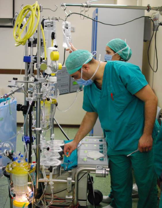
Cardiopulmonary bypass (CPB) or heart-lung machine, also called the pump or CPB pump, is a machine that temporarily takes over the function of the heart and lungs during open-heart surgery by maintaining the circulation of blood and oxygen throughout the body. As such it is an Extracorporeal, extracorporeal device. CPB is operated by a perfusionist. The machine mechanically circulates and oxygenates blood throughout the patient's body while bypassing the heart and lungs allowing the surgeon to work in a bloodless surgical field. Uses CPB is commonly used in operations or surgical procedures involving the heart. The technique allows the surgical team to oxygenate and circulate the patient's blood, thus allowing the surgeon to operate safely on the heart. In many operations, such as Coronary artery bypass surgery, coronary artery bypass grafting (CABG), the heart is Cardioplegia, arrested, due to the degree of the difficulty of operating on a beating heart. Operations requiring t ... [...More Info...] [...Related Items...] OR: [Wikipedia] [Google] [Baidu] |
Heart Lung Machine
Cardiopulmonary bypass (CPB) or heart-lung machine, also called the pump or CPB pump, is a machine that temporarily takes over the function of the heart and lungs during open-heart surgery by maintaining the circulation of blood and oxygen throughout the body. As such it is an extracorporeal device. CPB is operated by a perfusionist. The machine mechanically circulates and oxygenates blood throughout the patient's body while bypassing the heart and lungs allowing the surgeon to work in a bloodless surgical field. Uses CPB is commonly used in operations or surgical procedures involving the heart. The technique allows the surgical team to oxygenate and circulate the patient's blood, thus allowing the surgeon to operate safely on the heart. In many operations, such as coronary artery bypass grafting (CABG), the heart is arrested, due to the degree of the difficulty of operating on a beating heart. Operations requiring the opening of the chambers of the heart, for example mitra ... [...More Info...] [...Related Items...] OR: [Wikipedia] [Google] [Baidu] |
Coronary Artery Bypass Surgery
Coronary artery bypass surgery, also known as coronary artery bypass graft (CABG, pronounced "cabbage"), is a surgical procedure to treat coronary artery disease (CAD), the buildup of plaques in the arteries of the heart. It can relieve chest pain caused by CAD, slow the progression of CAD, and increase life expectancy. It aims to bypass narrowings in heart arteries by using arteries or veins harvested from other parts of the body, thus restoring adequate blood supply to the previously ischemic (deprived of blood) heart. There are two main approaches. The first uses a cardiopulmonary bypass machine, a machine which takes over the functions of the heart and lungs during surgery by circulating blood and oxygen. With the heart in cardioplegic arrest, harvested arteries and veins are used to connect across problematic regions—a construction known as surgical anastomosis. In the second approach, called the off-pump coronary artery bypass (OPCAB), these anastomoses are cons ... [...More Info...] [...Related Items...] OR: [Wikipedia] [Google] [Baidu] |
Tetralogy Of Fallot
Tetralogy of Fallot (TOF), formerly known as Steno-Fallot tetralogy, is a congenital heart defect characterized by four specific cardiac defects. Classically, the four defects are: * Pulmonary stenosis, which is narrowing of the exit from the right ventricle; * A ventricular septal defect, which is a hole allowing blood to flow between the two ventricles; * Right ventricular hypertrophy, which is thickening of the right ventricular muscle; and * an overriding aorta, which is where the aorta expands to allow blood from both ventricles to enter. At birth, children may be asymptomatic or present with many severe symptoms. Later in infancy, there are typically episodes of bluish colour to the skin due to a lack of sufficient oxygenation, known as cyanosis. When affected babies cry or have a bowel movement, they may undergo a "tet spell" where they turn cyanotic, have difficulty breathing, become limp, and occasionally lose consciousness. Other symptoms may include a heart mur ... [...More Info...] [...Related Items...] OR: [Wikipedia] [Google] [Baidu] |
Congenital Heart Defect
A congenital heart defect (CHD), also known as a congenital heart anomaly, congenital cardiovascular malformation, and congenital heart disease, is a defect in the structure of the heart or great vessels that is present at birth. A congenital heart defect is classed as a cardiovascular disease. Signs and symptoms depend on the specific type of defect. Symptoms can vary from none to life-threatening. When present, symptoms are variable and may include rapid breathing, bluish skin (cyanosis), poor weight gain, and feeling tired. CHD does not cause chest pain. Most congenital heart defects are not associated with other diseases. A complication of CHD is heart failure. Congenital heart defects are the most common birth defect. In 2015, they were present in 48.9 million people globally. They affect between 4 and 75 per 1,000 live births, depending upon how they are diagnosed. In about 6 to 19 per 1,000 they cause a moderate to severe degree of problems. Congenital heart defects are t ... [...More Info...] [...Related Items...] OR: [Wikipedia] [Google] [Baidu] |
Atrioventricular Septal Defect
Atrioventricular septal defect (AVSD) or atrioventricular canal defect (AVCD), also known as "common atrioventricular canal" or "endocardial cushion defect" (ECD), is characterized by a deficiency of the atrioventricular septum of the heart that creates connections between all four of its chambers. It is a very specific combination of 3 defects: 1) Atrial septal defect, Atrial Septal Defect (ASD), a hole in the wall between the right and left atria; 2) Ventricular septal defect, Ventricular Septal Defect (VSD), a hole in the wall between the right and left ventricles; and 3) Abnormalities of the mitral and/or tricuspid valves. AVCD is caused by an abnormal or inadequate fusion of the superior and inferior endocardial cushions with the mid portion of the atrial septum and the muscular portion of the ventricular septum. Unlike some heart defects, the condition will not resolve over time and most infants must undergo open heart surgery. The surgery to correct this defect is usual ... [...More Info...] [...Related Items...] OR: [Wikipedia] [Google] [Baidu] |
Ventricular Septal Defect
A ventricular septal defect (VSD) is a defect in the ventricular septum, the wall dividing the left and right ventricles of the heart. It's a common heart problem present at birth ( congenital heart defect). The extent of the opening may vary from pin size to complete absence of the ventricular septum, creating one common ventricle. The ventricular septum consists of an inferior muscular and superior membranous portion and is extensively innervated with conducting cardiomyocytes. The membranous portion, which is close to the atrioventricular node, is most commonly affected in adults and older children in the United States. It is also the type that will most commonly require surgical intervention, comprising over 80% of cases. Membranous ventricular septal defects are more common than muscular ventricular septal defects, and are the most common congenital cardiac anomaly. Signs and symptoms Ventricular septal defect is usually symptomless at birth. It usually manifests a fe ... [...More Info...] [...Related Items...] OR: [Wikipedia] [Google] [Baidu] |
Atrial Septal Defect
Atrial septal defect (ASD) is a congenital heart defect in which blood flows between the atrium (heart), atria (upper chambers) of the heart. Some flow is a normal condition both pre-birth and immediately post-birth via the Foramen ovale (heart), foramen ovale; however, when this does not naturally close after birth it is referred to as a patent (open) foramen ovale (PFO). It is common in patients with a congenital interatrial septum, atrial septal aneurysm (ASA). After PFO closure the atria normally are separated by a dividing wall, the interatrial septum. If this septum is defective or absent, then oxygen-rich blood can flow directly from the left side of the heart to mix with the oxygen-poor blood in the right side of the heart; or the opposite, depending on whether the left or right atrium has the higher blood pressure. In the absence of other heart defects, the left atrium has the higher pressure. This can lead to lower-than-normal oxygen levels in the arterial blood that su ... [...More Info...] [...Related Items...] OR: [Wikipedia] [Google] [Baidu] |
Septum
In biology, a septum (Latin language, Latin for ''something that encloses''; septa) is a wall, dividing a Body cavity, cavity or structure into smaller ones. A cavity or structure divided in this way may be referred to as septate. Examples Human anatomy * Interatrial septum, the wall of tissue that is a sectional part of the left and right atria of the heart * Interventricular septum, the wall separating the left and right ventricles of the heart * Lingual septum, a vertical layer of fibrous tissue that separates the halves of the tongue *Nasal septum: the cartilage wall separating the nostrils of the nose * Alveolar septum: the thin wall which separates the Pulmonary alveolus, alveoli from each other in the lungs * Orbital septum, a palpebral ligament in the upper and lower eyelids * Septum pellucidum or septum lucidum, a thin structure separating two fluid pockets in the brain * Uterine septum, a malformation of the uterus * Septum of the penis, Penile septum, a fibrous w ... [...More Info...] [...Related Items...] OR: [Wikipedia] [Google] [Baidu] |
Pulmonic Valve
The pulmonary valve (sometimes referred to as the pulmonic valve) is a valve of the heart that lies between the right ventricle and the pulmonary artery, and has three cusps. It is one of the four valves of the heart and one of the two semilunar valves, the other being the aortic valve. Similar to the aortic valve, the pulmonary valve opens in ventricular systole when the pressure in the right ventricle rises above the pressure in the pulmonary artery. At the end of ventricular systole, when the pressure in the right ventricle falls rapidly, the pressure in the pulmonary artery closes the pulmonary valve. The closure of the pulmonary valve contributes to the P2 component of the second heart sound (S2). Structure The pulmonary orifice lies nearly in the horizontal plane, and is situated at a superior level than the aortic orifice. Cusps File:Lunules of semilunar leaflets of pulmonary valve.png File:Anterior semilunar leaflet of pulmonary valve.png File:Right semilunar leafl ... [...More Info...] [...Related Items...] OR: [Wikipedia] [Google] [Baidu] |
Tricuspid Valve
The tricuspid valve, or right atrioventricular valve, is on the right dorsal side of the mammalian heart, at the superior portion of the right ventricle. The function of the valve is to allow blood to flow from the right atrium to the right ventricle during diastole, and to close to prevent backflow (Regurgitation (circulation), regurgitation) from the right ventricle into the right atrium during right ventricular contraction (systole). Structure The tricuspid valve usually has three Cusps of heart valves, cusps or leaflets, named the anterior, posterior, and septal cusps. Each leaflet is connected via chordae tendineae to the anterior, posterior, and septal papillary muscles of the right ventricle, respectively. Tricuspid valves may also occur with two or four leaflets; the number may change over a lifetime. Function The tricuspid valve functions as a one-way valve that closes during ventricular systole to prevent regurgitation of blood from the right ventricle back into th ... [...More Info...] [...Related Items...] OR: [Wikipedia] [Google] [Baidu] |
Mitral Valve
The mitral valve ( ), also known as the bicuspid valve or left atrioventricular valve, is one of the four heart valves. It has two Cusps of heart valves, cusps or flaps and lies between the atrium (heart), left atrium and the ventricle (heart), left ventricle of the heart. The heart valves are all one-way valves allowing blood flow in just one direction. The mitral valve and the tricuspid valve are known as the Heart valve#Atrioventricular valves, atrioventricular valves because they lie between the atria and the ventricles. In normal conditions, blood flows through an open mitral valve during diastole with contraction of the left atrium, and the mitral valve closes during systole with contraction of the left ventricle. The valve opens and closes because of pressure differences, opening when there is greater pressure in the left atrium than ventricle and closing when there is greater pressure in the left ventricle than atrium. In abnormal conditions, blood may flow backward thro ... [...More Info...] [...Related Items...] OR: [Wikipedia] [Google] [Baidu] |
Aortic Valve
The aortic valve is a valve in the heart of humans and most other animals, located between the left ventricle and the aorta. It is one of the four valves of the heart and one of the two semilunar valves, the other being the pulmonary valve. The aortic valve normally has three cusps or leaflets, although in 1–2% of the population it is found to congenitally have two leaflets. The aortic valve is the last structure in the heart the blood travels through before stopping the flow through the systemic circulation. Structure The aortic valve normally has three cusps however there is some discrepancy in their naming. They may be called the left coronary, right coronary and non-coronary cusp. Some sources also advocate they be named as a left, right and posterior cusp. Anatomists have traditionally named them the left posterior (origin of left coronary), anterior (origin of the right coronary) and right posterior. The three cusps, when the valve is closed, contain a sinus called ... [...More Info...] [...Related Items...] OR: [Wikipedia] [Google] [Baidu] |


