|
Volumetric Electron Microscopy
Volumetric Electron Microscopy (Volume EM) is an electron microscopy method used to generate 3D reconstructions of thick (>500 nm) samples. The initial role of electron microscopes in imaging two-dimensional slices (TEM) or a specimen surface (SEM with secondary electrons) has also increasingly expanded into the depth of samples. An early example of these volume EM workflows was simply to stack TEM images of serial sections cut through a sample. The next development was virtual reconstruction of a thick section (200-500 nm) volume by backprojection of a set of images taken at different tilt angles - TEM tomography. To acquire volume EM datasets of larger depths than TEM tomography (micrometers or millimeters in the z axis), a series of images taken through the sample depth can be used. For example, ribbons of serial sections can be imaged in a TEM as described above, and when thicker sections are used, serial TEM tomography can be used to increase the z-resolution. More rec ... [...More Info...] [...Related Items...] OR: [Wikipedia] [Google] [Baidu] |
Transmission Electron Microscopy
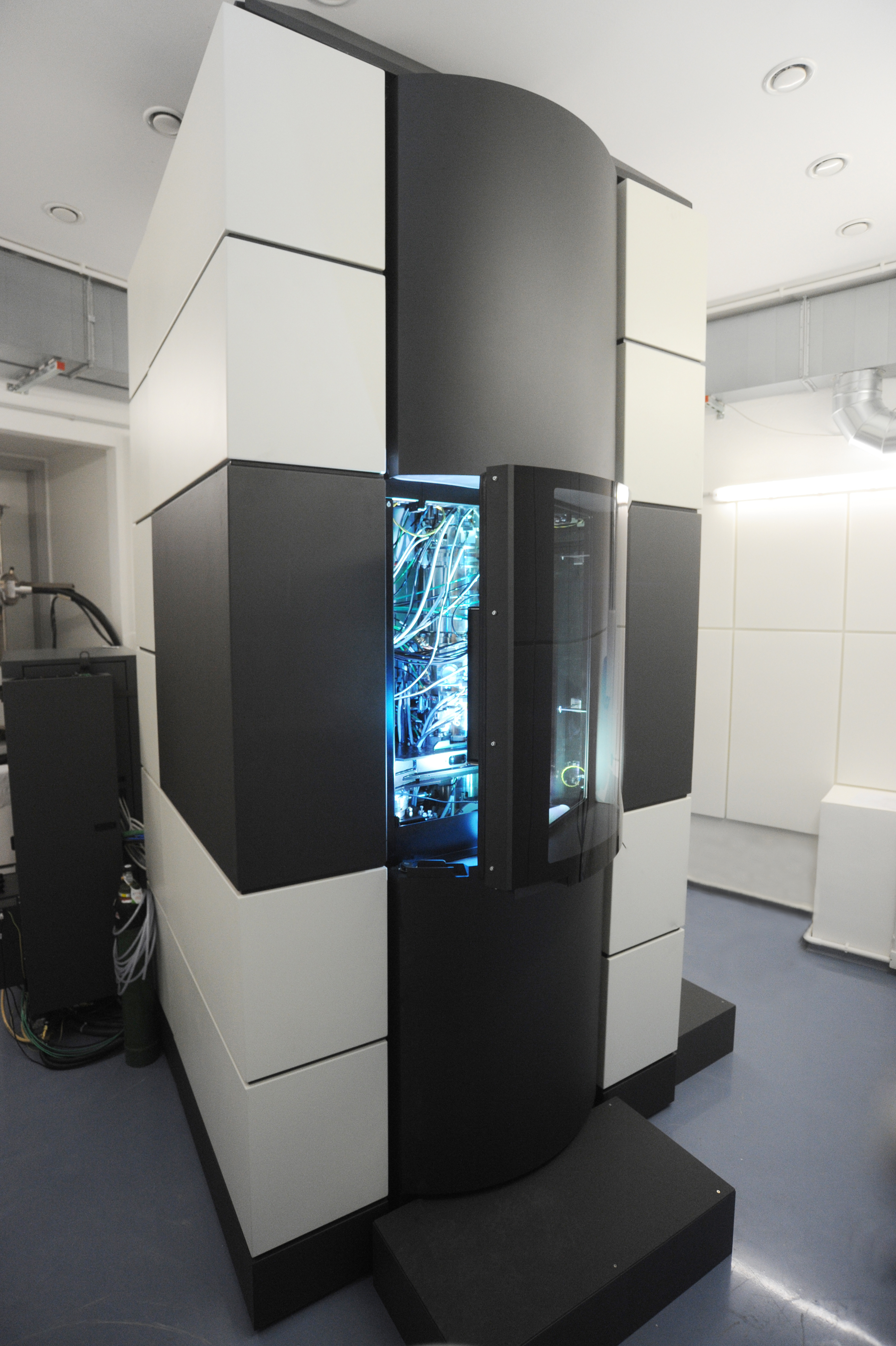
Transmission electron microscopy (TEM) is a microscopy technique in which a beam of electrons is transmitted through a specimen to form an image. The specimen is most often an ultrathin section less than 100 nm thick or a suspension on a grid. An image is formed from the interaction of the electrons with the sample as the beam is transmitted through the specimen. The image is then magnified and focused onto an imaging device, such as a fluorescent screen, a layer of photographic film, or a detector such as a scintillator attached to a charge-coupled device or a direct electron detector. Transmission electron microscopes are capable of imaging at a significantly higher resolution than light microscopes, owing to the smaller de Broglie wavelength of electrons. This enables the instrument to capture fine detail—even as small as a single column of atoms, which is thousands of times smaller than a resolvable object seen in a light microscope. Transmission electron micr ... [...More Info...] [...Related Items...] OR: [Wikipedia] [Google] [Baidu] |
Focused Ion Beam
Focused ion beam, also known as FIB, is a technique used particularly in the semiconductor industry, materials science and increasingly in the biological field for site-specific analysis, deposition, and ablation of materials. A FIB setup is a scientific instrument that resembles a scanning electron microscope (SEM). However, while the SEM uses a focused beam of electrons to image the sample in the chamber, a FIB setup uses a focused beam of ions instead. FIB can also be incorporated in a system with both electron and ion beam columns, allowing the same feature to be investigated using either of the beams. FIB should not be confused with using a beam of focused ions for direct write lithography (such as in proton beam writing). These are generally quite different systems where the material is modified by other mechanisms. Ion beam source Most widespread instruments are using liquid metal ion sources (LMIS), especially gallium ion sources. Ion sources based on elemental gold an ... [...More Info...] [...Related Items...] OR: [Wikipedia] [Google] [Baidu] |
Volume EM
Volumetric Electron Microscopy (Volume EM) is an electron microscopy method used to generate 3D reconstructions of thick (>500 nm) samples. The initial role of electron microscopes in imaging two-dimensional slices (TEM) or a specimen surface (SEM with secondary electrons) has also increasingly expanded into the depth of samples. An early example of these volume EM workflows was simply to stack TEM images of serial sections cut through a sample. The next development was virtual reconstruction of a thick section (200-500 nm) volume by backprojection of a set of images taken at different tilt angles - TEM tomography. To acquire volume EM datasets of larger depths than TEM tomography (micrometers or millimeters in the z axis), a series of images taken through the sample depth can be used. For example, ribbons of serial sections can be imaged in a TEM as described above, and when thicker sections are used, serial TEM tomography can be used to increase the z-resolution. More rec ... [...More Info...] [...Related Items...] OR: [Wikipedia] [Google] [Baidu] |
Electron Microscopy Techniques
The electron (, or in nuclear reactions) is a subatomic particle with a negative one elementary electric charge. It is a fundamental particle that comprises the ordinary matter that makes up the universe, along with up and down quarks. Electrons are extremely lightweight particles that orbit the positively charged nucleus of atoms. Their negative charge is balanced by the positive charge of protons in the nucleus, giving atoms their overall neutral charge. Ordinary matter is composed of atoms, each consisting of a positively charged nucleus surrounded by a number of orbiting electrons equal to the number of protons. The configuration and energy levels of these orbiting electrons determine the chemical properties of an atom. Electrons are bound to the nucleus to different degrees. The outermost or valence electrons are the least tightly bound and are responsible for the formation of chemical bonds between atoms to create molecules and crystals. These valence electrons als ... [...More Info...] [...Related Items...] OR: [Wikipedia] [Google] [Baidu] |
Electron Microscopy
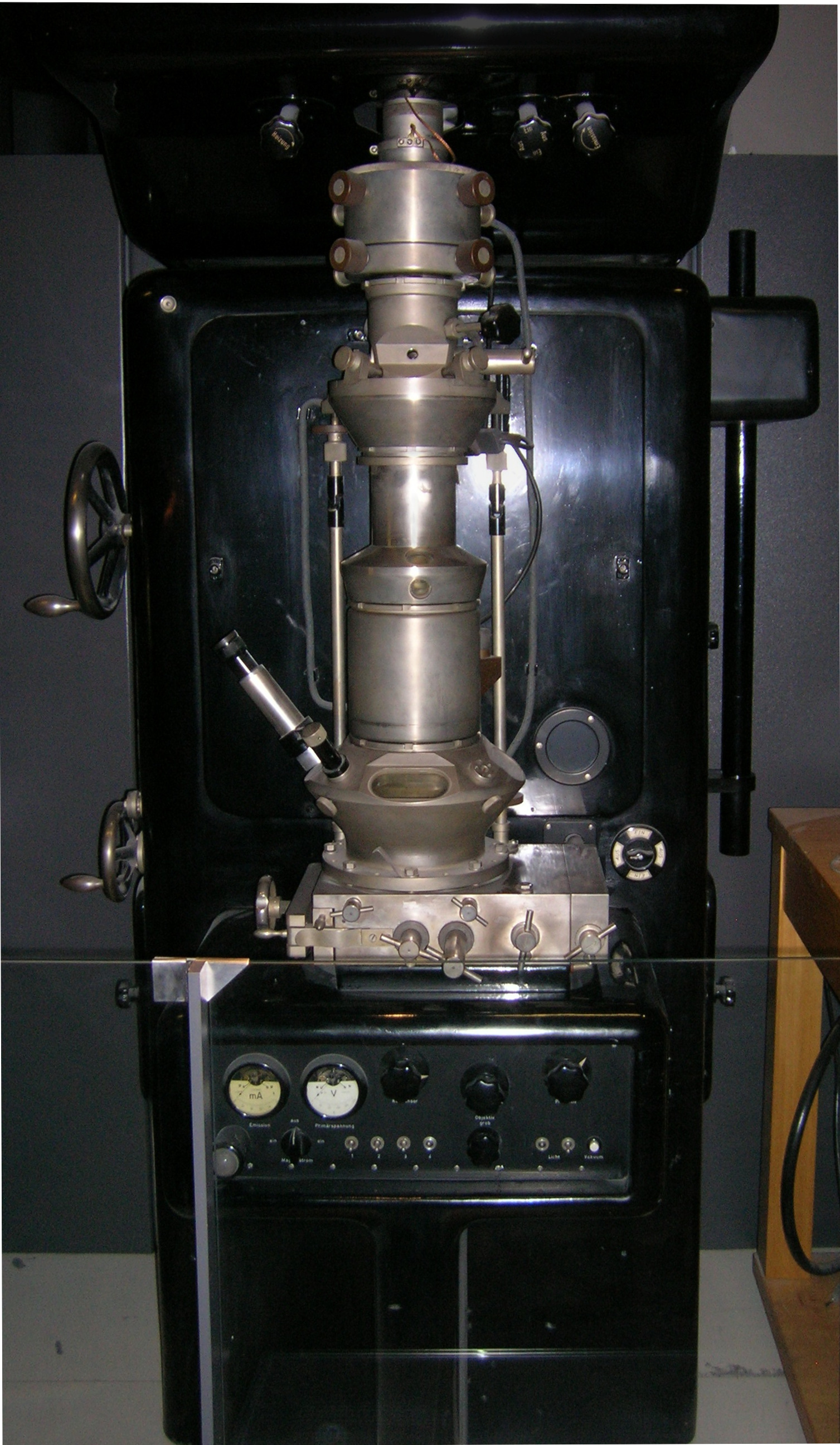
An electron microscope is a microscope that uses a beam of electrons as a source of illumination. It uses electron optics that are analogous to the glass lenses of an optical light microscope to control the electron beam, for instance focusing it to produce magnified images or electron diffraction patterns. As the wavelength of an electron can be up to 100,000 times smaller than that of visible light, electron microscopes have a much higher resolution of about 0.1 nm, which compares to about 200 nm for light microscopes. ''Electron microscope'' may refer to: * Transmission electron microscope (TEM) where swift electrons go through a thin sample * Scanning transmission electron microscope (STEM) which is similar to TEM with a scanned electron probe * Scanning electron microscope (SEM) which is similar to STEM, but with thick samples * Electron microprobe similar to a SEM, but more for chemical analysis * Low-energy electron microscope (LEEM), used to image surfaces * ... [...More Info...] [...Related Items...] OR: [Wikipedia] [Google] [Baidu] |


