|
Ventricle (brain)
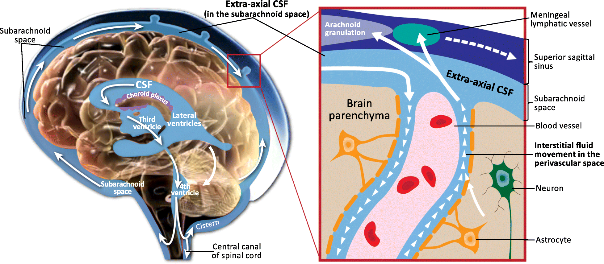
In neuroanatomy, the ventricular system is a set of four interconnected cavities known as cerebral ventricles in the brain. Within each ventricle is a region of choroid plexus which produces the circulating cerebrospinal fluid (CSF). The ventricular system is continuous with the central canal of the spinal cord from the fourth ventricle, allowing for the flow of CSF to circulate. All of the ventricular system and the central canal of the spinal cord are lined with ependyma, a specialised form of epithelium connected by tight junctions that make up the blood–cerebrospinal fluid barrier. Structure The system comprises four ventricles: * lateral ventricles right and left (one for each hemisphere) * third ventricle * fourth ventricle There are several foramina, openings acting as channels, that connect the ventricles. The interventricular foramina (also called the foramina of Monro) connect the lateral ventricles to the third ventricle through which the cerebrospinal f ... [...More Info...] [...Related Items...] OR: [Wikipedia] [Google] [Baidu] |
Neuroanatomy
Neuroanatomy is the study of the structure and organization of the nervous system. In contrast to animals with radial symmetry, whose nervous system consists of a distributed network of cells, animals with bilateral symmetry have segregated, defined nervous systems. Their neuroanatomy is therefore better understood. In vertebrates, the nervous system is segregated into the internal structure of the brain and spinal cord (together called the central nervous system, or CNS) and the series of nerves that connect the CNS to the rest of the body (known as the peripheral nervous system, or PNS). Breaking down and identifying specific parts of the nervous system has been crucial for figuring out how it operates. For example, much of what neuroscientists have learned comes from observing how damage or "lesions" to specific brain areas affects behavior or other neural functions. For information about the composition of non-human animal nervous systems, see nervous system. For information a ... [...More Info...] [...Related Items...] OR: [Wikipedia] [Google] [Baidu] |
Median Aperture
The median aperture (median aperture of fourth ventricle or foramen of Magendie) is an opening at the caudal portion of the roof of the fourth ventricle. It allows the flow of cerebrospinal fluid (CSF) from the fourth ventricle into the cisterna magna. The other openings of the fourth ventricle are the lateral apertures - one on either side. The median aperture varies in size but accounts for most of the outflow of CSF from the fourth ventricle. Structure Relations The median foramen on axial images is posterior to the pons and anterior to the caudal cerebellum. It is surrounded by the obex and gracile tubercles of the medulla, tela choroidea of the fourth ventricle and its choroid plexus, which is attached to the cerebellar vermis The cerebellar vermis (from Latin ''vermis,'' "worm") is located in the medial, cortico-nuclear zone of the cerebellum, which is in the posterior cranial fossa, posterior fossa of the cranium. The primary fissure in the vermis curves ventr ... [...More Info...] [...Related Items...] OR: [Wikipedia] [Google] [Baidu] |
Ectoderm
The ectoderm is one of the three primary germ layers formed in early embryonic development. It is the outermost layer, and is superficial to the mesoderm (the middle layer) and endoderm (the innermost layer). It emerges and originates from the outer layer of germ cells. The word ectoderm comes from the Greek language, Greek ''ektos'' meaning "outside", and ''derma'' meaning "skin".Gilbert, Scott F. Developmental Biology. 9th ed. Sunderland, MA: Sinauer Associates, 2010: 333-370. Print. Generally speaking, the ectoderm differentiates to form epithelial tissue, epithelial and nervous system, neural tissues (spinal cord, nerves and brain). This includes the Epidermis (skin), skin, linings of the mouth, anus, nostrils, sweat glands, hair and nails, and tooth enamel. Other types of epithelium are derived from the endoderm. In vertebrate embryos, the ectoderm can be divided into two parts: the dorsal surface ectoderm also known as the external ectoderm, and the neural plate, which inv ... [...More Info...] [...Related Items...] OR: [Wikipedia] [Google] [Baidu] |
Obex
The obex () is the point in the human brain at which the fourth ventricle narrows to become the central canal of the spinal cord. Cerebrospinal fluid can flow from the fourth ventricle into the obex. In anatomical studies, the obex has been found to occur approximately 10–12 mm above the level of the foramen magnum. In patients with low tonsillar position, the obex has been found at or below the plane of the foramen magnum. The obex occurs in the caudal medulla. The decussation of sensory fibers happens at this point. Clinical significance Lesions at the location can result in obstructive hydrocephalus. The most common lesion at this location is a subependymoma, a benign tumor. Hemangioblastoma has been observed in this location. Detection of prions Immunohistochemistry (IHC) to test brain, lymph, and neuroendocrine tissues for the presence of the abnormal prion protein to diagnose wasting diseases like chronic wasting disease in deer A deer (: deer) or ... [...More Info...] [...Related Items...] OR: [Wikipedia] [Google] [Baidu] |
Cerebral Aqueduct
The cerebral aqueduct (aqueduct of the midbrain, aqueduct of Sylvius, Sylvian aqueduct, mesencephalic duct) is a small, narrow tube connecting the third and fourth ventricles of the brain. The cerebral aqueduct is a midline structure that passes through the midbrain. It extends rostrocaudally through the entirety of the more posterior part of the midbrain. It is surrounded by the periaqueductal gray (central gray), a layer of gray matter. Congenital stenosis of the cerebral aqueduct is a cause of congenital hydrocephalus. It is named for Franciscus Sylvius. Anatomy The cerebral aqueduct is roughly circular in transverse section, and measures 1-2 mm in diameter. It is 15 mm long and is commonly subdivided into a pars anterior antrum, and pars posterior. Relations Rostrally, it is continuous with the third ventricle, commencing just inferior to the posterior commissure. Caudally, it is continuous with the fourth ventricle at the junction of the mesencephalon and pons. ... [...More Info...] [...Related Items...] OR: [Wikipedia] [Google] [Baidu] |
Brainstem
The brainstem (or brain stem) is the posterior stalk-like part of the brain that connects the cerebrum with the spinal cord. In the human brain the brainstem is composed of the midbrain, the pons, and the medulla oblongata. The midbrain is continuous with the thalamus of the diencephalon through the tentorial notch, and sometimes the diencephalon is included in the brainstem. The brainstem is very small, making up around only 2.6 percent of the brain's total weight. It has the critical roles of regulating heart and respiratory system, respiratory function, helping to control heart rate and breathing rate. It also provides the main motor and sensory nerve supply to the face and neck via the cranial nerves. Ten pairs of cranial nerves come from the brainstem. Other roles include the regulation of the central nervous system and the body's sleep cycle. It is also of prime importance in the conveyance of motor and sensory pathways from the rest of the brain to the body, and from the b ... [...More Info...] [...Related Items...] OR: [Wikipedia] [Google] [Baidu] |
Neural Tube
In the developing chordate (including vertebrates), the neural tube is the embryonic precursor to the central nervous system, which is made up of the brain and spinal cord. The neural groove gradually deepens as the neural folds become elevated, and ultimately the folds meet and coalesce in the middle line and convert the groove into the closed neural tube. In humans, neural tube closure usually occurs by the fourth week of pregnancy (the 28th day after conception). Development The neural tube develops in two ways: primary neurulation and secondary neurulation. Primary neurulation divides the ectoderm into three cell types: * The internally located neural tube * The externally located epidermis * The neural crest cells, which develop in the region between the neural tube and epidermis but then migrate to new locations # Primary neurulation begins after the neural plate forms. The edges of the neural plate start to thicken and lift upward, forming the neural folds. The center ... [...More Info...] [...Related Items...] OR: [Wikipedia] [Google] [Baidu] |
Embryogenesis
An embryo ( ) is the initial stage of development for a multicellular organism. In organisms that reproduce sexually, embryonic development is the part of the life cycle that begins just after fertilization of the female egg cell by the male sperm cell. The resulting fusion of these two cells produces a single-celled zygote that undergoes many cell divisions that produce cells known as blastomeres. The blastomeres (4-cell stage) are arranged as a solid ball that when reaching a certain size, called a morula, (16-cell stage) takes in fluid to create a cavity called a blastocoel. The structure is then termed a blastula, or a blastocyst in mammals. The mammalian blastocyst hatches before implantating into the endometrial lining of the womb. Once implanted the embryo will continue its development through the next stages of gastrulation, neurulation, and organogenesis. Gastrulation is the formation of the three germ layers that will form all of the different parts o ... [...More Info...] [...Related Items...] OR: [Wikipedia] [Google] [Baidu] |
Medulla Oblongata
The medulla oblongata or simply medulla is a long stem-like structure which makes up the lower part of the brainstem. It is anterior and partially inferior to the cerebellum. It is a cone-shaped neuronal mass responsible for autonomic (involuntary) functions, ranging from vomiting to sneezing. The medulla contains the cardiovascular center, the respiratory center, vomiting and vasomotor centers, responsible for the autonomic functions of breathing, heart rate and blood pressure as well as the sleep–wake cycle. "Medulla" is from Latin, ‘pith or marrow’. And "oblongata" is from Latin, ‘lengthened or longish or elongated'. During embryonic development, the medulla oblongata develops from the myelencephalon. The myelencephalon is a secondary brain vesicle which forms during the maturation of the rhombencephalon, also referred to as the hindbrain. The bulb is an archaic term for the medulla oblongata. In modern clinical usage, the word bulbar (as in bulbar palsy) is r ... [...More Info...] [...Related Items...] OR: [Wikipedia] [Google] [Baidu] |
Cistern Of Great Cerebral Vein
The quadrigeminal cistern (also cistern of great cerebral vein, vein of Galen cistern, superior cistern, Bichat's canal, or peripineal cistern) is a subarachnoid cistern situated between splenium of corpus callosum, and the superior surface of the cerebellum. It contains a part of the great cerebral vein, the posterior cerebral artery, quadrigeminal artery, glossopharyngeal nerve (CN IX), and the pineal gland. Structure The quadrigeminal cistern lies between the splenium of the corpus callosum (superiorly), the cerebellar vermis (inferiorly and posteriorly), and the tentorial margin. It is just superior to the tectum of the mesencephalon (midbrain). It lies medial to part of the medial occipital cortex. It is posterior to the brainstem and third ventricle; it extends between the layers of the tela choroidea of the third ventricle. The cistern may extend anterior-ward between the thalamus and corpus callosum to form the ''cistern of velum interpositum''. Contents The sup ... [...More Info...] [...Related Items...] OR: [Wikipedia] [Google] [Baidu] |


