|
Thigh Bone
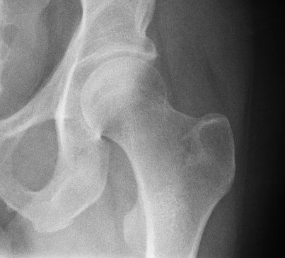
The femur (; ), or thigh bone, is the proximal bone of the hindlimb in tetrapod vertebrates. The head of the femur articulates with the acetabulum in the pelvic bone forming the hip joint, while the distal part of the femur articulates with the tibia (shinbone) and patella (kneecap), forming the knee joint. By most measures the two (left and right) femurs are the strongest bones of the body, and in humans, the largest and thickest. Structure The femur is the only bone in the upper leg. The two femurs converge medially toward the knees, where they articulate with the proximal ends of the tibiae. The angle of convergence of the femora is a major factor in determining the femoral-tibial angle. Human females have thicker pelvic bones, causing their femora to converge more than in males. In the condition ''genu valgum'' (knock knee) the femurs converge so much that the knees touch one another. The opposite extreme is ''genu varum'' (bow-leggedness). In the general population of ... [...More Info...] [...Related Items...] OR: [Wikipedia] [Google] [Baidu] |
Gastrocnemius
The gastrocnemius muscle (plural ''gastrocnemii'') is a superficial two-headed muscle that is in the back part of the lower leg of humans. It runs from its two heads just above the knee to the heel, a three joint muscle (knee, ankle and subtalar joints). The muscle is named via Latin, from Greek γαστήρ (''gaster'') 'belly' or 'stomach' and κνήμη (''knḗmē'') 'leg', meaning 'stomach of the leg' (referring to the bulging shape of the calf). Structure The gastrocnemius is located with the soleus in the posterior (back) compartment of the leg. The lateral head originates from the lateral condyle of the femur, while the medial head originates from the medial condyle of the femur. Its other end forms a common tendon with the soleus muscle; this tendon is known as the calcaneal tendon or Achilles tendon and inserts onto the posterior surface of the calcaneus, or heel bone. It is considered a superficial muscle as it is located directly under skin, and its shape may often b ... [...More Info...] [...Related Items...] OR: [Wikipedia] [Google] [Baidu] |
Hindlimb
A hindlimb or back limb is one of the paired articulated appendages (limbs) attached on the caudal ( posterior) end of a terrestrial tetrapod vertebrate's torso.http://www.merriam-webster.com/medical/hind%20limb, Merriam Webster Dictionary-Hindlimb With reference to quadrupeds, the term hindleg or back leg is often used instead. In bipedal animals with an upright posture (e.g. humans and some primates), the term lower limb is often used. Location It is located on the limb of an animal. Hindlimbs are present in a large number of quadrupeds. Though it is a posterior limb, it can cause lameness in some animals. The way of walking through hindlimbs are called bipedalism. Benefits of hindlimbs Hindlimbs are helpful in many ways, some examples are: Frogs Frogs can easily adapt at the surroundings using hindlimbs. The main reason is it can jump high to easily escape to its predator and also to catch prey. It can perform some tricks using the hindlimbs such as the somersault and h ... [...More Info...] [...Related Items...] OR: [Wikipedia] [Google] [Baidu] |
Anthropology
Anthropology is the scientific study of humanity, concerned with human behavior, human biology, cultures, societies, and linguistics, in both the present and past, including past human species. Social anthropology studies patterns of behavior, while cultural anthropology studies cultural meaning, including norms and values. A portmanteau term sociocultural anthropology is commonly used today. Linguistic anthropology studies how language influences social life. Biological or physical anthropology studies the biological development of humans. Archaeological anthropology, often termed as 'anthropology of the past', studies human activity through investigation of physical evidence. It is considered a branch of anthropology in North America and Asia, while in Europe archaeology is viewed as a discipline in its own right or grouped under other related disciplines, such as history and palaeontology. Etymology The abstract noun ''anthropology'' is first attested in reference t ... [...More Info...] [...Related Items...] OR: [Wikipedia] [Google] [Baidu] |
Ethnic Group
An ethnic group or an ethnicity is a grouping of people who identify with each other on the basis of shared attributes that distinguish them from other groups. Those attributes can include common sets of traditions, ancestry, language, history, society, culture, nation, religion, or social treatment within their residing area. The term ethnicity is often times used interchangeably with the term nation, particularly in cases of ethnic nationalism, and is separate from the related concept of races. Ethnicity may be construed as an inherited or as a societally imposed construct. Ethnic membership tends to be defined by a shared cultural heritage, ancestry, origin myth, history, homeland, language, or dialect, symbolic systems such as religion, mythology and ritual, cuisine, dressing style, art, or physical appearance. Ethnic groups may share a narrow or broad spectrum of genetic ancestry, depending on group identification, with many groups having mixed genetic ancestry. Ethnic ... [...More Info...] [...Related Items...] OR: [Wikipedia] [Google] [Baidu] |
Genu Varum
Genu varum (also called bow-leggedness, bandiness, bandy-leg, and tibia vara) is a varus deformity marked by (outward) bowing at the knee, which means that the lower leg is angled inward ( medially) in relation to the thigh's axis, giving the limb overall the appearance of an archer's bow. Usually medial angulation of both lower limb bones (femur and tibia) is involved. Causes If a child is sickly, either with rickets or any other ailment that prevents ossification of the bones or is improperly fed, the bowed condition may persist. Thus the chief cause of this deformity is rickets. Skeletal problems, infection, and tumors can also affect the growth of the leg, sometimes giving rise to a one-sided bow-leggedness. The remaining causes are occupational, especially among jockeys, and from physical trauma, the condition being very likely to supervene after accidents involving the condyles of the femur. Childhood Children until the age of 3 to 4 have a degree of genu varum. Th ... [...More Info...] [...Related Items...] OR: [Wikipedia] [Google] [Baidu] |
Genu Valgum
Genu, a Latin word for "knee," may refer to: * Genu of internal capsule * Genu of the corpus callosum * Genu recurvatum * Genu valgum * Genu varum Genu varum (also called bow-leggedness, bandiness, bandy-leg, and tibia vara) is a varus deformity marked by (outward) bowing at the knee, which means that the lower leg is angled inward ( medially) in relation to the thigh's axis, giving the ... * Genu, Iran (other), places in Iran {{dab ... [...More Info...] [...Related Items...] OR: [Wikipedia] [Google] [Baidu] |
Femoral-tibial Angle
The femoral-tibial angle is the angle between the femur and tibia. In humans, the two femurs converge medially toward the knees, where they articulate with the proximal ends of the tibiae. The angle of convergence of the femora is a major factor in determining the femoral-tibial angle. In human females the femora converge more than in males because the pelvic bone The hip bone (os coxae, innominate bone, pelvic bone or coxal bone) is a large flat bone, constricted in the center and expanded above and below. In some vertebrates (including humans before puberty) it is composed of three parts: the ilium, ischi ... is wider in females. In the condition ''genu valgum'' (knock knee) the femurs converge so much that the knees touch one another. The opposite extreme is ''genu varum'' (bow-leggedness). In the general population of people without either ''genu valgum'' or ''genu varum'', the femoral-tibial angle is about 175 degrees. References {{musculoskeletal-stub Knee ... [...More Info...] [...Related Items...] OR: [Wikipedia] [Google] [Baidu] |
Knee Joint
In humans and other primates, the knee joins the thigh with the leg and consists of two joints: one between the femur and tibia (tibiofemoral joint), and one between the femur and patella (patellofemoral joint). It is the largest joint in the human body. The knee is a modified hinge joint, which permits flexion and extension as well as slight internal and external rotation. The knee is vulnerable to injury and to the development of osteoarthritis. It is often termed a ''compound joint'' having tibiofemoral and patellofemoral components. (The fibular collateral ligament is often considered with tibiofemoral components.) Structure The knee is a modified hinge joint, a type of synovial joint, which is composed of three functional compartments: the patellofemoral articulation, consisting of the patella, or "kneecap", and the patellar groove on the front of the femur through which it slides; and the medial and lateral tibiofemoral articulations linking the femur, or thigh bone, ... [...More Info...] [...Related Items...] OR: [Wikipedia] [Google] [Baidu] |
Lower Extremity Of Femur
The lower extremity of femur (or distal extremity) is the lower end of the femur (thigh bone) in human and other animals, closer to the knee. It is larger than the upper extremity of femur, is somewhat cuboid in form, but its transverse diameter is greater than its antero-posterior; it consists of two oblong eminences known as the lateral condyle and medial condyle. Condyles Anteriorly, the condyles are slightly prominent and are separated by a smooth shallow articular depression called the patella surface. Posteriorly, they project considerably and a deep notch, the intercondylar fossa of femur, is present between them. The lateral condyle is the more prominent and is the broader both in its antero-posterior and transverse diameters, the medial condyle is the longer and, when the femur is held with its body perpendicular, projects to a lower level. When, however, the femur is in its natural oblique position the lower surfaces of the two condyles lie practically in the same ... [...More Info...] [...Related Items...] OR: [Wikipedia] [Google] [Baidu] |
Hip Joint
In vertebrate anatomy, hip (or "coxa"Latin ''coxa'' was used by Celsus in the sense "hip", but by Pliny the Elder in the sense "hip bone" (Diab, p 77) in medical terminology) refers to either an anatomical region or a joint. The hip region is located lateral and anterior to the gluteal region, inferior to the iliac crest, and overlying the greater trochanter of the femur, or "thigh bone". In adults, three of the bones of the pelvis have fused into the hip bone or acetabulum which forms part of the hip region. The hip joint, scientifically referred to as the acetabulofemoral joint (''art. coxae''), is the joint between the head of the femur and acetabulum of the pelvis and its primary function is to support the weight of the body in both static (e.g., standing) and dynamic (e.g., walking or running) postures. The hip joints have very important roles in retaining balance, and for maintaining the pelvic inclination angle. Pain of the hip may be the result of numerous cause ... [...More Info...] [...Related Items...] OR: [Wikipedia] [Google] [Baidu] |
Pelvic Bone
The hip bone (os coxae, innominate bone, pelvic bone or coxal bone) is a large flat bone, constricted in the center and expanded above and below. In some vertebrates (including humans before puberty) it is composed of three parts: the ilium, ischium, and the pubis. The two hip bones join at the pubic symphysis and together with the sacrum and coccyx (the pelvic part of the spine) comprise the skeletal component of the pelvis – the pelvic girdle which surrounds the pelvic cavity. They are connected to the sacrum, which is part of the axial skeleton, at the sacroiliac joint. Each hip bone is connected to the corresponding femur (thigh bone) (forming the primary connection between the bones of the lower limb and the axial skeleton) through the large ball and socket joint of the hip. Structure The hip bone is formed by three parts: the ilium, ischium, and pubis. At birth, these three components are separated by hyaline cartilage. They join each other in a Y-shaped portion of ca ... [...More Info...] [...Related Items...] OR: [Wikipedia] [Google] [Baidu] |
Articulation (anatomy)
A joint or articulation (or articular surface) is the connection made between bones, ossicles, or other hard structures in the body which link an animal's skeletal system into a functional whole.Saladin, Ken. Anatomy & Physiology. 7th ed. McGraw-Hill Connect. Webp.274/ref> They are constructed to allow for different degrees and types of movement. Some joints, such as the knee, elbow, and shoulder, are self-lubricating, almost frictionless, and are able to withstand compression and maintain heavy loads while still executing smooth and precise movements. Other joints such as sutures between the bones of the skull permit very little movement (only during birth) in order to protect the brain and the sense organs. The connection between a tooth and the jawbone is also called a joint, and is described as a fibrous joint known as a gomphosis. Joints are classified both structurally and functionally. Classification The number of joints depends on if sesamoids are included, age of the h ... [...More Info...] [...Related Items...] OR: [Wikipedia] [Google] [Baidu] |


