|
Transverse Plane
A transverse plane is a plane that is rotated 90° from two other planes. Anatomy The transverse plane is an anatomical plane that is perpendicular to the sagittal plane and the dorsal plane. It is also called the axial plane or horizontal plane, especially in human anatomy, but horizontal plane can be misleading with other animals. The plane splits the body into a cranial (head) side and caudal (tail) side, so in humans the plane will be horizontal (dividing the body into superior and inferior sections) but in quadrupeds it will be vertical. Human anatomy Clinically relevant anatomical planes * Transverse '' thoracic plane'' * '' Xiphosternal plane'' (or xiphosternal junction) * '' Transpyloric plane'' * '' Subcostal plane'' * '' Umbilical plane'' (or transumbilical plane) * '' Supracristal plane'' * '' Intertubercular plane'' (or transtubercular plane) * '' Interspinous plane'' Associated structures * The transverse '' thoracic plane'' ** Plane through T4 & T5 verteb ... [...More Info...] [...Related Items...] OR: [Wikipedia] [Google] [Baidu] [Amazon] |
Anatomical Plane
An anatomical plane is a hypothetical plane used to transect the body, in order to describe the location of structures or the direction of movements. In human anatomy and non-human anatomy, four principal planes are used: the median plane, sagittal plane, coronal plane, and transverse plane. * The median plane or midsagittal plane passes through the middle of the body, dividing it into left and right halves. * A para sagittal plane is any plane that runs parallel to the median plane, also dividing the body into left and right sections. * The dorsal plane divides the body into dorsal (towards the backbone) and ventral (towards the belly) parts. In human anatomy coronal plane is preferred, or sometimes the frontal plane, and the description may reference splitting the body into front and back parts, but this phrasing is not as clear for animals with a horizontal spine like quadrupeds or fish. * The transverse plane, also called the axial plane or horizontal plane, is perpendi ... [...More Info...] [...Related Items...] OR: [Wikipedia] [Google] [Baidu] [Amazon] |
Bifurcation Of The Trachea
The carina of trachea (also: "tracheal carina") is a ridge of cartilage at the base of the trachea separating the openings of the left and right main bronchi. Structure The carina is a cartilaginous ridge separating the left and right main bronchi that is formed by the inferior-ward and posterior-ward prolongation of the inferior-most tracheal cartilage. The carina occurs at the lower end of the trachea - usually at the level of the 4th to 5th thoracic vertebra. This is in line with the sternal angle, but the carina may raise or descend up to two vertebrae higher or lower with breathing. The carina lies to the left of the midline, and runs antero-posteriorly (front to back). Blood supply The bronchial arteries supply the carina and the rest of the lower trachea. Relations The carina is around the area posterior to where the aortic arch crosses to the left of the trachea. The azygos vein crosses right to the trachea above the carina. Physiology The mucous membrane of ... [...More Info...] [...Related Items...] OR: [Wikipedia] [Google] [Baidu] [Amazon] |
Costal Margin
The costal margin, also known as the costal arch, is the lower edge of the chest (thorax) formed by the bottom edge of the rib cage. Structure The costal margin is the medial margin formed by the cartilages of the seventh to tenth ribs. It attaches to the body and xiphoid process of the sternum. The thoracic diaphragm attaches to the costal margin. The costal angle is the angle between the left and right costal margins where they join the sternum. Function The costal margins somewhat protect the higher abdominal organs, such as the liver. Clinical significance The costal margin may be used for tissue harvesting of cartilage for use elsewhere in the body, such as to treat microtia. Different abdominal organs may be palpated just below the costal margin, such as the liver on the right side of the body. Pain across the costal margin is most commonly caused by costochondritis. The costal paradox, also known as Hoover's sign and the costal margin paradox, is a sign ... [...More Info...] [...Related Items...] OR: [Wikipedia] [Google] [Baidu] [Amazon] |
Subcostal Plane
The subcostal plane is a transverse plane which bisects the body at the level of the 10th costal margin and the upper border of the third lumbar vertebra. See also *Inferior mesenteric artery *Quadrants and regions of abdomen The human abdomen is divided into quadrants and regions by anatomy, anatomists and physicians for the purposes of study, medical diagnosis, diagnosis, and therapy, treatment. The division into four quadrants allows the localisation of pain and te ... * Supracristal plane * Transpyloric plane * Transtubercular plane References External links * http://www.liv.ac.uk/HumanAnatomy/phd/mbchb/travel/surface1.html * http://www.qub.ac.uk/cskills/Abd%20exam.htm Anatomical planes {{Anatomy-stub ... [...More Info...] [...Related Items...] OR: [Wikipedia] [Google] [Baidu] [Amazon] |
Costal Cartilages
Costal cartilage, also known as rib cartilage, are bars of hyaline cartilage that serve to prolong the ribs forward and contribute to the elasticity of the walls of the thorax. Costal cartilage is only found at the anterior ends of the ribs, providing medial extension. Differences from ribs 1-12 The first seven pairs are connected with the sternum; the next three are each articulated with the lower border of the cartilage of the preceding rib; the last two have pointed extremities, which end in the wall of the abdomen. Like the ribs, the costal cartilages vary in their length, breadth, and direction. They increase in length from the first to the seventh, then gradually decrease to the twelfth. Their breadth, as well as that of the intervals between them, diminishes from the first to the last. They are broad at their attachments to the ribs, and taper toward their sternal extremities, excepting the first two, which are of the same breadth throughout, and the sixth, seventh, and e ... [...More Info...] [...Related Items...] OR: [Wikipedia] [Google] [Baidu] [Amazon] |
Pylorus
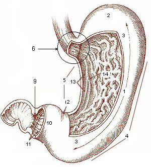
The pylorus ( or ) connects the stomach to the duodenum. The pylorus is considered as having two parts, the ''pyloric antrum'' (opening to the body of the stomach) and the ''pyloric canal'' (opening to the duodenum). The ''pyloric canal'' ends as the ''pyloric orifice'', which marks the junction between the stomach and the duodenum. The orifice is surrounded by a sphincter, a band of muscle, called the ''pyloric sphincter''. The word ''pylorus'' comes from Greek πυλωρός, via Latin. The word ''pylorus'' in Greek means "gatekeeper", related to "gate" () and is thus linguistically related to the word " pylon". Structure The pylorus is the furthest part of the stomach that connects to the duodenum. It is divided into two parts, the ''antrum'', which connects to the body of the stomach, and the ''pyloric canal'', which connects to the duodenum. Antrum The antrum also called the ''gastric antrum'' or the ''pyloric antrum'' is the initial portion of the pyloric region. ... [...More Info...] [...Related Items...] OR: [Wikipedia] [Google] [Baidu] [Amazon] |
Pubic Symphysis
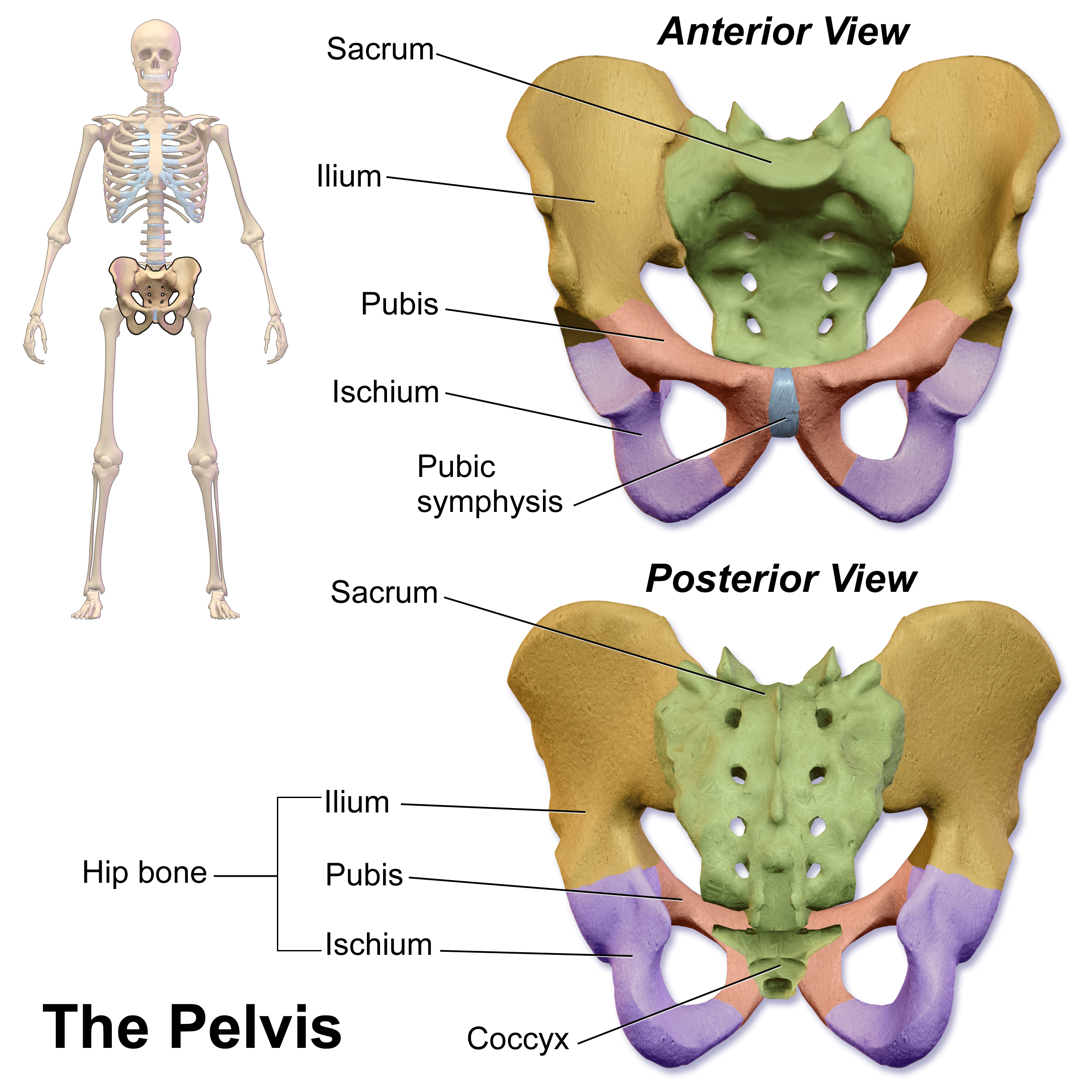
The pubic symphysis (: symphyses) is a secondary cartilaginous joint between the left and right superior rami of the pubis of the hip bones. It is in front of and below the urinary bladder. In males, the suspensory ligament of the penis attaches to the pubic symphysis. In females, the pubic symphysis is attached to the suspensory ligament of the clitoris. In most adults, it can be moved roughly 2 mm and with 1 degree rotation. This increases for women at the time of childbirth. The name comes from the Greek word ''symphysis'', meaning 'growing together'. Structure The pubic symphysis is a nonsynovial amphiarthrodial joint. The width of the pubic symphysis at the front is 3–5 mm greater than its width at the back. This joint is connected by fibrocartilage and may contain a fluid-filled cavity; the center is avascular, possibly due to the nature of the compressive forces passing through this joint, which may lead to harmful vascular disease. The ends of both pubi ... [...More Info...] [...Related Items...] OR: [Wikipedia] [Google] [Baidu] [Amazon] |
Transpyloric Plane
The transpyloric plane, also known as Addison's plane, is an imaginary transverse plane, horizontal plane, located halfway between the suprasternal notch of the manubrium and the upper border of the symphysis pubis at the level of the first lumbar vertebrae, L1. It lies roughly a hand's breadth beneath the xiphisternum or midway between the xiphisternum and the navel, umbilicus. The plane in most cases cuts through the pylorus of the stomach, the tips of the ninth costal cartilages and the lower border of the first lumbar vertebra. Structures crossed The transpyloric plane is clinically notable because it passes through several important abdominal structures. It also divides the Supracolic compartment, supracolic and infracolic compartments, with the liver, spleen and gastric fundus above it and the small intestine and colon below it. Lumbar vertebra and spinal cord The lower border of first Lumbar vertebrae, lumbar vertebra lies at the level of the transpyloric plane, according ... [...More Info...] [...Related Items...] OR: [Wikipedia] [Google] [Baidu] [Amazon] |
Heart
The heart is a muscular Organ (biology), organ found in humans and other animals. This organ pumps blood through the blood vessels. The heart and blood vessels together make the circulatory system. The pumped blood carries oxygen and nutrients to the tissue, while carrying metabolic waste such as carbon dioxide to the lungs. In humans, the heart is approximately the size of a closed fist and is located between the lungs, in the middle compartment of the thorax, chest, called the mediastinum. In humans, the heart is divided into four chambers: upper left and right Atrium (heart), atria and lower left and right Ventricle (heart), ventricles. Commonly, the right atrium and ventricle are referred together as the right heart and their left counterparts as the left heart. In a healthy heart, blood flows one way through the heart due to heart valves, which prevent cardiac regurgitation, backflow. The heart is enclosed in a protective sac, the pericardium, which also contains a sma ... [...More Info...] [...Related Items...] OR: [Wikipedia] [Google] [Baidu] [Amazon] |
Thoracic Diaphragm
The thoracic diaphragm, or simply the diaphragm (; ), is a sheet of internal Skeletal striated muscle, skeletal muscle in humans and other mammals that extends across the bottom of the thoracic cavity. The diaphragm is the most important Muscles of respiration, muscle of respiration, and separates the thoracic cavity, containing the heart and lungs, from the abdominal cavity: as the diaphragm contracts, the volume of the thoracic cavity increases, creating a negative pressure there, which draws air into the lungs. Its high oxygen consumption is noted by the many mitochondria and capillaries present; more than in any other skeletal muscle. The term ''diaphragm'' in anatomy, created by Gerard of Cremona, can refer to other flat structures such as the urogenital diaphragm or Pelvic floor, pelvic diaphragm, but "the diaphragm" generally refers to the thoracic diaphragm. In humans, the diaphragm is slightly asymmetric—its right half is higher up (superior) to the left half, since th ... [...More Info...] [...Related Items...] OR: [Wikipedia] [Google] [Baidu] [Amazon] |
Liver
The liver is a major metabolic organ (anatomy), organ exclusively found in vertebrates, which performs many essential biological Function (biology), functions such as detoxification of the organism, and the Protein biosynthesis, synthesis of various proteins and various other Biochemistry, biochemicals necessary for digestion and growth. In humans, it is located in the quadrants and regions of abdomen, right upper quadrant of the abdomen, below the thoracic diaphragm, diaphragm and mostly shielded by the lower right rib cage. Its other metabolic roles include carbohydrate metabolism, the production of a number of hormones, conversion and storage of nutrients such as glucose and glycogen, and the decomposition of red blood cells. Anatomical and medical terminology often use the prefix List of medical roots, suffixes and prefixes#H, ''hepat-'' from ἡπατο-, from the Greek language, Greek word for liver, such as hepatology, and hepatitis The liver is also an accessory digestive ... [...More Info...] [...Related Items...] OR: [Wikipedia] [Google] [Baidu] [Amazon] |
Thoracic Cavity
The thoracic cavity (or chest cavity) is the chamber of the body of vertebrates that is protected by the thoracic wall (rib cage and associated skin, muscle, and fascia). The central compartment of the thoracic cavity is the mediastinum. There are two openings of the thoracic cavity, a superior thoracic aperture known as the thoracic inlet and a lower inferior thoracic aperture known as the thoracic outlet. The thoracic cavity includes the tendons as well as the cardiovascular system which could be damaged from injury to the back, spine or the neck. Structure Structures within the thoracic cavity include: * structures of the cardiovascular system, including the heart and great vessels, which include the thoracic aorta, the pulmonary artery and all its branches, the superior and inferior vena cava, the pulmonary veins, and the azygos vein * structures of the respiratory system, including the diaphragm, trachea, bronchi and lungs * structures of the digestive system, incl ... [...More Info...] [...Related Items...] OR: [Wikipedia] [Google] [Baidu] [Amazon] |


