|
Tibial Nerve
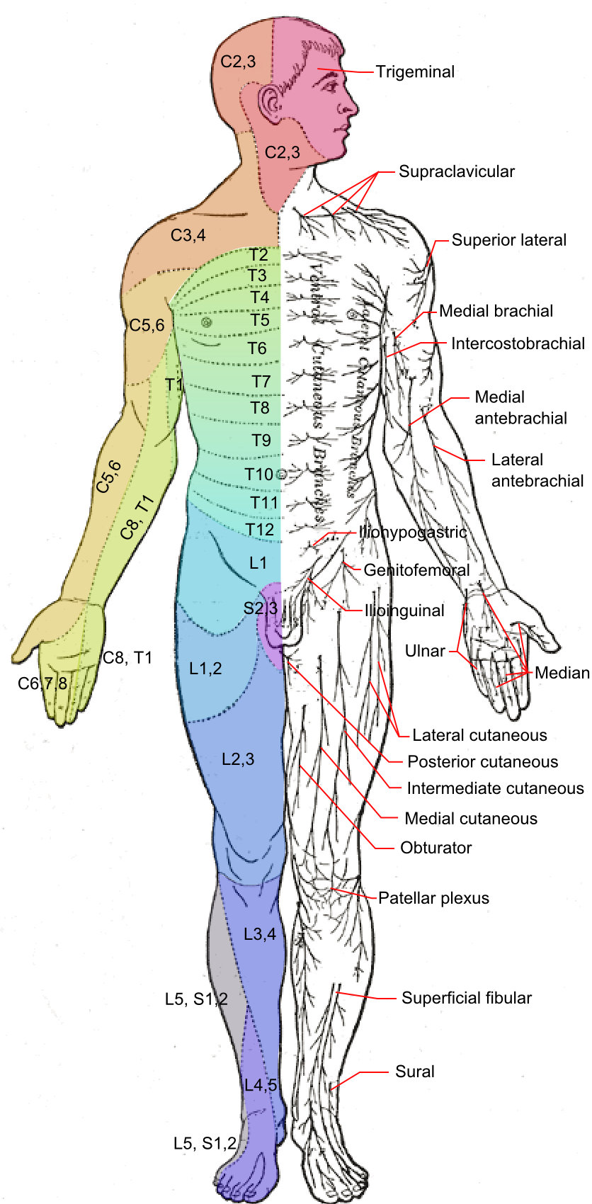
The tibial nerve is a branch of the sciatic nerve. The tibial nerve passes through the popliteal fossa to pass below the arch of soleus. Structure Popliteal fossa The tibial nerve is the larger terminal branch of the sciatic nerve with root values of L4, L5, S1, S2, and S3. It lies superficial (or posterior) to the popliteal vessels, extending from the superior angle to the inferior angle of the popliteal fossa, crossing the popliteal vessels from lateral to medial side. It gives off branches as shown below: * Muscular branches - Muscular branches arise from the distal part of the popliteal fossa. It supplies the medial and lateral heads of gastrocnemius, soleus, plantaris and popliteus muscles. Nerve to popliteus crosses the popliteus muscle, runs downwards and laterally, winds around the lower border of the popliteus to supply the deep (or anterior) surface of the popliteus. This nerve also supplies the tibialis posterior muscle, superior tibiofibular joint, tibia bone, ... [...More Info...] [...Related Items...] OR: [Wikipedia] [Google] [Baidu] |
Flexor Digitorum Longus
The flexor digitorum longus muscle or flexor digitorum communis longus is situated on the tibial side of the leg. At its origin it is thin and pointed, but it gradually increases in size as it descends. It serves to flex the second, third, fourth, and fifth toes. Structure The flexor digitorum longus muscle arises from the posterior surface of the body of the tibia, from immediately below the soleal line to within 7 or 8 cm of its lower extremity, medial to the tibial origin of the tibialis posterior muscle. It also arises from the fascia covering the tibialis posterior muscle. The fibers end in a tendon, which runs nearly the whole length of the posterior surface of the muscle. This tendon passes behind the medial malleolus, in a groove, common to it and the tibialis posterior, but separated from the latter by a fibrous septum, each tendon being contained in a special compartment lined by a separate mucous sheath. The tendon of the tibialis posterior and the tendon of the ... [...More Info...] [...Related Items...] OR: [Wikipedia] [Google] [Baidu] |
Lateral Plantar Nerve
The lateral plantar nerve (external plantar nerve) is a branch of the tibial nerve, in turn a branch of the sciatic nerve and supplies the skin of the fifth toe and lateral half of the fourth, as well as most of the deep muscles, its distribution being similar to that of the ulnar nerve in the hand. It passes obliquely forward with the lateral plantar artery to the lateral side of the foot, lying between the flexor digitorum brevis and quadratus plantae and, in the interval between the flexor muscle and the abductor digiti minimi, divides into a superficial and a deep branch. Before its division, it supplies the quadratus plantae and abductor digiti minimi. It divides into deep and superficial branches. Additional images File:Gray357.png, Coronal section through right talocrural and talocalcaneal joint In human anatomy, the subtalar joint, also known as the talocalcaneal joint, is a joint of the foot. It occurs at the meeting point of the talus and the calcaneus. Th ... [...More Info...] [...Related Items...] OR: [Wikipedia] [Google] [Baidu] |
Medial Condyle Of Femur
The medial condyle is one of the two projections on the lower extremity of femur, the other being the lateral condyle. The medial condyle is larger than the lateral (outer) condyle due to more weight bearing caused by the centre of mass being medial to the knee. On the posterior surface of the condyle the linea aspera The linea aspera () is a ridge of roughened surface on the posterior surface of the shaft of the femur. It is the site of attachments of muscles and the intermuscular septum. Its margins diverge above and below. The linea aspera is a prominent ... (a ridge with two lips: medial and lateral; running down the posterior shaft of the femur) turns into the medial and lateral supracondylar ridges, respectively. The outermost protrusion on the medial surface of the medial condyle is referred to as the "medial epicondyle" and can be palpated by running fingers medially from the patella with the knee in flexion. It is important to take into consideration the diff ... [...More Info...] [...Related Items...] OR: [Wikipedia] [Google] [Baidu] |
Medial Sural Cutaneous Nerve
The medial sural cutaneous nerve ''(L4-S3)'' is a sensory nerve of the leg. It supplies cutaneous innervation to the posteromedial leg. Structure The medial sural cutaneous nerve originates from the posterior aspect of the tibial nerve of the sciatic nerve. It descends between the two heads of the gastrocnemius muscle. Around the middle of the back of the leg, it pierces the deep fascia to become superficial. It unites with the lateral sural cutaneous nerve to form the sural nerve The sural nerve ''(L4-S1)'' is generally considered a pure cutaneous nerve of the posterolateral leg to the lateral ankle. The sural nerve originates from a combination of either the sural communicating branch and medial sural cutaneous nerve, .... Morphometric properties According to a large cadaveric study in which 208 sural nerves were dissected in their native position (bSteele et al. the medial sural cutaneous nerve was consistently present in most lower extremities. This information alig ... [...More Info...] [...Related Items...] OR: [Wikipedia] [Google] [Baidu] |
Cutaneous Nerve
A cutaneous nerve is a nerve that provides nerve supply to the skin. Human anatomy In human anatomy, cutaneous nerves are primarily responsible for providing cutaneous innervation, sensory innervation to the skin. In addition to sympathetic and autonomic afferent (sensory) fibers, most cutaneous nerves also contain sympathetic efferent (visceromotor) fibers, which innervate cutaneous blood vessels, sweat glands, and the arrector pilli muscles of hair follicles. These structures are important to the sympathetic nervous response. There are many cutaneous nerves in the human body, only some of which are named. Some of the larger cutaneous nerves are as follows: Upper body * In the arm (proper) ** Superior lateral cutaneous nerve of arm (Superior LCNOA) ** Inferior lateral cutaneous nerve of arm (Inferior LCNOA) ** Posterior cutaneous nerve of arm (PCNOA) ** Medial cutaneous nerve of arm (MCNOA) * In the forearm ** Lateral cutaneous nerve of forearm (LCNOF) ** Posterior c ... [...More Info...] [...Related Items...] OR: [Wikipedia] [Google] [Baidu] |
Inferior Tibiofibular Joint
The inferior tibiofibular joint, also known as the distal tibiofibular joint (tibiofibular syndesmosis), is formed by the rough, convex surface of the medial side of the distal end of the fibula, and a rough concave surface on the lateral side of the tibia. Below, to the extent of about 4 mm, these surfaces are smooth and covered with cartilage, which is continuous with that of the ankle joint. The ligaments are: * Anterior ligament of the lateral malleolus * Posterior ligament of the lateral malleolus * Interosseous membrane of leg The inferior transverse ligament of the tibiofibular syndesmosis is included in older versions of ''Gray's Anatomy ''Gray's Anatomy'' is a reference book of human anatomy written by Henry Gray, illustrated by Henry Vandyke Carter and first published in London in 1858. It has had multiple revised editions, and the current edition, the 42nd (October 2020 ...'', but not in '' Terminologia Anatomica''. However, it still appears in ... [...More Info...] [...Related Items...] OR: [Wikipedia] [Google] [Baidu] |
Interosseous Membrane Of Leg
The interosseous membrane of the leg (middle tibiofibular ligament) extends between the interosseous crests of the tibia and fibula, helps stabilize the Tib-Fib relationship and separates the muscles on the front from those on the back of the leg. It consists of a thin, aponeurotic joint lamina composed of oblique fibers, which for the most part run downward and lateralward; some few fibers, however, pass in the opposite direction. It is broader above than below. Its upper margin does not quite reach the tibiofibular joint, but presents a free concave border, above which is a large, oval aperture for the passage of the anterior tibial vessels to the front of the leg. In its lower part is an opening for the passage of the anterior peroneal vessels. It is continuous below with the interosseous ligament of the tibiofibular syndesmosis A syndesmosis (“fastened with a band”) is a type of fibrous joint in which two bones are united to each other by fibrous connective tissu ... [...More Info...] [...Related Items...] OR: [Wikipedia] [Google] [Baidu] |
Tibia
The tibia (; : tibiae or tibias), also known as the shinbone or shankbone, is the larger, stronger, and anterior (frontal) of the two Leg bones, bones in the leg below the knee in vertebrates (the other being the fibula, behind and to the outside of the tibia); it connects the knee with the ankle bones, ankle. The tibia is found on the anatomical terms of location#Medial, medial side of the leg next to the fibula and closer to the median plane. The tibia is connected to the fibula by the interosseous membrane of leg, forming a type of fibrous joint called a syndesmosis with very little movement. The tibia is named for the flute ''aulos, tibia''. It is the second largest bone in the human body, after the femur. The leg bones are the strongest long bones as they support the rest of the body. Structure In human anatomy, the tibia is the second largest bone next to the femur. As in other vertebrates the tibia is one of two bones in the lower leg, the other being the fibula, and is a ... [...More Info...] [...Related Items...] OR: [Wikipedia] [Google] [Baidu] |
Superior Tibiofibular Joint
The superior tibiofibular articulation (also called proximal tibiofibular joint) is an arthrodial joint between the lateral condyle of tibia and the head of the fibula. The contiguous surfaces of the bones present flat, oval facets covered with cartilage and connected together by an articular capsule and by anterior and posterior cruciate ligaments. When the term ''tibiofibular articulation'' is used without a modifier, it refers to the proximal, not the distal (i.e., inferior) tibiofibular articulation. Clinical significance Injuries to the proximal tibiofibular joint are uncommon and usually associated with other injuries to the lower leg. Dislocations can be classified into the following five types: * Anterolateral dislocation (most common) * Posteromedial dislocation * Superior dislocation (uncommon, associated with shortened tibia fractures or severe ankle injuries) * Inferior dislocation (rare, associated with lengthened tibia fractures or avulsion of the foot, us ... [...More Info...] [...Related Items...] OR: [Wikipedia] [Google] [Baidu] |
Tibialis Posterior
The tibialis posterior muscle is the most central of all the leg muscles, and is located in the deep posterior compartment of the leg. It is the key stabilizing muscle of the lower leg. Posterior tibial tendonitis Posterior tibial tendonitis is a condition that predominantly affects runners and active individuals. It involves inflammation or tearing of the posterior tibial tendon, which connects the calf muscle to the bones on the inside of the foot. It plays a vital role in supporting the arch and assisting in foot movement. This condition can cause pain, swelling, and potentially lead to flatfoot if left untreated. Structure The tibialis posterior muscle originates on the inner posterior border of the fibula laterally. It is also attached to the interosseous membrane medially, which attaches to the tibia and fibula. The tendon of the tibialis posterior muscle (sometimes called the posterior tibial tendon) descends posterior to the medial malleolus. It terminates by dividin ... [...More Info...] [...Related Items...] OR: [Wikipedia] [Google] [Baidu] |
Popliteus
The popliteus muscle in the leg is used for unlocking the knees when walking, by laterally rotating the femur on the tibia during the closed chain portion of the gait cycle (one with the foot in contact with the ground). In open chain movements (when the involved limb is not in contact with the ground), the popliteus muscle medially rotates the tibia on the femur. It is also used when sitting down and standing up. It is the only muscle in the posterior (back) compartment of the lower leg that acts just on the knee and not on the ankle. The gastrocnemius muscle acts on both joints. Structure The popliteus muscle originates from the lateral surface of the lateral condyle of the femur by a rounded tendon. Its fibers pass downward and medially. It inserts onto the posterior surface of tibia, above the soleal line. The popliteus tendon runs beneath the lateral collateral ligament and tendon of biceps femoris. The muscle also runs above the lateral meniscus but has no connection ... [...More Info...] [...Related Items...] OR: [Wikipedia] [Google] [Baidu] |
Plantaris
The plantaris is one of the superficial muscles of the superficial posterior compartment of the leg, one of the fascial compartments of the leg. It is composed of a thin muscle belly and a long thin tendon. While not as thick as the achilles tendon, the plantaris tendon (which tends to be between in length) is the longest tendon in the human body. Not including the tendon, the plantaris muscle is approximately long and is absent in 8-12% of the population. It is one of the plantar flexors in the posterior compartment of the leg, along with the gastrocnemius and soleus muscles. The plantaris is considered to have become an unimportant muscle when human ancestors switched from climbing trees to bipedalism and in anatomically modern humans it mainly acts with the gastrocnemius. Structure The plantaris muscle arises from the inferior part of the lateral supracondylar ridge of the femur at a position slightly superior to the origin of the lateral head of gastrocnemius. It passes ... [...More Info...] [...Related Items...] OR: [Wikipedia] [Google] [Baidu] |


