|
Tatton-Brown–Rahman Syndrome
Tatton-Brown–Rahman syndrome (TBRS) is a rare overgrowth syndrome, overgrowth and intellectual disability syndrome caused by autosomal dominant mutations in the ''DNMT3A'' gene. The syndrome was first recognized in 2014 by Katrina Tatton-Brown, Nazneen Rahman, and collaborators. Signs and symptoms TBRS is defined by overgrowth and mild-to-severe intellectual disability. All individuals with TBRS experience some degree of developmental delay and/or intellectual disability, with 86% of well-documented cases falling in the mild to moderate range. Most individuals with TBRS exhibit increased Human height, stature, Macrocephaly, head circumference, and Overweight, weight at least two standard deviations above the mean. Generalized Hypermobility (joints), joint hypermobility and hypotonia are observed in ~75% and ~55% of cases, respectively, and are often associated with musculoskeletal pain and joint instability. Approximately half of individuals exhibit behavioral or psychiatric i ... [...More Info...] [...Related Items...] OR: [Wikipedia] [Google] [Baidu] |
Medical Genetics
Medical genetics is the branch of medicine that involves the diagnosis and management of hereditary disorders. Medical genetics differs from human genetics in that human genetics is a field of scientific research that may or may not apply to medicine, while medical genetics refers to the application of genetics to medical care. For example, research on the causes and inheritance of genetic disorders would be considered within both human genetics and medical genetics, while the diagnosis, management, and counselling people with genetic disorders would be considered part of medical genetics. In contrast, the study of typically non-medical phenotypes such as the genetics of eye color would be considered part of human genetics, but not necessarily relevant to medical genetics (except in situations such as albinism). ''Genetic medicine'' is a newer term for medical genetics and incorporates areas such as gene therapy, personalized medicine, and the rapidly emerging new medical specia ... [...More Info...] [...Related Items...] OR: [Wikipedia] [Google] [Baidu] |
Hypotonia
Hypotonia is a state of low muscle tone (the amount of tension or resistance to stretch in a muscle), often involving reduced muscle strength. Hypotonia is not a specific medical disorder, but it is a potential manifestation of many different diseases and disorders that affect motor nerve control by the brain or muscle strength. Hypotonia is a lack of resistance to passive movement whereas muscle weakness results in impaired active movement. Central hypotonia originates from the central nervous system, while peripheral hypotonia is related to problems within the spinal cord, peripheral nerves, and/or skeletal muscles. Severe hypotonia in infancy is commonly known as floppy baby syndrome. Recognizing hypotonia, even in early infancy, is usually relatively straightforward, but medical diagnosis, diagnosing the underlying cause can be difficult and often unsuccessful. The long-term effects of hypotonia on a child's development and later life depend primarily on the severity of the mus ... [...More Info...] [...Related Items...] OR: [Wikipedia] [Google] [Baidu] |
Arcuate Fasciculus
In neuroanatomy, the arcuate fasciculus (AF; ) is a bundle of axons that generally connects Broca's area and Wernicke's area in the brain. It is an association fiber tract connecting caudal temporal lobe and inferior frontal lobe. Structure The arcuate fasciculus is a white matter tract that runs parallel to the superior longitudinal fasciculus. Due to their proximity, they are sometimes referred to interchangeably. They can be distinguished by the location and function of their endpoints in the frontal cortex. The arcuate fasciculus terminates in Broca's area (specifically BA 44) which is linked to processing complex syntax. However, the superior longitudinal fasciculus ends in the premotor cortex which is implicated in acoustic-motor mapping. Connection Historically, the arcuate fasciculus has been understood to connect two important areas for language use: Broca's area in the inferior frontal gyrus and Wernicke's area in the posterior superior temporal gyrus. It is mo ... [...More Info...] [...Related Items...] OR: [Wikipedia] [Google] [Baidu] |
Posterior Cranial Fossa
The posterior cranial fossa is the part of the cranial cavity located between the foramen magnum, and tentorium cerebelli. It is formed by the sphenoid bones, temporal bones, and occipital bone. It lodges the cerebellum, and parts of the brainstem. Anatomy The posterior cranial fossa is formed by the sphenoid bones, temporal bones, and occipital bone. It is the most inferior of the fossae. It houses the cerebellum, medulla oblongata, and pons. Boundaries Anteriorly, the posterior cranial fossa is bounded by the dorsum sellae, posterior aspect of the body of sphenoid bone, and the basilar part of occipital bone/ clivus. Laterally, it is bounded by the petrous parts and mastoid parts of the temporal bones, and the lateral parts of occipital bone. Posteriorly, it is bounded by the squamous part of occipital bone. Features Foramen magnum The foramen magnum is a large opening of the floor of the posterior cranial fossa, its most conspicuous feature. In ... [...More Info...] [...Related Items...] OR: [Wikipedia] [Google] [Baidu] |
Corpus Callosum
The corpus callosum (Latin for "tough body"), also callosal commissure, is a wide, thick nerve tract, consisting of a flat bundle of commissural fibers, beneath the cerebral cortex in the brain. The corpus callosum is only found in placental mammals. It spans part of the longitudinal fissure, connecting the left and right cerebral hemispheres, enabling communication between them. It is the largest white matter structure in the human brain, about in length and consisting of 200–300 million axonal projections. A number of separate nerve tracts, classed as subregions of the corpus callosum, connect different parts of the hemispheres. The main ones are known as the genu, the rostrum, the trunk or body, and the splenium. Structure The corpus callosum forms the floor of the longitudinal fissure that separates the two cerebral hemispheres. Part of the corpus callosum forms the roof of the lateral ventricles. The corpus callosum has four main parts – individual nerv ... [...More Info...] [...Related Items...] OR: [Wikipedia] [Google] [Baidu] |
Hypospadias
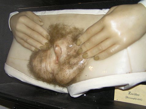
Hypospadias is a common malformation in fetal development of the penis in which the urethra does not open from its usual location on the head of the penis. It is the second-most common birth defect of the male reproductive system, affecting about one of every 250 males at birth, although when including milder cases, is found in up to 4% of newborn males. Roughly 90% of cases are the less serious distal hypospadias, in which the urethral opening (the Urinary meatus, meatus) is on or near the head of the penis (Glans penis, glans). The remainder have proximal hypospadias, in which the meatus is all the way back on the shaft of the penis, near or within the scrotum. Shiny tissue or anything that typically forms the urethra instead extends from the meatus to the tip of the glans; this tissue is called the urethral plate. In most cases, the foreskin is less developed and does not wrap completely around the penis, leaving the underside of the glans uncovered. Also, a downward bending of ... [...More Info...] [...Related Items...] OR: [Wikipedia] [Google] [Baidu] |
Vesicoureteral Reflux
Vesicoureteral reflux (VUR), also known as vesicoureteric reflux, is a condition in which urine flows retrograde, or backward, from the urinary bladder, bladder into one or both ureters and then to the renal calyx or kidneys. Urine normally travels in one direction (forward, or anterograde) from the kidneys to the bladder via the ureters, with a one-way valve at the vesicoureteral (ureteral-bladder) junction preventing backflow. The valve is formed by oblique tunneling of the distal ureter through the wall of the bladder, creating a short length of ureter (1–2 cm) that can be compressed as the bladder fills. Reflux occurs if the ureter enters the bladder without sufficient tunneling, i.e., too "end-on". Signs and symptoms Most children with vesicoureteral reflux are asymptomatic. Vesicoureteral reflux may be diagnosed as a result of further evaluation of hydronephrosis, dilation of the kidney or hydroureter, ureters draining urine from the kidney while in utero as well a ... [...More Info...] [...Related Items...] OR: [Wikipedia] [Google] [Baidu] |
Cryptorchidism
Cryptorchidism, also known as undescended testis, is the failure of one or both testes to descend into the scrotum. The word is . It is the most common birth defect of the male genital tract. About 3% of full-term and 30% of premature infant boys are born with at least one undescended testis. However, about 80% of cryptorchid testes descend by the first year of life (the majority within three months), making the true incidence of cryptorchidism around 1% overall. Cryptorchidism may develop after infancy, sometimes as late as young adulthood, but that is exceptional. Cryptorchidism is distinct from monorchism, the condition of having only one testicle. Though the condition may occur on one or both sides, it more commonly affects the right testis. A testis absent from the normal scrotal position may be: # Anywhere along the "path of descent" from high in the posterior (retroperitoneal) abdomen, just below the kidney, to the inguinal ring # In the inguinal canal # Ectopic, havin ... [...More Info...] [...Related Items...] OR: [Wikipedia] [Google] [Baidu] |
Aneurysm Of Sinus Of Valsalva
Aneurysm of the aortic sinus, also known as the sinus of Valsalva, is a rare abnormality of the aorta, the largest artery in the body. The aorta normally has three small pouches that sit directly above the aortic valve (the sinuses of Valsalva), and an aneurysm of one of these sinuses is a thin-walled swelling. Aneurysms may affect the right (65–85%), non-coronary (10–30%), or rarely the left (< 5%) coronary sinus. These aneurysms may not cause any symptoms but if large can cause , or blackouts. Aortic sinus aneurysms can burst or rupture into adjacent cardiac chambers, which can lead t ... [...More Info...] [...Related Items...] OR: [Wikipedia] [Google] [Baidu] |
Congenital Heart Defect
A congenital heart defect (CHD), also known as a congenital heart anomaly, congenital cardiovascular malformation, and congenital heart disease, is a defect in the structure of the heart or great vessels that is present at birth. A congenital heart defect is classed as a cardiovascular disease. Signs and symptoms depend on the specific type of defect. Symptoms can vary from none to life-threatening. When present, symptoms are variable and may include rapid breathing, bluish skin (cyanosis), poor weight gain, and feeling tired. CHD does not cause chest pain. Most congenital heart defects are not associated with other diseases. A complication of CHD is heart failure. Congenital heart defects are the most common birth defect. In 2015, they were present in 48.9 million people globally. They affect between 4 and 75 per 1,000 live births, depending upon how they are diagnosed. In about 6 to 19 per 1,000 they cause a moderate to severe degree of problems. Congenital heart defects are t ... [...More Info...] [...Related Items...] OR: [Wikipedia] [Google] [Baidu] |
Maxillary Central Incisor
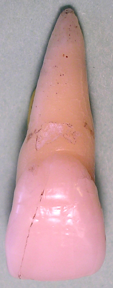
The maxillary central incisor is a human tooth in the front upper jaw, or maxilla, and is usually the most visible of all teeth in the mouth. It is located Commonly used terms of relationship and comparison in dentistry, mesial (closer to the midline of the face) to the maxillary lateral incisor. As with all incisors, their function is for Shearing (physics), shearing or cutting food during mastication (chewing). There is typically a single cusp (dentistry), cusp on each tooth, called an Commonly used terms of relationship and comparison in dentistry, incisal ridge or incisal edge. Formation of these teeth begins at 14 weeks in utero for the deciduous teeth, deciduous (baby) set and 3–4 months of age for the permanent teeth, permanent set. There are some minor differences between the deciduous maxillary central incisor and that of the permanent maxillary central incisor. The deciduous tooth appears in the mouth at 8–12 months of age and shed at 6–7 years, and is replaced b ... [...More Info...] [...Related Items...] OR: [Wikipedia] [Google] [Baidu] |
Palpebral Fissure
The palpebral fissure is the elliptic space between the medial and lateral canthi of the two open eyelids. In simple terms, it is the opening between the eyelids. In adult humans, this measures about 10 mm vertically and 30 mm horizontally. Variations Congenital dysmorphisms It can be reduced (short, "narrow") in horizontal size by fetal alcohol syndrome and in Williams syndrome. The chromosomal conditions trisomy 9 and trisomy 21 (Down syndrome) can cause the palpebral fissures to be upslanted, whereas Marfan syndrome can cause a downslant. An increase in vertical height can be seen in genetic disorders such as cri-du-chat syndrome. Acquired The fissure may be increased in vertical height in Graves' disease, which is manifested as Dalrymple's sign. It is seen in disorders such as cri-du-chat syndrome. In animal studies using four times the therapeutic concentration of the ophthalmic solution latanoprost, the size of the palpebral fissure can be increased. The condi ... [...More Info...] [...Related Items...] OR: [Wikipedia] [Google] [Baidu] |


