|
Schmerling Caves
The Schmerling Caves (also known as Grottes d'Engis, meaning Engis Caves) are a group of caves located in Wallonia on the right bank of the stream called the Awirs, near the village of Awirs in Flémalle, Belgium. The caves are notable for their past fossil finds, particularly of hominins. They were explored in 1829 by Philippe-Charles Schmerling, who discovered, in the lower cave, the remains of two individuals, one of which, now known as Engis 2, was a fossil of the first Neanderthal ever found; the other was a Neolithic homo sapiens. Also known as Trô Cwaheur or Trou Caheur, this lower cave has since collapsed. A third cave was destroyed because of work on the adjacent quarry, the Ancienne Carrière des Awirs. The caves have been classified on the list of sites since 1978, as well as since 2013, because of the Neanderthal fossil. Name, location, and finds Schmerling named the caves for Engis because he accessed them from the Plateau des Fagnes, which is in the Engis com ... [...More Info...] [...Related Items...] OR: [Wikipedia] [Google] [Baidu] |
Awirs
Awirs ( wa, Les Awires) is a village of Wallonia and a district of the municipality of Flémalle, located in the province of Liège, Belgium. The population on December 31, 2004 was 2,869. A notable building is the 18th century Château d'Aigremont. A Neanderthal skull, Engis 2, was found in the Schmerling Caves The Schmerling Caves (also known as Grottes d'Engis, meaning Engis Caves) are a group of caves located in Wallonia on the right bank of the stream called the Awirs, near the village of Awirs in Flémalle, Belgium. The caves are notable for their p .... Sub-municipalities of Flémalle Former municipalities of Liège Province {{Liege-geo-stub ... [...More Info...] [...Related Items...] OR: [Wikipedia] [Google] [Baidu] |
Thoracic Vertebrae
In vertebrates, thoracic vertebrae compose the middle segment of the vertebral column, between the cervical vertebrae and the lumbar vertebrae. In humans, there are twelve thoracic vertebra (anatomy), vertebrae and they are intermediate in size between the cervical and lumbar vertebrae; they increase in size going towards the lumbar vertebrae, with the lower ones being much larger than the upper. They are distinguished by the presence of Zygapophysial joint, facets on the sides of the bodies for Articulation (anatomy), articulation with the head of rib, heads of the ribs, as well as facets on the transverse processes of all, except the eleventh and twelfth, for articulation with the tubercle (rib), tubercles of the ribs. By convention, the human thoracic vertebrae are numbered T1–T12, with the first one (T1) located closest to the skull and the others going down the spine toward the lumbar region. General characteristics These are the general characteristics of the second throu ... [...More Info...] [...Related Items...] OR: [Wikipedia] [Google] [Baidu] |
Engis Skul01
Engis (; wa, Indji) is a municipality of Wallonia located in the province of Liège, Belgium. On 1 January 2006 Engis had a total population of 5,686. The total area is 27.74 km² which gives a population density of 205 inhabitants per km². The municipality consists of the following districts: Clermont-sous-Huy, Engis and Hermalle-sous-Huy. In 1829, in this village, Philippe-Charles Schmerling discovered the first Neanderthal ever, Engis 2, the damaged skull of a young child. This was before the 1856 discovery of the Neanderthal type specimen in the Neander Valley. Its importance was not recognised until 1936. Pollution fatalities In late 1930 and early 1931, several thousand cases of acute pulmonary attacks occurred in the Meuse valley, centered on Engis, and 60 people died. A commission of inquiry set up by the Belgian government concluded that the cause was poisonous waste gases, primarily sulfur dioxide, emitted by the many factories in the valley and the furnaces ... [...More Info...] [...Related Items...] OR: [Wikipedia] [Google] [Baidu] |
National Treasure
The idea of national treasure, like national epics and national anthems, is part of the language of romantic nationalism, which arose in the late 18th century and 19th centuries. Nationalism is an ideology that supports the nation as the fundamental unit of human social life, which includes shared language, values, and culture. Thus national treasure, part of the ideology of nationalism, is shared culture. A national treasure can be a shared cultural asset, which may or may not have monetary value; for example, a skilled banjo player would be a Living National Treasure. Or it may refer to a rare cultural object, such as the medieval manuscript Plan of St. Gall in Switzerland. The government of Japan designates the most famous of the nation's cultural properties as National Treasures of Japan. The National Treasures of Korea are a set of artifacts, sites, and buildings that are recognised by South Korea as having exceptional cultural value. Notable examples There are thousands o ... [...More Info...] [...Related Items...] OR: [Wikipedia] [Google] [Baidu] |
Collarbone
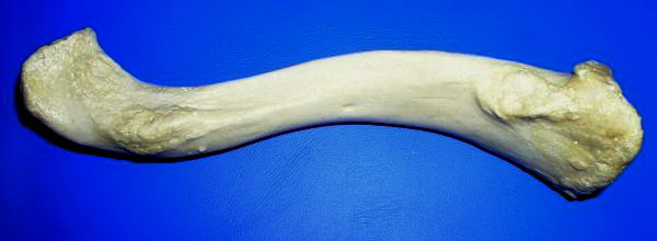
The clavicle, or collarbone, is a slender, S-shaped long bone approximately 6 inches (15 cm) long that serves as a strut between the shoulder blade and the sternum (breastbone). There are two clavicles, one on the left and one on the right. The clavicle is the only long bone in the body that lies horizontally. Together with the shoulder blade, it makes up the shoulder girdle. It is a palpable bone and, in people who have less fat in this region, the location of the bone is clearly visible. It receives its name from the Latin ''clavicula'' ("little key"), because the bone rotates along its axis like a key when the shoulder is abducted. The clavicle is the most commonly fractured bone. It can easily be fractured by impacts to the shoulder from the force of falling on outstretched arms or by a direct hit. Structure The collarbone is a thin doubly curved long bone that connects the arm to the trunk of the body. Located directly above the first rib, it acts as a strut to keep ... [...More Info...] [...Related Items...] OR: [Wikipedia] [Google] [Baidu] |
Milk Teeth
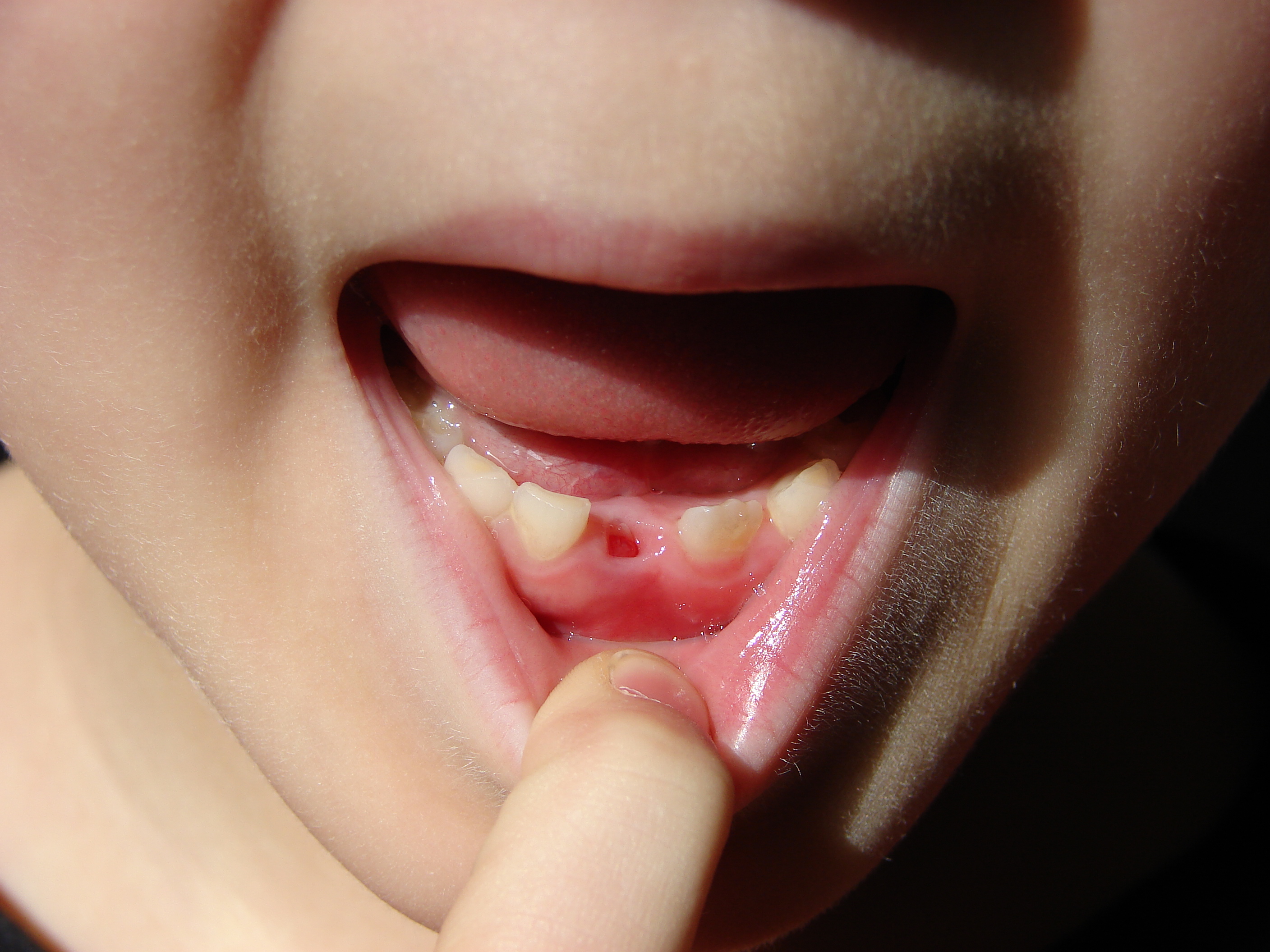
Deciduous teeth or primary teeth, also informally known as baby teeth, milk teeth, or temporary teeth,Illustrated Dental Embryology, Histology, and Anatomy, Bath-Balogh and Fehrenbach, Elsevier, 2011, page 255 are the first set of teeth in the growth and development of humans and other diphyodonts, which include most mammals but not elephants, kangaroos, or manatees which are polyphyodonts. Deciduous teeth develop during the embryonic stage of development and erupt (break through the gums and become visible in the mouth) during infancy. They are usually lost and replaced by permanent teeth, but in the absence of their permanent replacements, they can remain functional for many years into adulthood. Development Formation Primary teeth start to form during the embryonic phase of human life. The development of primary teeth starts at the sixth week of tooth development as the dental lamina. This process starts at the midline and then spreads back into the posterior region. ... [...More Info...] [...Related Items...] OR: [Wikipedia] [Google] [Baidu] |
Metatarsal Bone
The metatarsal bones, or metatarsus, are a group of five long bones in the foot, located between the tarsal bones of the hind- and mid-foot and the phalanges of the toes. Lacking individual names, the metatarsal bones are numbered from the medial side (the side of the great toe): the first, second, third, fourth, and fifth metatarsal (often depicted with Roman numerals). The metatarsals are analogous to the metacarpal bones of the hand. The lengths of the metatarsal bones in humans are, in descending order, second, third, fourth, fifth, and first. Structure The five metatarsals are dorsal convex long bones consisting of a shaft or body, a base ( proximally), and a head ( distally).Platzer 2004, p. 220 The body is prismoid in form, tapers gradually from the tarsal to the phalangeal extremity, and is curved longitudinally, so as to be concave below, slightly convex above. The base or posterior extremity is wedge-shaped, articulating proximally with the tarsal bones, ... [...More Info...] [...Related Items...] OR: [Wikipedia] [Google] [Baidu] |
Metacarpal Bone
In human anatomy, the metacarpal bones or metacarpus form the intermediate part of the skeletal hand located between the phalanges of the fingers and the carpal bones of the wrist, which forms the connection to the forearm. The metacarpal bones are analogous to the metatarsal bones in the foot. Structure The metacarpals form a transverse arch to which the rigid row of distal carpal bones are fixed. The peripheral metacarpals (those of the thumb and little finger) form the sides of the cup of the palmar gutter and as they are brought together they deepen this concavity. The index metacarpal is the most firmly fixed, while the thumb metacarpal articulates with the trapezium and acts independently from the others. The middle metacarpals are tightly united to the carpus by intrinsic interlocking bone elements at their bases. The ring metacarpal is somewhat more mobile while the fifth metacarpal is semi-independent.Tubiana ''et al'' 1998, p 11 Each metacarpal bone consists of a body o ... [...More Info...] [...Related Items...] OR: [Wikipedia] [Google] [Baidu] |
Ulna
The ulna (''pl''. ulnae or ulnas) is a long bone found in the forearm that stretches from the elbow to the smallest finger, and when in anatomical position, is found on the medial side of the forearm. That is, the ulna is on the same side of the forearm as the little finger. It runs parallel to the radius, the other long bone in the forearm. The ulna is usually slightly longer than the radius, but the radius is thicker. Therefore, the radius is considered to be the larger of the two. Structure The ulna is a long bone found in the forearm that stretches from the elbow to the smallest finger, and when in anatomical position, is found on the medial side of the forearm. It is broader close to the elbow, and narrows as it approaches the wrist. Close to the elbow, the ulna has a bony process, the olecranon process, a hook-like structure that fits into the olecranon fossa of the humerus. This prevents hyperextension and forms a hinge joint with the trochlea of the humerus. There is ... [...More Info...] [...Related Items...] OR: [Wikipedia] [Google] [Baidu] |
Radius (bone)
The radius or radial bone is one of the two large bones of the forearm, the other being the ulna. It extends from the lateral side of the elbow to the thumb side of the wrist and runs parallel to the ulna. The ulna is usually slightly longer than the radius, but the radius is thicker. Therefore the radius is considered to be the larger of the two. It is a long bone, prism-shaped and slightly curved longitudinally. The radius is part of two joints: the elbow and the wrist. At the elbow, it joins with the capitulum of the humerus, and in a separate region, with the ulna at the radial notch. At the wrist, the radius forms a joint with the ulna bone. The corresponding bone in the lower leg is the fibula. Structure The long narrow medullary cavity is enclosed in a strong wall of compact bone. It is thickest along the interosseous border and thinnest at the extremities, same over the cup-shaped articular surface (fovea) of the head. The trabeculae of the spongy tissue are some ... [...More Info...] [...Related Items...] OR: [Wikipedia] [Google] [Baidu] |
Vertebrae
The spinal column, a defining synapomorphy shared by nearly all vertebrates,Hagfish are believed to have secondarily lost their spinal column is a moderately flexible series of vertebrae (singular vertebra), each constituting a characteristic irregular bone whose complex structure is composed primarily of bone, and secondarily of hyaline cartilage. They show variation in the proportion contributed by these two tissue types; such variations correlate on one hand with the cerebral/caudal rank (i.e., location within the vertebral column, backbone), and on the other with phylogenetic differences among the vertebrate taxon, taxa. The basic configuration of a vertebra varies, but the bone is its ''body'', with the central part of the body constituting the ''centrum''. The upper (closer to) and lower (further from), respectively, the cranium and its central nervous system surfaces of the vertebra body support attachment to the intervertebral discs. The posterior part of a vertebra fo ... [...More Info...] [...Related Items...] OR: [Wikipedia] [Google] [Baidu] |
Stalactite
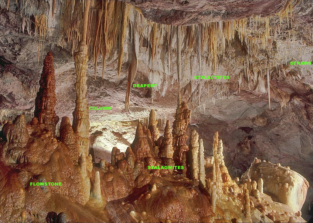
A stalactite (, ; from the Greek 'stalaktos' ('dripping') via ''stalassein'' ('to drip') is a mineral formation that hangs from the ceiling of caves, hot springs, or man-made structures such as bridges and mines. Any material that is soluble and that can be deposited as a colloid, or is in suspension, or is capable of being melted, may form a stalactite. Stalactites may be composed of lava, minerals, mud, peat, pitch, sand, sinter, and amberat (crystallized urine of pack rats). A stalactite is not necessarily a speleothem, though speleothems are the most common form of stalactite because of the abundance of limestone caves. The corresponding formation on the floor of the cave is known as a stalagmite. Mnemonics have been developed for which word refers to which type of formation; one is that ''stalactite'' has a C for "ceiling", and ''stalagmite'' has a G for "ground". Another example is that ''stalactites'' "hang on ''T''ight" and ''stalagmites'' "''M''ight grow up" †... [...More Info...] [...Related Items...] OR: [Wikipedia] [Google] [Baidu] |


_dorsal_view.png)

