|
Supratrochlear Artery
The supratrochlear artery (or frontal artery) is one of the terminal branches of the ophthalmic artery. It arises within the orbit. It exits the orbit alongside the supratrochlear nerve. It contributes arterial supply to the skin, muscles and pericranium of the forehead. Anatomy It branches from the ophthalmic artery near the trochlea of the superior oblique muscle in the orbit. Origin The supratrochlear artery branches from the ophthalmic artery in the orbit near the trochlea of the superior oblique muscle. Course After branching from the ophthalmic artery, it passes anteriorly through the superomedial orbit. It travels medial to the trochlear nerve. With the supratrochlear nerve, the supratrochlear artery exits the orbit through the supratrochlear notch (variably present), medial to the supraorbital foramen. It then ascends on the forehead. Anastomoses The supratrochlear artery anastomoses with the contralateral supratrochlear artery, and the ipsilateral supraorbit ... [...More Info...] [...Related Items...] OR: [Wikipedia] [Google] [Baidu] |
Ophthalmic Artery
The ophthalmic artery (OA) is an artery of the head. It is the first branch of the internal carotid artery distal to the cavernous sinus. Branches of the ophthalmic artery supply all the structures in the orbit around the eye, as well as some structures in the nose, face, and meninges. Occlusion of the ophthalmic artery or its branches can produce sight-threatening conditions. Structure The ophthalmic artery emerges from the internal carotid artery. This is usually just after the internal carotid artery emerges from the cavernous sinus. In some cases, the ophthalmic artery branches just before the internal carotid exits the carotid sinus. The ophthalmic artery emerges along the medial side of the anterior clinoid process. It runs anteriorly, passing through the optic canal inferolaterally to the optic nerve. It can also pass superiorly to the optic nerve in a minority of cases. In the posterior third of the cone of the orbit, the ophthalmic artery turns sharply and med ... [...More Info...] [...Related Items...] OR: [Wikipedia] [Google] [Baidu] |
Forehead
In human anatomy, the forehead is an area of the head bounded by three features, two of the skull and one of the scalp. The top of the forehead is marked by the hairline, the edge of the area where hair on the scalp grows. The bottom of the forehead is marked by the supraorbital ridge, the bone feature of the skull above the eyes. The two sides of the forehead are marked by the temporal ridge, a bone feature that links the supraorbital ridge to the coronal suture line and beyond. However, the eyebrows do not form part of the forehead. In '' Terminologia Anatomica'', ''sinciput'' is given as the Latin equivalent to "forehead" (etymology of ''sinciput'': from ''semi-'' "half" and ''caput'' "head".). Structure The bone of the forehead is the squamous part of the frontal bone. The overlying muscles are the occipitofrontalis, procerus, and corrugator supercilii muscles, all of which are controlled by the temporal branch of the facial nerve. The sensory nerves of the forehea ... [...More Info...] [...Related Items...] OR: [Wikipedia] [Google] [Baidu] |
Scalp
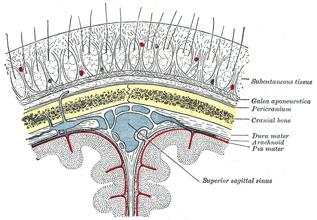
The scalp is the area of the head where head hair grows. It is made up of skin, layers of connective and fibrous tissues, and the membrane of the skull. Anatomically, the scalp is part of the epicranium, a collection of structures covering the cranium. The scalp is bordered by the face at the front, and by the neck at the sides and back. The scientific study of hair and scalp is called trichology. Structure Layers The scalp is usually described as having five layers, which can be remembered using the mnemonic 'SCALP': * S: Skin. The skin of the scalp contains numerous hair follicles and sebaceous glands. * C: Connective tissue. A dense subcutaneous layer of fat and fibrous tissue that lies beneath the skin, containing the nerves and vessels of the scalp. * A: Aponeurosis. The epicranial aponeurosis or galea aponeurotica is a tough layer of dense fibrous tissue which anchors the above layers in place. It runs from the frontalis muscle anteriorly to the occipitalis ... [...More Info...] [...Related Items...] OR: [Wikipedia] [Google] [Baidu] |
Pericranium
The periosteum is a membrane that covers the outer surface of all bones, except at the articular surfaces (i.e. the parts within a joint space) of long bones. (At the joints of long bones the bone's outer surface is lined with "articular cartilage", a type of hyaline cartilage.) Endosteum lines the inner surface of the medullary cavity of all long bones. Structure The periosteum consists of an outer fibrous layer, and an inner ''cambium layer'' (or osteogenic layer). The fibrous layer is of dense irregular connective tissue, containing fibroblasts, while the cambium layer is highly cellular containing progenitor cells that develop into osteoblasts. These osteoblasts are responsible for increasing the width of a long bone (the length of a long bone is controlled by the epiphyseal plate) and the overall size of the other bone types. After a bone fracture, the progenitor cells develop into osteoblasts and chondroblasts, which are essential to the healing process. The outer fi ... [...More Info...] [...Related Items...] OR: [Wikipedia] [Google] [Baidu] |
Frontalis Muscle
The frontalis muscle () is a muscle which covers parts of the forehead of the skull. Some sources consider the frontalis muscle to be a distinct muscle. However, Terminologia Anatomica currently classifies it as part of the occipitofrontalis muscle along with the occipitalis muscle. In humans, the frontalis muscle only serves for facial expressions. The frontalis muscle is supplied by the facial nerve and receives blood from the supraorbital and supratrochlear arteries. Structure The frontalis muscle is thin, of a quadrilateral form, and intimately adherent to the superficial fascia. It is broader than the occipitalis and its fibers are longer and paler in color. It is located on the front of the head. The muscle has no bony attachments. Its medial fibers are continuous with those of the procerus; its intermediate fibers blend with the corrugator and orbicularis oculi muscles, thus attached to the skin of the eyebrows; and its lateral fibers are also blended with the latte ... [...More Info...] [...Related Items...] OR: [Wikipedia] [Google] [Baidu] |
Orbit (anatomy)
In anatomy Anatomy () is the branch of morphology concerned with the study of the internal structure of organisms and their parts. Anatomy is a branch of natural science that deals with the structural organization of living things. It is an old scien ..., the orbit is the Body cavity, cavity or socket/hole of the skull in which the eye and Accessory visual structures, its appendages are situated. "Orbit" can refer to the bony socket, or it can also be used to imply the contents. In the adult human, the volume of the orbit is about , of which the eye occupies . The orbital contents comprise the eye, the Orbital fascia, orbital and retrobulbar fascia, extraocular muscles, cranial nerves optic nerve, II, oculomotor nerve, III, trochlear nerve, IV, trigeminal nerve, V, and abducens nerve, VI, blood vessels, fat, the lacrimal gland with its Lacrimal sac, sac and nasolacrimal duct, duct, the eyelids, Medial palpebral ligament, medial and Lateral palpebral raphe, lateral palpebr ... [...More Info...] [...Related Items...] OR: [Wikipedia] [Google] [Baidu] |
Supratrochlear Nerve
The supratrochlear nerve is a branch of the frontal nerve, itself a branch of the ophthalmic nerve (CN V1) from the trigeminal nerve (CN V). It provides sensory innervation to the skin of the forehead and the upper eyelid. Structure Origin The supratrochlear nerve is the smaller of the two terminal branches of the frontal nerve (the other being the supraorbital nerve). It arises midway between the base and apex of the orbit where the frontal nerve splits into said terminal branches. Course The supratrochlear nerve passes medially above the trochlea of the superior oblique muscle. It then travels anteriorly above the levator palpebrae superioris muscle. It exits the orbit through the supratrochlear notch or foramen. It then ascends onto the forehead beneath the corrugator supercilii muscle and frontalis muscle. It finally divides into sensory branches. The supratrochlear nerve travels with the supratrochlear artery, a branch of the ophthalmic artery. Branches Be ... [...More Info...] [...Related Items...] OR: [Wikipedia] [Google] [Baidu] |
Trochlea Of Superior Oblique
{{wiktionary Trochlea (Latin for pulley) is a term in anatomy. It refers to a grooved structure reminiscent of a pulley's wheel. Related to joints Most commonly, trochleae bear the articular surface of saddle and other joints: * Trochlea of humerus (part of the elbow hinge joint with the ulna) * Trochlea of femur The femur (; : femurs or femora ), or thigh bone is the only long bone, bone in the thigh — the region of the lower limb between the hip and the knee. In many quadrupeds, four-legged animals the femur is the upper bone of the hindleg. The Femo ... (forming the knee hinge joint with the patella) * The trochlea tali in the superior surface of the body of talus (part of the ankle hinge joint with the tibia) * Trochlear process of the calcaneus * In quadrupeds, the trochlea of Radius (bone) * The "knuckles" of the tarsometatarsus which articulate with the proximal phalanges in a bird's foot Related to muscles It also can refer to structures which serve as a guid ... [...More Info...] [...Related Items...] OR: [Wikipedia] [Google] [Baidu] |
Superior Oblique Muscle
The superior oblique muscle or obliquus oculi superior is a fusiform muscle originating in the upper, medial side of the orbit (anatomy), orbit (i.e. from beside the nose) which abducts, depresses and internally rotates the eye. It is the only extraocular muscle innervated by the trochlear nerve (the fourth Cranial nerves, cranial nerve). Structure The superior oblique muscle loops through a pulley-like structure (the trochlea of superior oblique) and inserts into the sclera on the posterotemporal surface of the eyeball. It is the pulley system that gives superior oblique its actions, causing depression of the eyeball despite being inserted on the superior surface. The superior oblique arises immediately above the margin of the Optic canal, optic foramen, superior and medial to the origin of the superior rectus, and, passing forward, ends in a rounded tendon, which plays in a fibrocartilaginous ring or pulley attached to the trochlear fossa of the frontal bone. The contiguous ... [...More Info...] [...Related Items...] OR: [Wikipedia] [Google] [Baidu] |
Trochlear Nerve
The trochlear nerve (), ( lit. ''pulley-like'' nerve) also known as the fourth cranial nerve, cranial nerve IV, or CN IV, is a cranial nerve that innervates a single muscle - the superior oblique muscle of the eye (which operates through the pulley-like trochlea). Unlike most other cranial nerves, the trochlear nerve is exclusively a motor nerve ( somatic efferent nerve). The trochlear nerve is unique among the cranial nerves in several respects: * It is the ''smallest'' nerve in terms of the number of axons it contains. * It has the greatest intracranial length. * It is the only cranial nerve that exits from the dorsal (rear) aspect of the brainstem. * It innervates a muscle, the superior oblique muscle, on the opposite side (contralateral) from its nucleus. The trochlear nerve decussates within the brainstem before emerging on the contralateral side of the brainstem (at the level of the inferior colliculus). An injury to the trochlear nucleus in the brainstem will result i ... [...More Info...] [...Related Items...] OR: [Wikipedia] [Google] [Baidu] |
Supraorbital Foramen
The supraorbital foramen, is a bony elongated opening located above the orbit (eye socket) and under the forehead. It is part of the frontal bone of the skull. The supraorbital foramen lies directly under the eyebrow. In some people this foramen is incomplete and is then known as the supraorbital notch. Structure The supraorbital foramen is a small groove at superior and medial margin of the orbit in the frontal bone. It is part of the frontal bone of the skull. It arches transversely below the superciliary arches and is the upper part of the brow ridge. It is thin and prominent in its lateral two-thirds, but rounded in its medial third. Between these two parts, the supraorbital nerve, the supraorbital artery, and the supraorbital vein pass. The supraorbital nerve divides into superficial and deep branches after it has left the supraorbital foramen. Additional images File:Gray135.png, Frontal bone. Inner surface. File:Gray1193.svg, Side view of head, showing surface relations ... [...More Info...] [...Related Items...] OR: [Wikipedia] [Google] [Baidu] |


