|
Superior Mesenteric Lymph Nodes
The superior mesenteric lymph nodes may be divided into three principal groups: * mesenteric lymph nodes * ileocolic lymph nodes * mesocolic lymph nodes Structure Mesenteric lymph nodes The mesenteric lymph nodes or mesenteric glands are one of the three principal groups of superior mesenteric lymph nodes and lie between the layers of the mesentery. They number from one hundred to one hundred and fifty, and are sited as two main groups: * one ileocolic group lying close to the wall of the small intestine, among the terminal twigs of the superior mesenteric artery; * a second larger mesocolic group placed in relation to the loops and primary branches of the vessels. Ileocolic lymph nodes The ileocolic lymph nodes, from ten to twenty in number, form a chain around the ileocolic artery, but tend to subdivide into two groups, one near the duodenum and the other on the lower part of the trunk of the artery. Where the vessel divides into its terminal branches the chain is broken ... [...More Info...] [...Related Items...] OR: [Wikipedia] [Google] [Baidu] |
Preaortic Lymph Node
The preaortic lymph nodes lie in front of the aorta, and may be divided into celiac lymph nodes, superior mesenteric lymph nodes, and inferior mesenteric lymph nodes groups, arranged around the origins of the corresponding arteries. The celiac lymph nodes are grouped into three sets: the gastric lymph nodes, gastric, hepatic lymph nodes, hepatic and splenic lymph nodes. These groups also form their own subgroups. The superior mesenteric lymph nodes are grouped into three sets: the mesenteric lymph nodes, mesenteric, ileocolic lymph nodes, ileocolic and mesocolic lymph nodes. The inferior mesenteric lymph nodes have a subgroup of pararectal lymph nodes. The preaortic lymph nodes receive a few vessels from the lateral aortic lymph nodes, but their principal afferents are derived from the organs supplied by the three arteries with which they are associated–the celiac artery, celiac, superior mesenteric artery, superior and inferior mesenteric artery, inferior mesenteric arteries. ... [...More Info...] [...Related Items...] OR: [Wikipedia] [Google] [Baidu] |
Large Intestine
The large intestine, also known as the large bowel, is the last part of the gastrointestinal tract and of the Digestion, digestive system in tetrapods. Water is absorbed here and the remaining waste material is stored in the rectum as feces before being removed by defecation. The Colon (anatomy), colon (progressing from the ascending colon to the transverse colon, transverse, the descending colon, descending and finally the sigmoid colon) is the longest portion of the large intestine, and the terms "large intestine" and "colon" are often used interchangeably, but most sources define the large intestine as the combination of the cecum, colon, rectum, and anal canal. Some other sources exclude the anal canal. In humans, the large intestine begins in the right iliac region of the pelvis, just at or below the waist, where it is joined to the end of the small intestine at the cecum, via the ileocecal valve. It then continues as the colon ascending colon, ascending the abdomen, across t ... [...More Info...] [...Related Items...] OR: [Wikipedia] [Google] [Baidu] |
Vermiform Process
The appendix (: appendices or appendixes; also vermiform appendix; cecal (or caecal, cæcal) appendix; vermix; or vermiform process) is a finger-like, blind-ended tube connected to the cecum, from which it develops in the embryo. The cecum is a pouch-like structure of the large intestine, located at the junction of the small and the large intestines. The term "vermiform" comes from Latin and means "worm-shaped". The appendix was once considered a vestigial organ, but this view has changed since the early 2000s. Research suggests that the appendix may serve as a reservoir for beneficial gut bacteria. Structure The human appendix averages in length, ranging from . The diameter of the appendix is , and more than is considered a thickened or inflamed appendix. The longest appendix ever removed was long. The appendix is usually located in the lower right quadrant of the abdomen, near the right hip bone. The base of the appendix is located beneath the ileocecal valve that s ... [...More Info...] [...Related Items...] OR: [Wikipedia] [Google] [Baidu] |
Jejunum
The jejunum is the second part of the small intestine in humans and most higher vertebrates, including mammals, reptiles, and birds. Its lining is specialized for the absorption by enterocytes of small nutrient molecules which have been previously digested by enzymes in the duodenum. The jejunum lies between the duodenum and the ileum and is considered to start at the suspensory muscle of the duodenum, a location called the duodenojejunal flexure. The division between the jejunum and ileum is not anatomically distinct. In adult humans, the small intestine is usually long (post mortem), about two-fifths of which (about ) is the jejunum. Structure The interior surface of the jejunum—which is exposed to ingested food—is covered in finger–like projections of mucosa, called villi, which increase the surface area of tissue available to absorb nutrients from ingested foodstuffs. The epithelial cells which line these villi have microvilli. The transport of nutrien ... [...More Info...] [...Related Items...] OR: [Wikipedia] [Google] [Baidu] |
Middle Colic Artery
The middle colic artery is an artery of the abdomen; a branch of the superior mesenteric artery distributed to parts of the ascending and transverse colon. It usually divides into two terminal branches - a left one and a right one - which go on to form anastomoses with the left colic artery, and right colic artery (respectively), thus participating in the formation of the marginal artery of the colon. Parts of the artery may be removed in different types of hemicolectomy. Structure The middle colic artery supplies the superior/distal part of the ascending colon and right/proximal two-thirds of the transverse colon. Origin The middle colic artery is a branch of the superior mesenteric artery, branching off from its right aspect. Its origin is situated just inferior the neck of the pancreas. It may share a common origin with the right colic artery. Course The middle colic artery passes anterosuperiorly between the layers of the transverse mesocolon just right of the midl ... [...More Info...] [...Related Items...] OR: [Wikipedia] [Google] [Baidu] |
Right Colic Artery
The right colic artery is an artery of the abdomen, a branch of the superior mesenteric artery supplying the ascending colon. It divides into two terminal branches - an ascending branch and a descending branch - which form anastomoses with the middle colic artery, and ileocolic artery (respectively). The right colic artery may be removed during a right hemicolectomy. Structure The right colic artery is a relatively small and variable artery. It affords arterial supply to the ascending colon. Origin The right colic artery is a branch of the superior mesenteric artery. It usually arises from a common trunk with the middle colic artery, but may also arise directly from the superior mesenteric artery, or from the ileocolic artery. Course It passes right-ward posterior to the peritoneum, and anterior to the right gonadal vessels, the right ureter, the psoas major muscle, passing toward the middle of the ascending colon. Sometimes, it lies at a higher level, and crosses the de ... [...More Info...] [...Related Items...] OR: [Wikipedia] [Google] [Baidu] |
Colic Flexures
In the anatomy of the human digestive tract, there are two colic flexures, or curvatures in the transverse colon. The right colic flexure is also known as the hepatic flexure, and the left colic flexure is also known as the splenic flexure. Structure Right colic flexure The right colic flexure or hepatic flexure (as it is next to the liver) is the sharp bend between the ascending colon and the transverse colon. The hepatic flexure lies in the right upper quadrant of the human abdomen. It receives blood supply from the superior mesenteric artery. Left colic flexure The left colic flexure or splenic flexure (as it is close to the spleen) is the sharp bend between the transverse colon and the descending colon. The splenic flexure receives dual blood supply from the terminal branches of the superior mesenteric artery and the inferior mesenteric artery. Clinical significance The splenic flexure is the last and highest positioned flexure in the colon. Gas can build up at this flex ... [...More Info...] [...Related Items...] OR: [Wikipedia] [Google] [Baidu] |
Transverse Mesocolon
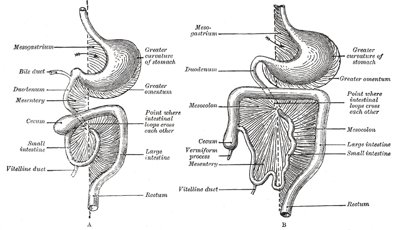
In human anatomy, the mesentery is an organ that attaches the intestines to the posterior abdominal wall, consisting of a double fold of the peritoneum. It helps (among other functions) in storing fat and allowing blood vessels, lymphatics, and nerves to supply the intestines. The (the part of the mesentery that attaches the colon to the abdominal wall) was formerly thought to be a fragmented structure, with all named parts—the ascending, transverse, descending, and sigmoid mesocolons, the mesoappendix, and the mesorectum—separately terminating their insertion into the posterior abdominal wall. However, in 2012, new microscopic and electron microscopic examinations showed the mesocolon to be a single structure derived from the duodenojejunal flexure and extending to the distal mesorectal layer. Thus the mesentery is an internal organ. Structure The mesentery of the small intestine arises from the root of the mesentery (or mesenteric root) and is the part connected wit ... [...More Info...] [...Related Items...] OR: [Wikipedia] [Google] [Baidu] |
Ascending Colon
In the anatomy of humans and homologous primates, the ascending colon is the part of the colon located between the cecum and the transverse colon. Characteristics and structure The ascending colon is smaller in calibre than the cecum from where it starts. It passes upward, opposite the colic valve, to the under surface of the right lobe of the liver, on the right of the gall-bladder, where it is lodged in a shallow depression, the colic impression; here it bends abruptly forward and to the left, forming the right colic flexure (hepatic) where it becomes the transverse colon. It is retained in contact with the posterior wall of the abdomen by the peritoneum, which covers its anterior surface and sides, its posterior surface being connected by loose areolar tissue with the iliacus, quadratus lumborum, aponeurotic origin of transversus abdominis, and with the front of the lower and lateral part of the right kidney. Sometimes the peritoneum completely invests it and f ... [...More Info...] [...Related Items...] OR: [Wikipedia] [Google] [Baidu] |
Appendix (anatomy)
The appendix (: appendices or appendixes; also vermiform appendix; cecal (or caecal, cæcal) appendix; vermix; or vermiform process) is a finger-like, blind-ended tube connected to the cecum, from which it develops in the embryo. The cecum is a pouch-like structure of the large intestine, located at the junction of the small and the large intestines. The term " vermiform" comes from Latin and means "worm-shaped". The appendix was once considered a vestigial organ, but this view has changed since the early 2000s. Research suggests that the appendix may serve as a reservoir for beneficial gut bacteria. Structure The human appendix averages in length, ranging from . The diameter of the appendix is , and more than is considered a thickened or inflamed appendix. The longest appendix ever removed was long. The appendix is usually located in the lower right quadrant of the abdomen, near the right hip bone. The base of the appendix is located beneath the ileocecal valve tha ... [...More Info...] [...Related Items...] OR: [Wikipedia] [Google] [Baidu] |
Ileum
The ileum () is the final section of the small intestine in most higher vertebrates, including mammals, reptiles, and birds. In fish, the divisions of the small intestine are not as clear and the terms posterior intestine or distal intestine may be used instead of ileum. Its main function is to absorb vitamin B12, vitamin B12, bile salts, and whatever products of digestion that were not absorbed by the jejunum. The ileum follows the duodenum and jejunum and is separated from the cecum by the ileocecal valve (ICV). In humans, the ileum is about 2–4 m long, and the pH is usually between 7 and 8 (neutral or slightly base (chemistry), basic). ''Ileum'' is derived from the Greek word εἰλεός (eileós), referring to a medical condition known as ileus. Structure The ileum is the third and final part of the small intestine. It follows the jejunum and ends at the ileocecal junction, where the wikt:terminal, terminal ileum communicates with the cecum of the large intestine thro ... [...More Info...] [...Related Items...] OR: [Wikipedia] [Google] [Baidu] |
Mesentery
In human anatomy, the mesentery is an Organ (anatomy), organ that attaches the intestines to the posterior abdominal wall, consisting of a double fold of the peritoneum. It helps (among other functions) in storing Adipose tissue, fat and allowing blood vessels, lymphatics, and nerves to supply the intestines. The (the part of the mesentery that attaches the colon to the abdominal wall) was formerly thought to be a fragmented structure, with all named parts—the ascending, transverse, descending, and sigmoid mesocolons, the mesoappendix, and the mesorectum—separately terminating their insertion into the posterior abdominal wall. However, in 2012, new microscopy, microscopic and electron microscope, electron microscopic histology, examinations showed the mesocolon to be a single structure derived from the duodenojejunal flexure and extending to the distal mesorectal layer. Thus the mesentery is an internal organ. Structure The mesentery of the small intestine arises from th ... [...More Info...] [...Related Items...] OR: [Wikipedia] [Google] [Baidu] |


