|
Ascending Colon
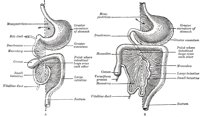
In the anatomy of humans and homologous primates, the ascending colon is the part of the colon located between the cecum and the transverse colon. Characteristics and structure The ascending colon is smaller in calibre than the cecum from where it starts. It passes upward, opposite the colic valve, to the under surface of the right lobe of the liver, on the right of the gall-bladder, where it is lodged in a shallow depression, the colic impression; here it bends abruptly forward and to the left, forming the right colic flexure (hepatic) where it becomes the transverse colon. It is retained in contact with the posterior wall of the abdomen by the peritoneum, which covers its anterior surface and sides, its posterior surface being connected by loose areolar tissue with the iliacus, quadratus lumborum, aponeurotic origin of transversus abdominis, and with the front of the lower and lateral part of the right kidney. Sometimes the peritoneum completely invests it and f ... [...More Info...] [...Related Items...] OR: [Wikipedia] [Google] [Baidu] [Amazon] |
Colon (anatomy)
The large intestine, also known as the large bowel, is the last part of the gastrointestinal tract and of the digestive system in tetrapods. Water is absorbed here and the remaining waste material is stored in the rectum as feces before being removed by defecation. The colon (progressing from the ascending colon to the transverse, the descending and finally the sigmoid colon) is the longest portion of the large intestine, and the terms "large intestine" and "colon" are often used interchangeably, but most sources define the large intestine as the combination of the cecum, colon, rectum, and anal canal. Some other sources exclude the anal canal. In humans, the large intestine begins in the right iliac region of the pelvis, just at or below the waist, where it is joined to the end of the small intestine at the cecum, via the ileocecal valve. It then continues as the colon ascending the abdomen, across the width of the abdominal cavity as the transverse colon, and then desce ... [...More Info...] [...Related Items...] OR: [Wikipedia] [Google] [Baidu] [Amazon] |
Colic Impression
The liver is a major metabolic organ (anatomy), organ exclusively found in vertebrates, which performs many essential biological Function (biology), functions such as detoxification of the organism, and the Protein biosynthesis, synthesis of various proteins and various other Biochemistry, biochemicals necessary for digestion and growth. In humans, it is located in the quadrants and regions of abdomen, right upper quadrant of the abdomen, below the thoracic diaphragm, diaphragm and mostly shielded by the lower right rib cage. Its other metabolic roles include carbohydrate metabolism, the production of a number of hormones, conversion and storage of nutrients such as glucose and glycogen, and the decomposition of red blood cells. Anatomical and medical terminology often use the prefix List of medical roots, suffixes and prefixes#H, ''hepat-'' from ἡπατο-, from the Greek language, Greek word for liver, such as hepatology, and hepatitis The liver is also an accessory digestive ... [...More Info...] [...Related Items...] OR: [Wikipedia] [Google] [Baidu] [Amazon] |
Splanchnic Nerves
The splanchnic nerves are paired visceral nerves (nerves that contribute to the innervation of the internal organs), carrying fibers of the autonomic nervous system ( visceral efferent fibers) as well as sensory fibers from the organs ( visceral afferent fibers). All carry sympathetic fibers except for the pelvic splanchnic nerves, which carry parasympathetic fibers. Types The term ''splanchnic nerves'' can refer to: * Cardiopulmonary nervesEssential Clinical Anatomy. K.L. Moore & A.M. Agur. Lippincott, 3 ed. 2007. Page 181 * Thoracic splanchnic nerves (greater, lesser, and least) * Lumbar splanchnic nerves * Sacral splanchnic nerves * Pelvic splanchnic nerves See also * Terminal cisterna * Rexed lamina * Preganglionic nerve fiber * Postganglionic nerve fiber In the autonomic nervous system, nerve fibers from the ganglion to the effector organ are called postganglionic nerve fibers. Neurotransmitters The neurotransmitters of postganglionic fibers differ: * In t ... [...More Info...] [...Related Items...] OR: [Wikipedia] [Google] [Baidu] [Amazon] |
Sympathetic Nervous System
The sympathetic nervous system (SNS or SANS, sympathetic autonomic nervous system, to differentiate it from the somatic nervous system) is one of the three divisions of the autonomic nervous system, the others being the parasympathetic nervous system and the enteric nervous system. The enteric nervous system is sometimes considered part of the autonomic nervous system, and sometimes considered an independent system. The autonomic nervous system functions to regulate the body's unconscious actions. The sympathetic nervous system's primary process is to stimulate the body's fight or flight response. It is, however, constantly active at a basic level to maintain homeostasis. The sympathetic nervous system is described as being antagonistic to the parasympathetic nervous system. The latter stimulates the body to "feed and breed" and to (then) "rest-and-digest". The SNS has a major role in various physiological processes such as blood glucose levels, body temperature, cardiac output, ... [...More Info...] [...Related Items...] OR: [Wikipedia] [Google] [Baidu] [Amazon] |
Vagus Nerve
The vagus nerve, also known as the tenth cranial nerve (CN X), plays a crucial role in the autonomic nervous system, which is responsible for regulating involuntary functions within the human body. This nerve carries both sensory and motor fibers and serves as a major pathway that connects the brain to various organs, including the heart, lungs, and digestive tract. As a key part of the parasympathetic nervous system, the vagus nerve helps regulate essential involuntary functions like heart rate, breathing, and digestion. By controlling these processes, the vagus nerve contributes to the body's "rest and digest" response, helping to calm the body after stress, lower heart rate, improve digestion, and maintain homeostasis. The vagus nerve consists of two branches: the right and left vagus nerves. In the neck, the right vagus nerve contains approximately 105,000 fibers, while the left vagus nerve has about 87,000 fibers, according to one source. However, other sources report sl ... [...More Info...] [...Related Items...] OR: [Wikipedia] [Google] [Baidu] [Amazon] |
Parasympathetic Nervous System
The parasympathetic nervous system (PSNS) is one of the three divisions of the autonomic nervous system, the others being the sympathetic nervous system and the enteric nervous system. The autonomic nervous system is responsible for regulating the body's unconscious actions. The parasympathetic system is responsible for stimulation of "rest-and-digest" or "feed-and-breed" activities that occur when the body is at rest, especially after eating, including sexual arousal, salivation, lacrimation (tears), urination, digestion, and defecation. Its action is described as being complementary to that of the sympathetic nervous system, which is responsible for stimulating activities associated with the fight-or-flight response. Nerve fibres of the parasympathetic nervous system arise from the central nervous system. Specific nerves include several cranial nerves, specifically the oculomotor nerve, facial nerve, glossopharyngeal nerve, and vagus nerve. Three spinal nerves ... [...More Info...] [...Related Items...] OR: [Wikipedia] [Google] [Baidu] [Amazon] |
Ileum
The ileum () is the final section of the small intestine in most higher vertebrates, including mammals, reptiles, and birds. In fish, the divisions of the small intestine are not as clear and the terms posterior intestine or distal intestine may be used instead of ileum. Its main function is to absorb vitamin B12, vitamin B12, bile salts, and whatever products of digestion that were not absorbed by the jejunum. The ileum follows the duodenum and jejunum and is separated from the cecum by the ileocecal valve (ICV). In humans, the ileum is about 2–4 m long, and the pH is usually between 7 and 8 (neutral or slightly base (chemistry), basic). ''Ileum'' is derived from the Greek word εἰλεός (eileós), referring to a medical condition known as ileus. Structure The ileum is the third and final part of the small intestine. It follows the jejunum and ends at the ileocecal junction, where the wikt:terminal, terminal ileum communicates with the cecum of the large intestine thro ... [...More Info...] [...Related Items...] OR: [Wikipedia] [Google] [Baidu] [Amazon] |
Mesocolon
In human anatomy, the mesentery is an organ that attaches the intestines to the posterior abdominal wall, consisting of a double fold of the peritoneum. It helps (among other functions) in storing fat and allowing blood vessels, lymphatics, and nerves to supply the intestines. The (the part of the mesentery that attaches the colon to the abdominal wall) was formerly thought to be a fragmented structure, with all named parts—the ascending, transverse, descending, and sigmoid mesocolons, the mesoappendix, and the mesorectum—separately terminating their insertion into the posterior abdominal wall. However, in 2012, new microscopic and electron microscopic examinations showed the mesocolon to be a single structure derived from the duodenojejunal flexure and extending to the distal mesorectal layer. Thus the mesentery is an internal organ. Structure The mesentery of the small intestine arises from the root of the mesentery (or mesenteric root) and is the part connected ... [...More Info...] [...Related Items...] OR: [Wikipedia] [Google] [Baidu] [Amazon] |
Transversus Abdominis
The transverse abdominal muscle (TVA), also known as the transverse abdominis, transversalis muscle and transversus abdominis muscle, is a muscle layer of the anterior and lateral (front and side) abdominal wall, deep to (layered below) the internal oblique muscle. It serves to compress and retain the contents of the abdomen as well as assist in exhalation. Structure The transverse abdominal, so called for the direction of its fibers, is the innermost of the flat muscles of the abdomen. It is positioned immediately deep to the internal oblique muscle. The transverse abdominal arises as fleshy fibers, from the lateral third of the inguinal ligament, from the anterior three-fourths of the inner lip of the iliac crest, from the inner surfaces of the cartilages of the lower six ribs, interdigitating with the diaphragm, and from the thoracolumbar fascia. It ends anteriorly in a broad aponeurosis (the Spigelian fascia), the lower fibers of which curve inferomedially (medially and do ... [...More Info...] [...Related Items...] OR: [Wikipedia] [Google] [Baidu] [Amazon] |
Aponeurotic
An aponeurosis (; : aponeuroses) is a flattened tendon by which muscle attaches to bone or fascia. Aponeuroses exhibit an ordered arrangement of collagen fibres, thus attaining high tensile strength in a particular direction while being vulnerable to tensional or shear forces in other directions. They have a shiny, whitish-silvery color, are histologically similar to tendons, and are very sparingly supplied with blood vessels and nerves. When dissected, aponeuroses are papery and peel off by sections. The primary regions with thick aponeuroses are in the ventral abdominal region, the dorsal lumbar region, the ventriculus in birds, and the palmar (palms) and plantar (soles) regions. Anatomy Anterior abdominal aponeuroses The anterior abdominal aponeuroses are located just superficial to the rectus abdominis muscle. It has for its borders the external oblique, pectoralis muscles, and the latissimus dorsi. Posterior lumbar aponeuroses The posterior lumbar aponeuroses ar ... [...More Info...] [...Related Items...] OR: [Wikipedia] [Google] [Baidu] [Amazon] |
Quadratus Lumborum
The quadratus lumborum muscle, informally called the ''QL'', is a paired muscle of the left and right posterior abdominal wall. It is the deepest abdominal muscle, and commonly referred to as a back muscle. Each muscle of the pair is an irregular quadrilateral in shape, hence the name. The quadratus lumborum muscles originate from the wings of the ilium; their insertions are on the transverse processes of the upper four lumbar vertebrae plus the lower posterior border of the twelfth rib. Contraction of one of the pair of muscles causes lateral flexion of the lumbar spine, ''elevation'' of the pelvis, or both. Contraction of both causes ''extension'' of the lumbar spine. A disorder of the quadratus lumborum muscles is pain due to muscle fatigue from constant contraction due to prolonged sitting, such as at a computer or in a car.Core Topics in Pain, p. 131, Anita Holdcraft and Sian Jaggar, 2005. Kyphosis and weak gluteal muscles can also contribute to the likelihood of quadratu ... [...More Info...] [...Related Items...] OR: [Wikipedia] [Google] [Baidu] [Amazon] |
Iliacus
The iliacus is a flat, triangular muscle which fills the iliac fossa. It forms the lateral portion of iliopsoas, providing flexion of the thigh and lower limb at the acetabulofemoral joint. Structure The iliacus arises from the iliac fossa on the interior side of the hip bone, and also from the region of the anterior inferior iliac spine (AIIS). It joins the psoas major to form the iliopsoas. It proceeds across the iliopubic eminence through the muscular lacuna to its insertion on the lesser trochanter of the femur. Its fibers are often inserted in front of those of the psoas major and extend distally over the lesser trochanter.Platzer (2004), p 234 Nerve supply The iliopsoas is innervated by the femoral nerve and direct branches from the lumbar plexus.''Thieme Atlas of Anatomy'' (2006), p 422 Function In open-chain exercises, as part of the iliopsoas, the iliacus is important for lifting (flexing) the femur forward (e.g. front scale). In closed-chain exercises, the iliops ... [...More Info...] [...Related Items...] OR: [Wikipedia] [Google] [Baidu] [Amazon] |


