|
Subparabrachial Nucleus
The parabrachial nuclei, also known as the parabrachial complex, are a group of nuclei in the dorsolateral pons that surrounds the superior cerebellar peduncle as it enters the brainstem from the cerebellum. They are named from the Latin term for the superior cerebellar peduncle, the ''brachium conjunctivum''. In the human brain, the expansion of the superior cerebellar peduncle expands the parabrachial nuclei, which form a thin strip of grey matter over most of the peduncle. The parabrachial nuclei are typically divided along the lines suggested by Baxter and Olszewski in humans, into a medial parabrachial nucleus and lateral parabrachial nucleus. These have in turn been subdivided into a dozen subnuclei: the superior, dorsal, ventral, internal, external and extreme lateral subnuclei; the lateral crescent and subparabrachial nucleus (Kolliker-Fuse nucleus) along the ventrolateral margin of the lateral parabrachial complex; and the medial and external medial subnuclei Anatomy ... [...More Info...] [...Related Items...] OR: [Wikipedia] [Google] [Baidu] |
Brainstem
The brainstem (or brain stem) is the posterior stalk-like part of the brain that connects the cerebrum with the spinal cord. In the human brain the brainstem is composed of the midbrain, the pons, and the medulla oblongata. The midbrain is continuous with the thalamus of the diencephalon through the tentorial notch, and sometimes the diencephalon is included in the brainstem. The brainstem is very small, making up around only 2.6 percent of the brain's total weight. It has the critical roles of regulating heart and respiratory system, respiratory function, helping to control heart rate and breathing rate. It also provides the main motor and sensory nerve supply to the face and neck via the cranial nerves. Ten pairs of cranial nerves come from the brainstem. Other roles include the regulation of the central nervous system and the body's sleep cycle. It is also of prime importance in the conveyance of motor and sensory pathways from the rest of the brain to the body, and from the b ... [...More Info...] [...Related Items...] OR: [Wikipedia] [Google] [Baidu] |
Human Anatomical Terms
Anatomical terminology is a specialized system of terms used by anatomists, zoologists, and health professionals, such as doctors, surgeons, and pharmacists, to describe the structures and functions of the body. This terminology incorporates a range of unique terms, prefixes, and suffixes derived primarily from Ancient Greek and Latin. While these terms can be challenging for those unfamiliar with them, they provide a level of precision that reduces ambiguity and minimizes the risk of errors. Because anatomical terminology is not commonly used in everyday language, its meanings are less likely to evolve or be misinterpreted. For example, everyday language can lead to confusion in descriptions: the phrase "a scar above the wrist" could refer to a location several inches away from the hand, possibly on the forearm, or it could be at the base of the hand, either on the palm or dorsal (back) side. By using precise anatomical terms, such as "proximal," "distal," "palmar," or "do ... [...More Info...] [...Related Items...] OR: [Wikipedia] [Google] [Baidu] |
Hypoxia (medical)
Hypoxia is a condition in which the body or a region of the body is deprived of an adequate oxygen supply at the tissue (biology), tissue level. Hypoxia may be classified as either ''Generalized hypoxia, generalized'', affecting the whole body, or ''local'', affecting a region of the body. Although hypoxia is often a pathological condition, variations in arterial oxygen concentrations can be part of the normal physiology, for example, during strenuous physical exercise. Hypoxia differs from hypoxemia and anoxemia, in that hypoxia refers to a state in which oxygen present in a tissue or the whole body is insufficient, whereas hypoxemia and anoxemia refer specifically to states that have low or no Oxygen saturation (medicine), oxygen in the blood. Hypoxia in which there is complete absence of oxygen supply is referred to as anoxia. Hypoxia can be due to external causes, when the breathing gas is hypoxic, or internal causes, such as reduced effectiveness of gas transfer in the lung ... [...More Info...] [...Related Items...] OR: [Wikipedia] [Google] [Baidu] |
Calcitonin Gene-related Peptide
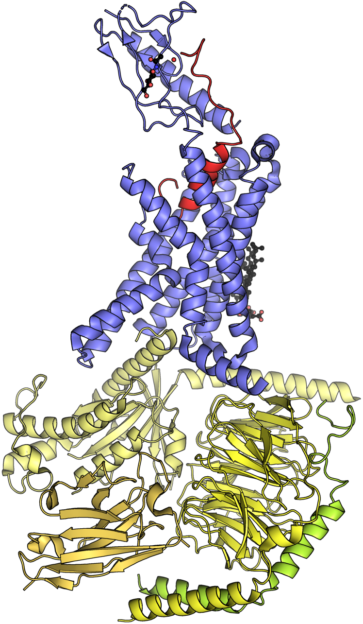
Calcitonin gene-related peptide (CGRP) is a neuropeptide that belongs to the calcitonin family. Human CGRP consists of two Protein isoform, isoforms, CGRP alpha (α-CGRP, also known as CGRP I) and CGRP beta (β-CGRP, also known as CGRP II). α-CGRP is a 37-amino acid neuropeptide formed by alternative splicing of the calcitonin/CGRP gene located on chromosome 11. β-CGRP is less studied. In humans, β-CGRP differs from α-CGRP by three amino acids and is encoded in a separate, nearby gene. The CGRP family includes calcitonin (CT), adrenomedullin (AM), and amylin (AMY). Function CGRP is produced in both peripheral and central neurons. It is a potent peptide vasodilator and can function in the transmission of nociception. In the spinal cord, the function and expression of CGRP may differ depending on the location of synthesis. CGRP is derived mainly from the cell bodies of motor neurons when synthesized in the Ventral horn of spinal cord, ventral horn of the spinal cord and may c ... [...More Info...] [...Related Items...] OR: [Wikipedia] [Google] [Baidu] |
Neuropeptide
Neuropeptides are chemical messengers made up of small chains of amino acids that are synthesized and released by neurons. Neuropeptides typically bind to G protein-coupled receptors (GPCRs) to modulate neural activity and other tissues like the gut, muscles, and heart. Neuropeptides are synthesized from large precursor proteins which are cleaved and post-translationally processed then packaged into large dense core vesicles. Neuropeptides are often co-released with other neuropeptides and neurotransmitters in a single neuron, yielding a multitude of effects. Once released, neuropeptides can diffuse widely to affect a broad range of targets. Neuropeptides are extremely ancient and highly diverse chemical messengers. Placozoa, Placozoans such as ''Trichoplax'', extremely basal animals which do not possess neurons, use peptides for cell-to-cell communication in a way similar to the neuropeptides of higher animals. Examples Peptide signals play a role in information processing t ... [...More Info...] [...Related Items...] OR: [Wikipedia] [Google] [Baidu] |
Forebrain
In the anatomy of the brain of vertebrates, the forebrain or prosencephalon is the rostral (forward-most) portion of the brain. The forebrain controls body temperature, reproductive functions, eating, sleeping, and the display of emotions. Vesicles of the forebrain (prosencephalon), the midbrain (mesencephalon), and hindbrain (rhombencephalon) are the three primary brain vesicles during the early development of the nervous system. At the five-vesicle stage, the forebrain separates into the diencephalon (thalamus, hypothalamus, subthalamus, and epithalamus) and the telencephalon which develops into the cerebrum. The cerebrum consists of the cerebral cortex, underlying white matter, and the basal ganglia. In humans, by 5 weeks in utero it is visible as a single portion toward the front of the fetus. At 8 weeks in utero, the forebrain splits into the left and right cerebral hemispheres. When the embryonic forebrain fails to divide the brain into two lobes, it results i ... [...More Info...] [...Related Items...] OR: [Wikipedia] [Google] [Baidu] |
Ventrolateral Medulla
The ventrolateral medulla, part of the medulla oblongata of the brainstem, plays a major role in regulating arterial blood pressure and breathing. It regulates blood pressure by regulating the activity of the sympathetic nerves that target the heart and peripheral blood vessels. The ventrolateral medulla consists of a rostral ventrolateral medulla (RVLM) and a caudal ventrolateral medulla (CVLM). Neurons in the RVLM project directly to preganglionic neurons in the spinal cord and maintain tonic activity in the sympathetic vasomotor nerves. This activity is inhibited by GABA GABA (gamma-aminobutyric acid, γ-aminobutyric acid) is the chief inhibitory neurotransmitter in the developmentally mature mammalian central nervous system. Its principal role is reducing neuronal excitability throughout the nervous system. GA ... output from the CVLM. References Sympathetic nervous system Reflexes Medulla oblongata Cardiovascular physiology {{neuroanatomy-stub ... [...More Info...] [...Related Items...] OR: [Wikipedia] [Google] [Baidu] |
Intralaminar Thalamic Nuclei
The intralaminar thalamic nuclei (ITN) are collections of neurons in the medullary laminae of thalamus, internal medullary lamina of the thalamus.Mancall, E., Brock, D. & Gray, H. (2011). Gray's clinical neuroanatomy the anatomic basis for clinical neuroscience. Philadelphia: Elsevier/Saunders. Anatomy Structure The ITN are generally divided in two groups as follows: * anterior (rostral) group ** central lateral nucleus ** central medial nucleus (''not'' referred to as "centromedial") ** paracentral nucleus * posterior (caudal) intralaminar group ** centromedian nucleus ** parafascicular nucleus Some sources also include a "central dorsal" nucleus. Afferents Midline intralaminar nuclei receive afferents from the brain stem, spinal cord, and cerebellum. Connections with the cerebral cortex and basal nuclei are reciprocal. Afferents from the spinothalamic tract as well as periaqueductal gray are part of a pathway involved in pain processing. Efferents The intralaminar nucl ... [...More Info...] [...Related Items...] OR: [Wikipedia] [Google] [Baidu] |
Taste
The gustatory system or sense of taste is the sensory system that is partially responsible for the perception of taste. Taste is the perception stimulated when a substance in the mouth biochemistry, reacts chemically with taste receptor cells located on taste buds in the oral cavity, mostly on the tongue. Taste, along with olfaction, the sense of smell and trigeminal nerve stimulation (registering texture, pain, and temperature), determines Flavoring, flavors of food and other substances. Humans have taste receptors on taste buds and other areas, including the upper surface of the tongue and the epiglottis. The gustatory cortex is responsible for the perception of taste. The tongue is covered with thousands of small bumps called lingual papillae, papillae, which are visible to the naked eye. Within each papilla are hundreds of taste buds. The exceptions to this is the filiform papillae that do not contain taste buds. There are between 2000 and 5000Boron, W.F., E.L. Boulpaep. 200 ... [...More Info...] [...Related Items...] OR: [Wikipedia] [Google] [Baidu] |
Special Visceral Afferent
Special visceral afferent fibers (SVA) are afferent fibers that develop in association with the gastrointestinal tract. They carry the special sense of taste (gustation). The cranial nerves containing SVA fibers are the facial nerve (VII), the glossopharyngeal nerve (IX), and the vagus nerve (X). The facial nerve receives taste from the anterior 2/3 of the tongue; the glossopharyngeal from the posterior 1/3, and the vagus nerve from the epiglottis. The sensory processes, using their primary cell bodies from the inferior ganglion, send projections to the medulla, from which they travel in the tractus solitarius, later terminating at the rostral nucleus solitarius.Bhatnagar C. Subhash. ''Neuroscience for the study of communicative disorders''. Philadelphia, Pennsylvania. Lippincott Williams & Wilkins, 2002 See also * General somatic afferent fiber (GSA) * General visceral afferent fiber The general visceral afferent (GVA) fibers conduct sensory impulses (usually pain or reflex sen ... [...More Info...] [...Related Items...] OR: [Wikipedia] [Google] [Baidu] |
Amygdala
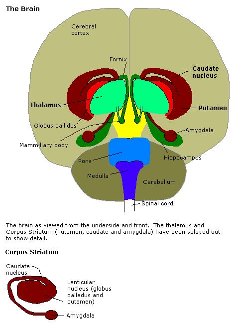
The amygdala (; : amygdalae or amygdalas; also '; Latin from Greek language, Greek, , ', 'almond', 'tonsil') is a paired nucleus (neuroanatomy), nuclear complex present in the Cerebral hemisphere, cerebral hemispheres of vertebrates. It is considered part of the limbic system. In Primate, primates, it is located lateral and medial, medially within the temporal lobes. It consists of many nuclei, each made up of further subnuclei. The subdivision most commonly made is into the Basolateral amygdala, basolateral, Central nucleus of the amygdala, central, cortical, and medial nuclei together with the intercalated cells of the amygdala, intercalated cell clusters. The amygdala has a primary role in the processing of memory, decision making, decision-making, and emotions, emotional responses (including fear, anxiety, and aggression). The amygdala was first identified and named by Karl Friedrich Burdach in 1822. Structure Thirteen Nucleus (neuroanatomy), nuclei have been identif ... [...More Info...] [...Related Items...] OR: [Wikipedia] [Google] [Baidu] |
Spinal Cord
The spinal cord is a long, thin, tubular structure made up of nervous tissue that extends from the medulla oblongata in the lower brainstem to the lumbar region of the vertebral column (backbone) of vertebrate animals. The center of the spinal cord is hollow and contains a structure called the central canal, which contains cerebrospinal fluid. The spinal cord is also covered by meninges and enclosed by the neural arches. Together, the brain and spinal cord make up the central nervous system. In humans, the spinal cord is a continuation of the brainstem and anatomically begins at the occipital bone, passing out of the foramen magnum and then enters the spinal canal at the beginning of the cervical vertebrae. The spinal cord extends down to between the first and second lumbar vertebrae, where it tapers to become the cauda equina. The enclosing bony vertebral column protects the relatively shorter spinal cord. It is around long in adult men and around long in adult women. The diam ... [...More Info...] [...Related Items...] OR: [Wikipedia] [Google] [Baidu] |


