|
Spiral Arteries
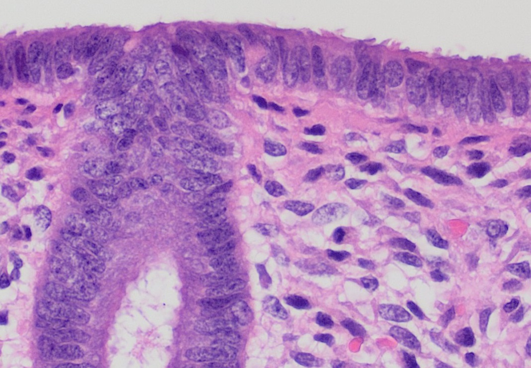
Spiral arteries are small arteries which temporarily supply blood to the endometrium of the uterus during the luteal phase of the menstrual cycle. In histology, identifying the presence of these arteries is one of the most useful techniques in identifying the phase of the cycle. The spiral arteries are converted for uteroplacental blood flow during pregnancy, involving: * Loss of smooth muscle and elastic lamina from the vessel wall. * 5-10 fold dilation at the mouth of the vessel. Failure of the physiological conversion of the spiral arteries can cause a number of complications, including intrauterine growth restriction and pre-eclampsia Pre-eclampsia is a multi-system disorder specific to pregnancy, characterized by the new onset of hypertension, high blood pressure and often a significant amount of proteinuria, protein in the urine or by the new onset of high blood pressure a .... References Arteries of the abdomen articles {{circulatory-stub ... [...More Info...] [...Related Items...] OR: [Wikipedia] [Google] [Baidu] |
Uterine Arterial Vasculature
The uterus (from Latin ''uterus'', : uteri or uteruses) or womb () is the organ in the reproductive system of most female mammals, including humans, that accommodates the embryonic and fetal development of one or more fertilized eggs until birth. The uterus is a hormone-responsive sex organ that contains glands in its lining that secrete uterine milk for embryonic nourishment. (The term ''uterus'' is also applied to analogous structures in some non-mammalian animals.) In humans, the lower end of the uterus is a narrow part known as the isthmus that connects to the cervix, the anterior gateway leading to the vagina. The upper end, the body of the uterus, is connected to the fallopian tubes at the uterine horns; the rounded part, the fundus, is above the openings to the fallopian tubes. The connection of the uterine cavity with a fallopian tube is called the uterotubal junction. The fertilized egg is carried to the uterus along the fallopian tube. It will have divided on its jou ... [...More Info...] [...Related Items...] OR: [Wikipedia] [Google] [Baidu] |
Endometrium
The endometrium is the inner epithelium, epithelial layer, along with its mucous membrane, of the mammalian uterus. It has a basal layer and a functional layer: the basal layer contains stem cells which regenerate the functional layer. The functional layer thickens and then is shed during menstruation in humans and some other mammals, including other apes, Old World monkeys, some species of bat, the elephant shrew and the Cairo spiny mouse. In most other mammals, the endometrium is reabsorbed in the estrous cycle. During pregnancy, the glands and blood vessels in the endometrium further increase in size and number. Vascular spaces fuse and become interconnected, forming the placenta, which supplies oxygen and nutrition to the embryo and fetus.Blue Histology - Female Reproductive System ... [...More Info...] [...Related Items...] OR: [Wikipedia] [Google] [Baidu] |
Uterus
The uterus (from Latin ''uterus'', : uteri or uteruses) or womb () is the hollow organ, organ in the reproductive system of most female mammals, including humans, that accommodates the embryonic development, embryonic and prenatal development, fetal development of one or more Fertilized egg, fertilized eggs until birth. The uterus is a hormone-responsive sex organ that contains uterine gland, glands in its endometrium, lining that secrete uterine milk for embryonic nourishment. (The term ''uterus'' is also applied to analogous structures in some non-mammalian animals.) In humans, the lower end of the uterus is a narrow part known as the Uterine isthmus, isthmus that connects to the cervix, the anterior gateway leading to the vagina. The upper end, the body of the uterus, is connected to the fallopian tubes at the uterine horns; the rounded part, the fundus, is above the openings to the fallopian tubes. The connection of the uterine cavity with a fallopian tube is called the utero ... [...More Info...] [...Related Items...] OR: [Wikipedia] [Google] [Baidu] |
Luteal Phase
The menstrual cycle is on average 28 days in length. It begins with Menstruation, menses (day 1–7) during the follicular phase (day 1–14), followed by ovulation (day 14) and ending with the luteal phase (day 14–28). While historically, medical experts believed the luteal phase to be relatively fixed at approximately 14 days (i.e. days 14–28), recent research suggests that there can be wide variability in luteal phase lengths not just from person to person, but from cycle to cycle within one person. The luteal phase is characterized by changes to hormone levels, such as an increase in progesterone and estrogen levels, decrease in gonadotropins such as follicle-stimulating hormone (FSH) and luteinizing hormone (LH), changes to the Endometrium, endometrial lining to promote Implantation (embryology), implantation of the fertilized egg, and development of the corpus luteum. In the absence of fertilization by sperm, the corpus luteum degenerates leading to a decrease in progeste ... [...More Info...] [...Related Items...] OR: [Wikipedia] [Google] [Baidu] |
Menstrual Cycle
The menstrual cycle is a series of natural changes in hormone production and the structures of the uterus and ovaries of the female reproductive system that makes pregnancy possible. The ovarian cycle controls the production and release of eggs and the cyclic release of estrogen and progesterone. The uterine cycle governs the preparation and maintenance of the lining of the uterus (womb) to receive an embryo. These cycles are concurrent and coordinated, normally last between 21 and 35 days, with a median length of 28 days. Menarche (the onset of the first period) usually occurs around the age of 12 years; menstrual cycles continue for about 30–45 years. Naturally occurring hormones drive the cycles; the cyclical rise and fall of the follicle stimulating hormone prompts the production and growth of oocytes (immature egg cells). The hormone estrogen stimulates the uterus lining ( endometrium) to thicken to accommodate an embryo should fertilization occur. The blood suppl ... [...More Info...] [...Related Items...] OR: [Wikipedia] [Google] [Baidu] |
Histology
Histology, also known as microscopic anatomy or microanatomy, is the branch of biology that studies the microscopic anatomy of biological tissue (biology), tissues. Histology is the microscopic counterpart to gross anatomy, which looks at larger structures visible without a microscope. Although one may divide microscopic anatomy into ''organology'', the study of organs, ''histology'', the study of tissues, and ''cytology'', the study of cell (biology), cells, modern usage places all of these topics under the field of histology. In medicine, histopathology is the branch of histology that includes the microscopic identification and study of diseased tissue. In the field of paleontology, the term paleohistology refers to the histology of fossil organisms. Biological tissues Animal tissue classification There are four basic types of animal tissues: muscle tissue, nervous tissue, connective tissue, and epithelial tissue. All animal tissues are considered to be subtypes of these ... [...More Info...] [...Related Items...] OR: [Wikipedia] [Google] [Baidu] |
Elastic Lamina
The internal elastic lamina or internal elastic lamella is a layer of elastic tissue that forms the outermost part of the tunica intima of blood vessels. It separates tunica intima from tunica media. Histology It is readily visualized with light microscopy in sections of muscular arteries, where it is thick and prominent, and arterioles, where it is slightly less prominent and often incomplete. It is very thin in veins and venules. In elastic arteries such as the aorta, which have very regular elastic laminae between layers of smooth muscle cells in their tunica media, the internal elastic lamina is approximately the same thickness as the other elastic laminae that are normally present. There is small amount of subendothelial connective tissue between basement membrane of endothelial cells and internal elastic lamina. Reduplication of internal elastic lamina can be seen in elderly individuals due to intimal fibroplasia, which is part of the aging process. Associated pathologic ... [...More Info...] [...Related Items...] OR: [Wikipedia] [Google] [Baidu] |
Intrauterine Growth Restriction
Intrauterine growth restriction (IUGR), or fetal growth restriction, is the poor growth of a fetus while in the womb during pregnancy. IUGR is defined by clinical features of malnutrition and evidence of reduced growth regardless of an infant's birth weight percentile. The causes of IUGR are broad and may involve maternal, fetal, or placental complications. At least 60% of the 4 million neonatal deaths that occur worldwide every year are associated with low birth weight, caused by intrauterine growth restriction (IUGR), preterm delivery, and genetic abnormalities, demonstrating that under-nutrition is already a leading health problem at birth. Intrauterine growth restriction can result in a baby being small for gestational age (SGA), which is most commonly defined as a weight below the 10th percentile for the gestational age. At the end of pregnancy, it can result in a low birth weight. Types There are two major categories of IUGR: pseudo IUGR and true IUGR With pseudo IU ... [...More Info...] [...Related Items...] OR: [Wikipedia] [Google] [Baidu] |
Pre-eclampsia
Pre-eclampsia is a multi-system disorder specific to pregnancy, characterized by the new onset of hypertension, high blood pressure and often a significant amount of proteinuria, protein in the urine or by the new onset of high blood pressure along with significant end-organ damage, with or without the proteinuria. When it arises, the condition begins after 20 Gestational age (obstetrics), weeks of pregnancy. In severe cases of the disease there may be hemolysis, red blood cell breakdown, a thrombocytopenia, low blood platelet count, impaired liver function, kidney dysfunction, edema, swelling, pulmonary edema, shortness of breath due to fluid in the lungs, or visual disturbances. Pre-eclampsia increases the risk of undesirable as well as lethal outcomes for both the mother and the fetus including preterm labor. If left untreated, it may result in seizures at which point it is known as eclampsia. Risk factors for pre-eclampsia include obesity, prior hypertension, older age, and ... [...More Info...] [...Related Items...] OR: [Wikipedia] [Google] [Baidu] |
Arteries Of The Abdomen
An artery () is a blood vessel in humans and most other animals that takes oxygenated blood away from the heart in the systemic circulation to one or more parts of the body. Exceptions that carry deoxygenated blood are the pulmonary arteries in the pulmonary circulation that carry blood to the lungs for oxygenation, and the umbilical arteries in the fetal circulation that carry deoxygenated blood to the placenta. It consists of a multi-layered artery wall wrapped into a tube-shaped channel. Arteries contrast with veins, which carry deoxygenated blood back towards the heart; or in the pulmonary and fetal circulations carry oxygenated blood to the lungs and fetus respectively. Structure The anatomy of arteries can be separated into gross anatomy, at the macroscopic level, and microanatomy, which must be studied with a microscope. The arterial system of the human body is divided into systemic arteries, carrying blood from the heart to the whole body, and pulmonary arteries, c ... [...More Info...] [...Related Items...] OR: [Wikipedia] [Google] [Baidu] |


