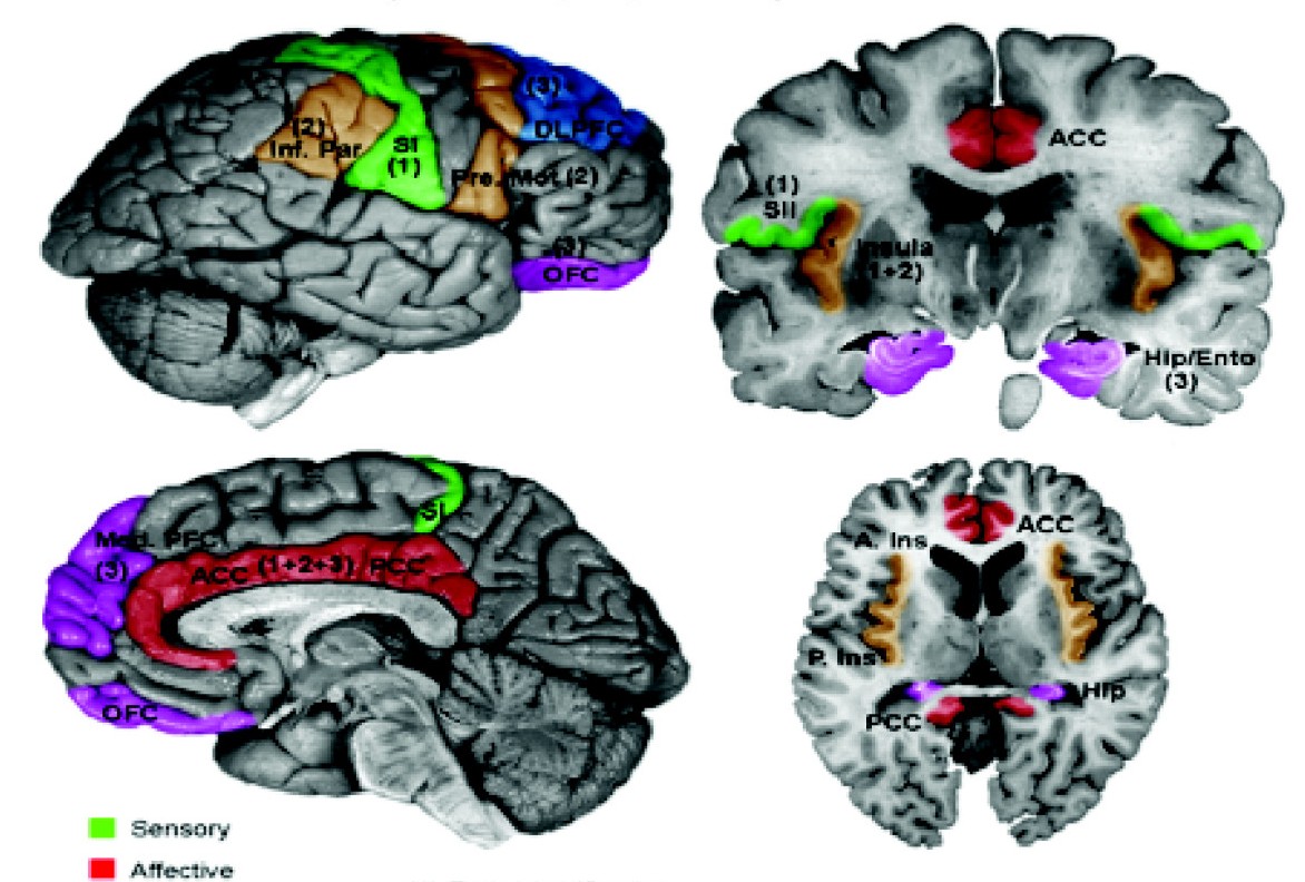
|
Spinoreticular Tract
The spinoreticular tract (also paleospinothalamic pathway, or indirect pathway of the anterolateral system) is a partially decussating (crossed-over) four-neuron sensory pathway of the central nervous system. The tract transmits slow nociceptive/pain information (but thermal, and crude touch information as well) from the spinal cord to reticular formation which in turn relays the information to the thalamus via reticulothalamic fibers as well as to other parts of the brain (as opposed to the spinothalamic tract - the direct pathway of the anterolateral system - which projects from the spinal cord to the thalamus directly without such "layovers"). Most (85%) second-order axons arising from sensory C first-order fibers ascend in the spinoreticular tract - it is consequently responsible for transmitting "slow", dull, poorly-localised pain. By projecting to the reticular activating system (RAS), the tract also mediates arousal/alertness (including wakefulness) in response to noxious ... [...More Info...] [...Related Items...] OR: [Wikipedia] [Google] [Baidu] |
Anterolateral System
The spinal cord is a long, thin, tubular structure made up of nervous tissue that extends from the medulla oblongata in the lower brainstem to the lumbar region of the vertebral column (backbone) of vertebrate animals. The center of the spinal cord is hollow and contains a structure called the central canal, which contains cerebrospinal fluid. The spinal cord is also covered by meninges and enclosed by the neural arches. Together, the brain and spinal cord make up the central nervous system. In humans, the spinal cord is a continuation of the brainstem and anatomically begins at the occipital bone, passing out of the foramen magnum and then enters the spinal canal at the beginning of the cervical vertebrae. The spinal cord extends down to between the first and second lumbar vertebrae, where it tapers to become the cauda equina. The enclosing bony vertebral column protects the relatively shorter spinal cord. It is around long in adult men and around long in adult women. The dia ... [...More Info...] [...Related Items...] OR: [Wikipedia] [Google] [Baidu] |
Autonomic Nervous System
The autonomic nervous system (ANS), sometimes called the visceral nervous system and formerly the vegetative nervous system, is a division of the nervous system that operates viscera, internal organs, smooth muscle and glands. The autonomic nervous system is a control system that acts largely unconsciously and regulates bodily functions, such as the heart rate, its Myocardial contractility, force of contraction, digestion, respiratory rate, pupillary dilation, pupillary response, Micturition, urination, and Animal sexual behaviour, sexual arousal. The fight-or-flight response, also known as the acute stress response, is set into action by the autonomic nervous system. The autonomic nervous system is regulated by integrated reflexes through the brainstem to the spinal cord and organ (anatomy), organs. Autonomic functions include control of respiration, heart rate, cardiac regulation (the cardiac control center), vasomotor activity (the vasomotor center), and certain reflex, reflex ... [...More Info...] [...Related Items...] OR: [Wikipedia] [Google] [Baidu] |
Hypothalamus
The hypothalamus (: hypothalami; ) is a small part of the vertebrate brain that contains a number of nucleus (neuroanatomy), nuclei with a variety of functions. One of the most important functions is to link the nervous system to the endocrine system via the pituitary gland. The hypothalamus is located below the thalamus and is part of the limbic system. It forms the Basal (anatomy), basal part of the diencephalon. All vertebrate brains contain a hypothalamus. In humans, it is about the size of an Almond#Nut, almond. The hypothalamus has the function of regulating certain metabolic biological process, processes and other activities of the autonomic nervous system. It biosynthesis, synthesizes and secretes certain neurohormones, called releasing hormones or hypothalamic hormones, and these in turn stimulate or inhibit the secretion of hormones from the pituitary gland. The hypothalamus controls thermoregulation, body temperature, hunger (physiology), hunger, important aspects o ... [...More Info...] [...Related Items...] OR: [Wikipedia] [Google] [Baidu] |
Enkephalin
An enkephalin is a pentapeptide involved in regulating nociception (pain sensation) in the body. The enkephalins are termed endogenous ligands, as they are internally derived (and therefore endogenous) and bind as ligands to the body's opioid receptors. Discovered in 1975, two forms of enkephalin have been found, one containing leucine ("leu"), and the other containing methionine ("met"). Both are products of the proenkephalin gene. * Met-enkephalin is Tyr-Gly-Gly-Phe-Met. * Leu-enkephalin is Tyr-Gly-Gly-Phe-Leu. Endogenous opioid peptides There are three well-characterized families of opioid peptides produced by the body: enkephalins, β-endorphin, and dynorphins. The met-enkephalin peptide sequence is coded for by the enkephalin gene; the leu-enkephalin peptide sequence is coded for by both the enkephalin gene and the dynorphin gene. The proopiomelanocortin gene ( POMC) also contains the met-enkephalin sequence on the N-terminus of beta-endorphin, but the endorphin pept ... [...More Info...] [...Related Items...] OR: [Wikipedia] [Google] [Baidu] |
Serotonin
Serotonin (), also known as 5-hydroxytryptamine (5-HT), is a monoamine neurotransmitter with a wide range of functions in both the central nervous system (CNS) and also peripheral tissues. It is involved in mood, cognition, reward, learning, memory, and physiological processes such as vomiting and vasoconstriction. In the CNS, serotonin regulates mood, appetite, and sleep. Most of the body's serotonin—about 90%—is synthesized in the gastrointestinal tract by enterochromaffin cells, where it regulates intestinal movements. It is also produced in smaller amounts in the brainstem's raphe nuclei, the skin's Merkel cells, pulmonary neuroendocrine cells, and taste receptor cells of the tongue. Once secreted, serotonin is taken up by platelets in the blood, which release it during clotting to promote vasoconstriction and platelet aggregation. Around 8% of the body's serotonin is stored in platelets, and 1–2% is found in the CNS. Serotonin acts as both a vasoconstrictor and vas ... [...More Info...] [...Related Items...] OR: [Wikipedia] [Google] [Baidu] |
Spinal Trigeminal Nucleus
The spinal trigeminal nucleus is a nucleus in the medulla that receives information about deep/crude touch, pain, and temperature from the ipsilateral face. In addition to the trigeminal nerve (CN V), the facial (CN VII), glossopharyngeal (CN IX), and vagus nerves (CN X) also convey pain information from their areas to the spinal trigeminal nucleus. Thus the spinal trigeminal nucleus receives afferents from V, VII, [...More Info...] [...Related Items...] OR: [Wikipedia] [Google] [Baidu] |
Raphespinal Tract
The raphespinal tract is a descending spinal cord tract located in the medulla oblongata. It consists of two tracts an anterior raphespinal tract, and a lateral raphespinal tract that mainly descend in the lateral funiculus. Fibers descend in the ventral portion of the lateral funiculus, mainly bilaterally to terminate in laminae I, II, and IV. The tract emerges from three of the raphe nuclei, the magnus, obscurus, and pallidus. The fibers of the raphespinal tract are mainly serotonergic. When raphe nuclei are stimulated they release serotonin which modulates the transmission of pain. Pathways Pain pathways converging upon the raphe nuclei to modulate pain via the raphespinal tract include: ** Laminae I and V of spinal cord→ spinomesencephalic tract → periaqueductal gray → nucleus raphe magnus → ** Laminae I and V of spinal cord → spinomesencephalic tract → mesencephalon raphe nuclei → ** Nociceptive group C first-order nerve fiber → interneurons of lami ... [...More Info...] [...Related Items...] OR: [Wikipedia] [Google] [Baidu] |
Gigantocellular Reticular Nucleus
The gigantocellular reticular nucleus (also magnocellular reticular nucleus) is the (efferent/motor) medial zone of the reticular formation of the caudal pons and rostral medulla oblongata. It consists of a substantial number of giant neurons, but also contains small and medium sized neurons. It gives rise to the lateral (medullary) reticulospinal tract which influences muscle tone of limb and trunk muscles, is involved in coordination of head-eye movements, promotes parasympathetic reduction of heart rate to decrease blood pressure, induces inspiration, and participates in the descending pain-inhibiting pathway. Anatomy Afferents It receives connections from the periaqueductal gray, the paraventricular hypothalamic nucleus, central nucleus of the amygdala, lateral hypothalamic area, and parvocellular reticular nucleus. It receives afferent corticoreticular fibers from the premotor cortex and supplementary motor area which modulate the activity of reticulospinal and reti ... [...More Info...] [...Related Items...] OR: [Wikipedia] [Google] [Baidu] |
Nucleus Raphe Magnus
The nucleus raphe magnus (NRM) is one of the seven raphe nuclei. It is situated in the pons in the brainstem, just rostral to the nucleus raphe obscurus. The NRM receives afferent stimuli from the enkephalinergic neurons of the periaqueductal gray; the serotonergic neurons of the NRM then bilaterally project efferents to the enkephalinergic and dynorphin-containing interneurons of the substantia gelatinosa of the posterior grey column of the spinal cord. This neural path thus mediates pain perception through pre-synaptic inhibition of first-order afferent (sensory) neurons. Anatomy Afferents It receives afferents from the spinal cord and cerebellum. It receives descending afferents from the periaqueductal grey matter (PAG), the paraventricular hypothalamic nucleus, central nucleus of the amygdala, lateral hypothalamic area, parvocellular reticular nucleus and the prelimbic, infralimbic, medial and lateral precentral cortices. It is one of the afferent targets of th ... [...More Info...] [...Related Items...] OR: [Wikipedia] [Google] [Baidu] |
Medial Dorsal Nucleus
The medial dorsal nucleus (or mediodorsal nucleus of thalamus, dorsomedial nucleus, dorsal medial nucleus, or medial nucleus group) is a large nucleus in the thalamus. It is separated from the other thalamic nuclei by the internal medullary lamina. The medial dorsal nucleus is interconnected with the prefrontal cortex, therefore involved in prefrontal functions. Damage to the interconnected tract or the nucleus itself will result in similar damage to the prefrontal cortex. It is also believed to play a role in memory. Structure The medial dorsal nucleus relays inputs from the amygdala and olfactory cortex and projects to the prefrontal cortex and the limbic system, and in turn relays them to the prefrontal association cortex. As a result, it plays a crucial role in attention, planning, organization, abstract thinking, multi-tasking, and active memory. The connections of the medial dorsal nucleus have even been used to delineate the prefrontal cortex of the Göttingen minip ... [...More Info...] [...Related Items...] OR: [Wikipedia] [Google] [Baidu] |
Primary Somatosensory Cortex
In neuroanatomy, the primary somatosensory cortex is located in the postcentral gyrus of the brain's parietal lobe, and is part of the somatosensory system. It was initially defined from surface stimulation studies of Wilder Penfield, and parallel surface potential studies of Bard, Woolsey, and Marshall. Although initially defined to be roughly the same as Brodmann areas 3, 1 and 2, more recent work by Kaas has suggested that for homogeny with other sensory fields only area 3 should be referred to as "primary somatosensory cortex", as it receives the bulk of the thalamocortical projections from the sensory input fields. At the primary somatosensory cortex, tactile representation is orderly arranged (in an inverted fashion) from the toe (at the top of the cerebral hemisphere) to mouth (at the bottom). However, some body parts may be controlled by partially overlapping regions of cortex. Each cerebral hemisphere of the primary somatosensory cortex only contains a tactile representatio ... [...More Info...] [...Related Items...] OR: [Wikipedia] [Google] [Baidu] |


