|
Scaphoid Bone
The scaphoid bone is one of the carpal bones of the wrist. It is situated between the hand and forearm on the thumb side of the wrist (also called the lateral or radial side). It forms the radial border of the carpal tunnel. The scaphoid bone is the largest bone of the proximal row of wrist bones, its long axis being from above downward, lateralward, and forward. It is approximately the size and shape of a medium cashew nut. Structure The scaphoid is situated between the proximal and distal rows of carpal bones. It is located on the radial side of the wrist, adjacent to the styloid process of the radius. It articulates with the radius, lunate, trapezoid, trapezium, and capitate. Over 80% of the bone is covered in articular cartilage. Bone The palmar surface of the scaphoid is concave, and forming a distal tubercle, giving attachment to the transverse carpal ligament. The proximal surface is triangular, smooth and convex. The lateral surface is narrow and gives ... [...More Info...] [...Related Items...] OR: [Wikipedia] [Google] [Baidu] |
Radius (bone)
The radius or radial bone (: radii or radiuses) is one of the two large bones of the forearm, the other being the ulna. It extends from the Anatomical terms of location, lateral side of the Elbow-joint, elbow to the thumb side of the wrist and runs parallel to the ulna. The ulna is longer than the radius, but the radius is thicker. The radius is a long bone, Prism (geometry), prism-shaped and slightly curved longitudinally. The radius is part of two joint (anatomy), joints: the elbow and the wrist. At the elbow, it joins with the capitulum of the humerus, and in a separate region, with the ulna at the radial notch. At the wrist, the radius forms a joint with the ulna bone. The corresponding bone in the human leg, lower leg is the tibia. Structure The long narrow medullary cavity is enclosed in a strong wall of compact bone. It is thickest along the interosseous border and thinnest at the extremities, same over the cup-shaped articular surface (fovea) of the head. The tra ... [...More Info...] [...Related Items...] OR: [Wikipedia] [Google] [Baidu] |
Cartilage
Cartilage is a resilient and smooth type of connective tissue. Semi-transparent and non-porous, it is usually covered by a tough and fibrous membrane called perichondrium. In tetrapods, it covers and protects the ends of long bones at the joints as articular cartilage, and is a structural component of many body parts including the rib cage, the neck and the bronchial tubes, and the intervertebral discs. In other taxa, such as chondrichthyans and cyclostomes, it constitutes a much greater proportion of the skeleton. It is not as hard and rigid as bone, but it is much stiffer and much less flexible than muscle. The matrix of cartilage is made up of glycosaminoglycans, proteoglycans, collagen fibers and, sometimes, elastin. It usually grows quicker than bone. Because of its rigidity, cartilage often serves the purpose of holding tubes open in the body. Examples include the rings of the trachea, such as the cricoid cartilage and carina. Cartilage is composed of specialized c ... [...More Info...] [...Related Items...] OR: [Wikipedia] [Google] [Baidu] |
Post-traumatic Arthritis
Post-traumatic arthritis (PTAr) is a form of osteoarthritis following an injury to a joint. Classification Post-traumatic arthritis is a form of osteoarthritis and the former can occur after the latter. However, post-traumatic arthritis can also occur after the development of chronic inflammatory arthritis. Generally, post-traumatic arthritis is classified in two groups: post-traumatic osteoarthritis and post-traumatic inflammatory arthritis. Post-traumatic osteoarthritis Post-traumatic osteoarthritis is the most common variation of post-traumatic arthritis. Between 20 and 50% of all osteoarthritis cases are preceded by post-traumatic arthritis. Patients having post-traumatic osteoarthritis are usually younger than osteoarthritis patients without any previous physical injuries. Post-traumatic inflammatory arthritis Less common is post-traumatic inflammatory arthritis, accounting for between 2 and 25% of all post-traumatic arthritis cases. There are reports about a connecti ... [...More Info...] [...Related Items...] OR: [Wikipedia] [Google] [Baidu] |
Herbert Screw
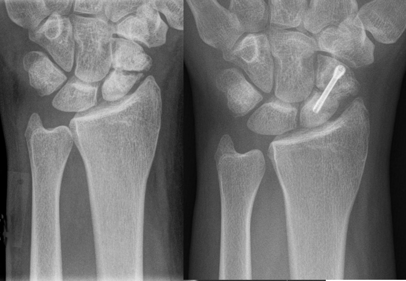
The Herbert screw (invented by Timothy Herbert) is a variable pitch cannulated screw typically made from titanium for its biocompatible properties as the screw is normally intended to remain in the patient indefinitely. It became generally available in 1978. It is one of the earliest designs of headless compression screws which are used to achieve interfragmentary compression through its differential pitch between the threads at each end of the screw (distance between adjacent threads of screw). It is used in scaphoid The scaphoid bone is one of the carpal bones of the wrist. It is situated between the hand and forearm on the thumb side of the wrist (also called the lateral or radial side). It forms the radial border of the carpal tunnel. The scaphoid bone ..., capitellum, radial head and in osteochondral fractures. Other uses include osteochondritis dissecans & small joint arthrodesis. References Orthopedic screws {{orthopedics-stub ... [...More Info...] [...Related Items...] OR: [Wikipedia] [Google] [Baidu] |
Osteoarthritis
Osteoarthritis is a type of degenerative joint disease that results from breakdown of articular cartilage, joint cartilage and underlying bone. A form of arthritis, it is believed to be the fourth leading cause of disability in the world, affecting 1 in 7 adults in the United States alone. The most common symptoms are joint pain and Joint stiffness, stiffness. Usually the symptoms progress slowly over years. Other symptoms may include joint effusion, joint swelling, decreased range of motion, and, when the back is affected, weakness or numbness of the arms and legs. The most commonly involved joints are the two near the ends of the fingers and the joint at the base of the thumbs, the knee and hip joints, and the joints of the neck and lower back. The symptoms can interfere with work and normal daily activities. Unlike some other types of arthritis, only the joints, not internal organs, are affected. Possible causes include previous joint injury, abnormal joint or limb development ... [...More Info...] [...Related Items...] OR: [Wikipedia] [Google] [Baidu] |
Scaphoid Fracture
A scaphoid fracture is a bone fracture, break of the scaphoid bone in the wrist. Symptoms generally includes pain at the base of the thumb which is worse with use of the hand. The Anatomical snuff box, anatomic snuffbox is generally tender and swelling may occur. Complications may include nonunion of the fracture, avascular necrosis of the proximal part of the bone, and arthritis. Scaphoid fractures are most commonly caused by a fall on an outstretched hand. Diagnosis is generally based on a combination of clinical examination and medical imaging. Some fractures may not be visible on plain radiography, X-rays. In such cases the affected area may be immobilised in a splint or cast and reviewed with repeat X-rays in two weeks, or alternatively an magnetic resonance imaging, MRI or bone scan may be performed. The fracture may be preventable by using wrist guards during certain activities. In those in whom the fracture remains well aligned a Orthopedic cast, cast is generally suffi ... [...More Info...] [...Related Items...] OR: [Wikipedia] [Google] [Baidu] |
Ulna
The ulna or ulnar bone (: ulnae or ulnas) is a long bone in the forearm stretching from the elbow to the wrist. It is on the same side of the forearm as the little finger, running parallel to the Radius (bone), radius, the forearm's other long bone. Longer and thinner than the radius, the ulna is considered to be the smaller long bone of the lower arm. The corresponding bone in the Human leg#Structure, lower leg is the fibula. Structure The ulna is a long bone found in the forearm that stretches from the elbow to the wrist, and when in standard anatomical position, is found on the Medial (anatomy), medial side of the forearm. It is broader close to the elbow, and narrows as it approaches the wrist. Close to the elbow, the ulna has a bony Process (anatomy), process, the olecranon process, a hook-like structure that fits into the olecranon fossa of the humerus. This prevents hyperextension and forms a hinge joint with the trochlea of the humerus. There is also a radial notch for ... [...More Info...] [...Related Items...] OR: [Wikipedia] [Google] [Baidu] |
Radius (bone)
The radius or radial bone (: radii or radiuses) is one of the two large bones of the forearm, the other being the ulna. It extends from the Anatomical terms of location, lateral side of the Elbow-joint, elbow to the thumb side of the wrist and runs parallel to the ulna. The ulna is longer than the radius, but the radius is thicker. The radius is a long bone, Prism (geometry), prism-shaped and slightly curved longitudinally. The radius is part of two joint (anatomy), joints: the elbow and the wrist. At the elbow, it joins with the capitulum of the humerus, and in a separate region, with the ulna at the radial notch. At the wrist, the radius forms a joint with the ulna bone. The corresponding bone in the human leg, lower leg is the tibia. Structure The long narrow medullary cavity is enclosed in a strong wall of compact bone. It is thickest along the interosseous border and thinnest at the extremities, same over the cup-shaped articular surface (fovea) of the head. The tra ... [...More Info...] [...Related Items...] OR: [Wikipedia] [Google] [Baidu] |
Abductor Pollicis Brevis
The abductor pollicis brevis is a muscle in the hand that functions as an abductor of the thumb. Structure The abductor pollicis brevis is a flat, thin muscle located just under the skin. It is a thenar muscle, and therefore contributes to the bulk of the palm's thenar eminence. It originates from the flexor retinaculum of the hand, the tubercle of the scaphoid bone, and additionally sometimes from the tubercle of the trapezium. Running lateralward and downward, it is inserted by a thin, flat tendon into the lateral side of the base of the first phalanx of the thumb, and the capsule of the metacarpophalangeal joint. Nerve supply The abductor pollicis brevis is supplied by the recurrent branch of the median nerve (Roots C8-T1). Function Abduction of the thumb is defined as the movement of the thumb anteriorly, a direction perpendicular to the palm. The abductor pollicis brevis does this by acting across both the carpometacarpal joint and the metacarpophalangeal joint ... [...More Info...] [...Related Items...] OR: [Wikipedia] [Google] [Baidu] |
Radial Artery
In human anatomy, the radial artery is the main artery of the lateral aspect of the forearm. Structure The radial artery arises from the bifurcation of the brachial artery in the antecubital fossa. It runs distally on the anterior part of the forearm. There, it serves as a landmark for the division between the anterior compartment of the forearm, anterior and posterior compartment of the forearm, posterior compartments of the forearm, with the posterior compartment beginning just lateral to the artery. The artery winds laterally around the wrist, passing through the anatomical snuff box and between the heads of the first dorsal interossei of the hand, dorsal interosseous muscle. It passes anteriorly between the heads of the adductor pollicis, and becomes the deep palmar arch, which joins with the deep branch of the ulnar artery. Along its course, it is accompanied by a similarly named vein, the radial vein. Branches The named branches of the radial artery may be divided int ... [...More Info...] [...Related Items...] OR: [Wikipedia] [Google] [Baidu] |
Ligament
A ligament is a type of fibrous connective tissue in the body that connects bones to other bones. It also connects flight feathers to bones, in dinosaurs and birds. All 30,000 species of amniotes (land animals with internal bones) have ligaments. It is also known as ''articular ligament'', ''articular larua'', ''fibrous ligament'', or ''true ligament''. Comparative anatomy Ligaments are similar to tendons and fasciae as they are all made of connective tissue. The differences among them are in the connections that they make: ligaments connect one bone to another bone, tendons connect muscle to bone, and fasciae connect muscles to other muscles. These are all found in the skeletal system of the human body. Ligaments cannot usually be regenerated naturally; however, there are periodontal ligament stem cells located near the periodontal ligament which are involved in the adult regeneration of periodontist ligament. The study of ligaments is known as . Humans Other ligame ... [...More Info...] [...Related Items...] OR: [Wikipedia] [Google] [Baidu] |


