|
Sarcolemma
The sarcolemma (''sarco'' (from ''sarx'') from Greek; flesh, and ''lemma'' from Greek; sheath), also called the myolemma, is the cell membrane surrounding a skeletal muscle fibre or a cardiomyocyte. It consists of a lipid bilayer and a thin outer coat of polysaccharide material ( glycocalyx) that contacts the basement membrane. The basement membrane contains numerous thin collagen fibrils and specialized proteins such as laminin that provide a scaffold to which the muscle fibre can adhere. Through transmembrane proteins in the plasma membrane, the actin skeleton inside the cell is connected to the basement membrane and the cell's exterior. At each end of the muscle fibre, the surface layer of the sarcolemma fuses with a tendon fibre, and the tendon fibres, in turn, collect into bundles to form the muscle tendons that adhere to bones. The sarcolemma generally maintains the same function in muscle cells as the plasma membrane does in other eukaryote cells. It acts as a barrie ... [...More Info...] [...Related Items...] OR: [Wikipedia] [Google] [Baidu] |
T-tubules
T-tubules (transverse tubules) are extensions of the cell membrane that penetrate into the center of skeletal and cardiac muscle cells. With membranes that contain large concentrations of ion channels, transporters, and pumps, T-tubules permit rapid transmission of the action potential into the cell, and also play an important role in regulating cellular calcium concentration. Through these mechanisms, T-tubules allow heart muscle cells to contract more forcefully by synchronising calcium release from the sarcoplasmic reticulum throughout the cell. T-tubule structure and function are affected beat-by-beat by cardiomyocyte contraction, as well as by diseases, potentially contributing to heart failure and arrhythmias. Although these structures were first seen in 1897, research into T-tubule biology is ongoing. Structure T-tubules are tubules formed from the same phospholipid bilayer as the surface membrane or sarcolemma of skeletal or cardiac muscle cells. They connect directly ... [...More Info...] [...Related Items...] OR: [Wikipedia] [Google] [Baidu] |
Skeletal Muscle
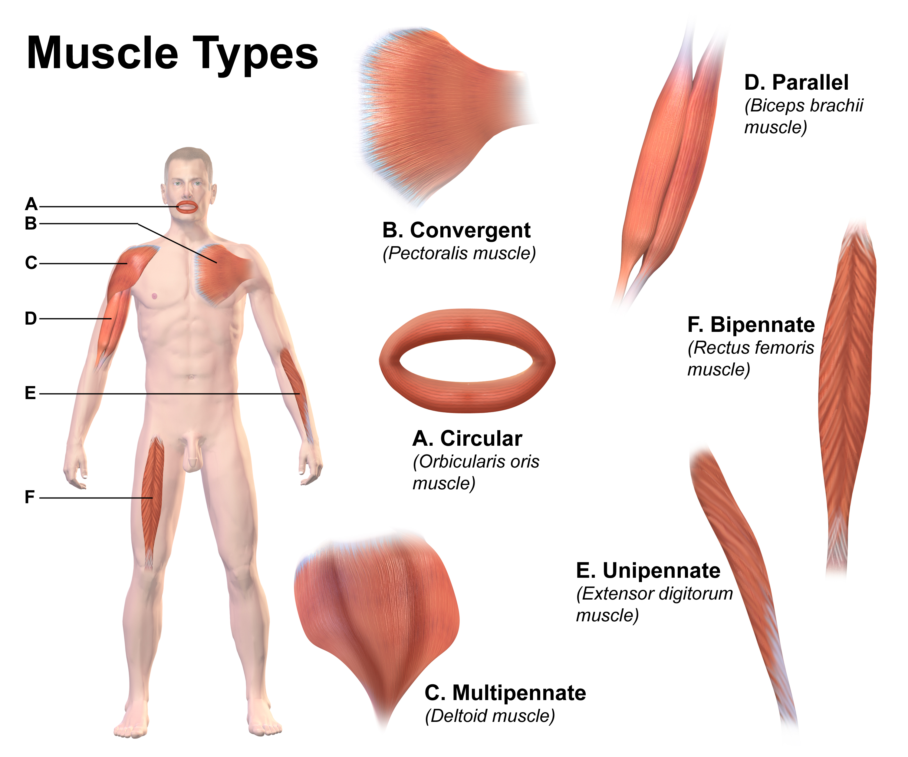
Skeletal muscle (commonly referred to as muscle) is one of the three types of vertebrate muscle tissue, the others being cardiac muscle and smooth muscle. They are part of the somatic nervous system, voluntary muscular system and typically are attached by tendons to bones of a skeleton. The skeletal muscle cells are much longer than in the other types of muscle tissue, and are also known as ''muscle fibers''. The tissue of a skeletal muscle is striated muscle tissue, striated – having a striped appearance due to the arrangement of the sarcomeres. A skeletal muscle contains multiple muscle fascicle, fascicles – bundles of muscle fibers. Each individual fiber and each muscle is surrounded by a type of connective tissue layer of fascia. Muscle fibers are formed from the cell fusion, fusion of developmental myoblasts in a process known as myogenesis resulting in long multinucleated cells. In these cells, the cell nucleus, nuclei, termed ''myonuclei'', are located along the inside ... [...More Info...] [...Related Items...] OR: [Wikipedia] [Google] [Baidu] |
Cell Membrane
The cell membrane (also known as the plasma membrane or cytoplasmic membrane, and historically referred to as the plasmalemma) is a biological membrane that separates and protects the interior of a cell from the outside environment (the extracellular space). The cell membrane consists of a lipid bilayer, made up of two layers of phospholipids with cholesterols (a lipid component) interspersed between them, maintaining appropriate membrane fluidity at various temperatures. The membrane also contains membrane proteins, including integral proteins that span the membrane and serve as membrane transporters, and peripheral proteins that loosely attach to the outer (peripheral) side of the cell membrane, acting as enzymes to facilitate interaction with the cell's environment. Glycolipids embedded in the outer lipid layer serve a similar purpose. The cell membrane controls the movement of substances in and out of a cell, being selectively permeable to ions and organic mole ... [...More Info...] [...Related Items...] OR: [Wikipedia] [Google] [Baidu] |
Action Potentials
An action potential (also known as a nerve impulse or "spike" when in a neuron) is a series of quick changes in voltage across a cell membrane. An action potential occurs when the membrane potential of a specific cell rapidly rises and falls. This depolarization then causes adjacent locations to similarly depolarize. Action potentials occur in several types of excitable cells, which include animal cells like neurons and muscle cells, as well as some plant cells. Certain endocrine cells such as pancreatic beta cells, and certain cells of the anterior pituitary gland are also excitable cells. In neurons, action potentials play a central role in cell–cell communication by providing for—or with regard to saltatory conduction, assisting—the propagation of signals along the neuron's axon toward synaptic boutons situated at the ends of an axon; these signals can then connect with other neurons at synapses, or to motor cells or glands. In other types of cells, their mai ... [...More Info...] [...Related Items...] OR: [Wikipedia] [Google] [Baidu] |
Striated Muscle Tissue
Striated muscle tissue is a muscle tissue that features repeating functional units called sarcomeres. Under the microscope, sarcomeres are visible along muscle fibers, giving a striated appearance to the tissue. The two types of striated muscle are skeletal muscle and cardiac muscle. Structure Striated muscle tissue contains T-tubules which enables the release of calcium ions from the sarcoplasmic reticulum. Skeletal muscle Skeletal muscle includes skeletal muscle fibers, blood vessels, nerve fibers, and connective tissue. Skeletal muscle is wrapped in epimysium, allowing structural integrity of the muscle despite contractions. The perimysium organizes the muscle fibers, which are encased in collagen and endomysium, into Muscle fascicle, fascicles. Each muscle fiber contains sarcolemma, sarcoplasm, and sarcoplasmic reticulum. The functional unit of a muscle fiber is called a sarcomere. Each muscle cell contains myofibrils composed of actin and myosin myofilaments repeated as a sa ... [...More Info...] [...Related Items...] OR: [Wikipedia] [Google] [Baidu] |
Cardiomyocyte
Cardiac muscle (also called heart muscle or myocardium) is one of three types of vertebrate muscle tissues, the others being skeletal muscle and smooth muscle. It is an involuntary, striated muscle that constitutes the main tissue of the wall of the heart. The cardiac muscle (myocardium) forms a thick middle layer between the outer layer of the heart wall (the pericardium) and the inner layer (the endocardium), with blood supplied via the coronary circulation. It is composed of individual cardiac muscle cells joined by intercalated discs, and encased by collagen fibers and other substances that form the extracellular matrix. Cardiac muscle contracts in a similar manner to skeletal muscle, although with some important differences. Electrical stimulation in the form of a cardiac action potential triggers the release of calcium from the cell's internal calcium store, the sarcoplasmic reticulum. The rise in calcium causes the cell's myofilaments to slide past each other in a p ... [...More Info...] [...Related Items...] OR: [Wikipedia] [Google] [Baidu] |
Sarcoplasmic Reticulum
The sarcoplasmic reticulum (SR) is a membrane-bound structure found within muscle cells that is similar to the smooth endoplasmic reticulum in other cells. The main function of the SR is to store calcium ions (Ca2+). Calcium ion levels are kept relatively constant, with the concentration of calcium ions within a cell being 10,000 times smaller than the concentration of calcium ions outside the cell. This means that small increases in calcium ions within the cell are easily detected and can bring about important cellular changes (the calcium is said to be a second messenger). Calcium is used to make calcium carbonate (found in chalk) and calcium phosphate, two compounds that the body uses to make teeth and bones. This means that too much calcium within the cells can lead to hardening (calcification) of certain intracellular structures, including the mitochondria, leading to cell death. Therefore, it is vital that calcium ion levels are controlled tightly, and can be released int ... [...More Info...] [...Related Items...] OR: [Wikipedia] [Google] [Baidu] |
Sarcoplasm
Sarcoplasm is the cytoplasm of a muscle cell. It is comparable to the cytoplasm of other cells, but it contains unusually large amounts of glycogen (a polymer of glucose), myoglobin, a red-colored protein necessary for binding oxygen molecules that diffuse into muscle fibers, and mitochondria.Roberts, Michael D.; Haun, Cody T.; Vann, Christopher G.; Osburn, Shelby C.; Young, Kaelin C. (2020). "Sarcoplasmic Hypertrophy in Skeletal Muscle: A Scientific "Unicorn" or Resistance Training Adaptation?". Frontiers in Physiology. 11. . . . . The calcium ion concentration in sarcoplasm is also a special element of the muscle fiber; it is the means by which muscle contractions take place and are regulated. The sarcoplasm plays a critical role in muscle contraction as an increase in Ca2+ concentration in the sarcoplasm begins the process of filament sliding. The decrease in Ca2+ in the sarcoplasm subsequently ceases filament sliding.Shahinpoor, Mohsen (2013). Muscular Biomimicry. Elsevier. pp. ... [...More Info...] [...Related Items...] OR: [Wikipedia] [Google] [Baidu] |
Co-transport
In cellular biology, active transport is the movement of molecules or ions across a cell membrane from a region of lower concentration to a region of higher concentration—against the concentration gradient. Active transport requires cellular energy to achieve this movement. There are two types of active transport: primary active transport that uses adenosine triphosphate (ATP), and secondary active transport that uses an electrochemical gradient. This process is in contrast to passive transport, which allows molecules or ions to move down their concentration gradient, from an area of high concentration to an area of low concentration, with energy. Active transport is essential for various physiological processes, such as nutrient uptake, hormone secretion, and nig impulse transmission. For example, the sodium-potassium pump uses ATP to pump sodium ions out of the cell and potassium ions into the cell, maintaining a concentration gradient essential for cellular function. A ... [...More Info...] [...Related Items...] OR: [Wikipedia] [Google] [Baidu] |
Terminal Cisterna
Terminal cisternae (singular: terminal cisterna) are enlarged areas of the sarcoplasmic reticulum surrounding the transverse tubules. Function Terminal cisternae are discrete regions within the muscle cell. They store calcium (increasing the capacity of the sarcoplasmic reticulum to release calcium) and release it when an action potential courses down the transverse tubules, eliciting muscle contraction. Because terminal cisternae ensure rapid calcium delivery, they are well developed in muscles that contract quickly, such as fast twitch skeletal muscle. Terminal cisternae then go on to release calcium, which binds to troponin. This releases tropomyosin, exposing active sites of the thin filament, actin. There are several mechanisms directly linked to the terminal cisternae which facilitate excitation-contraction coupling. When excitation of the membrane arrives at the T-tubule nearest the muscle fiber, a dihydropyridine channel ( DHP channel) is activated. This is simil ... [...More Info...] [...Related Items...] OR: [Wikipedia] [Google] [Baidu] |
Invagination
Invagination is the process of a surface folding in on itself to form a cavity, pouch or tube. In developmental biology, invagination of Epithelium, epithelial sheets occurs in many contexts during Animal embryonic development, embryonic development. Invagination is critical for making the Archenteron, primitive gut during gastrulation in many organisms, forming the neural tube in Vertebrate, vertebrates, and in the morphogenesis of countless Organ (biology), organs and sensory structures. Models of invagination that have been most thoroughly studied include the ventral furrow in Drosophila melanogaster, ''Drosophila'' ''melanogaster'', neural tube formation, and gastrulation in many marine organisms. The cellular mechanisms of invagination vary from one context to another but at their core they involve changing the mechanics of one side of a sheet of cells such that this pressure induces a bend in the tissue. The term, originally used in embryology, has been adopted in other disc ... [...More Info...] [...Related Items...] OR: [Wikipedia] [Google] [Baidu] |


