|
Right Bundle Branch
The bundle branches, or Tawara branches, are offshoots of the bundle of His in the heart's ventricle. They play an integral role in the electrical conduction system of the heart by transmitting cardiac action potentials from the bundle of His to the Purkinje fibers. Structure There are two branches of the bundle of His: the left bundle branch and the right bundle branch, both of which are located along the interventricular septum. The left bundle branch further divides into the left anterior fascicle and the left posterior fascicle. These structures lead to a network of thin filaments known as Purkinje fibers. They play an integral role in the electrical conduction system of the heart by transmitting cardiac action potentials to the Purkinje fibers. Clinical significance When a bundle branch or their fascicles becomes injured (by underlying heart disease, myocardial infarction, or cardiac surgery), it may cease to conduct electrical impulses appropriately, resulting in alte ... [...More Info...] [...Related Items...] OR: [Wikipedia] [Google] [Baidu] |
Bundle Of His
The bundle of His (BH) or His bundle (HB) ( "hiss"Medical Terminology for Health Professions, Spiral bound Version'. Cengage Learning; 2016. . pp. 129–.) is a collection of heart muscle cells specialized for electrical conduction. As part of the electrical conduction system of the heart, it transmits the electrical impulses from the atrioventricular node (located between the atria and the ventricles) to the point of the apex of the fascicular branches via the bundle branches. The fascicular branches then lead to the Purkinje fibers, which provide electrical conduction to the ventricles, causing the cardiac muscle of the ventricles to contract at a paced interval. Function The bundle of His is an important part of the electrical conduction system of the heart, as it transmits impulses from the atrioventricular node, located at the anterior-inferior end of the interatrial septum, to the ventricles of the heart. The bundle of His branches into the left and the right bundle br ... [...More Info...] [...Related Items...] OR: [Wikipedia] [Google] [Baidu] |
Ventricle (heart)
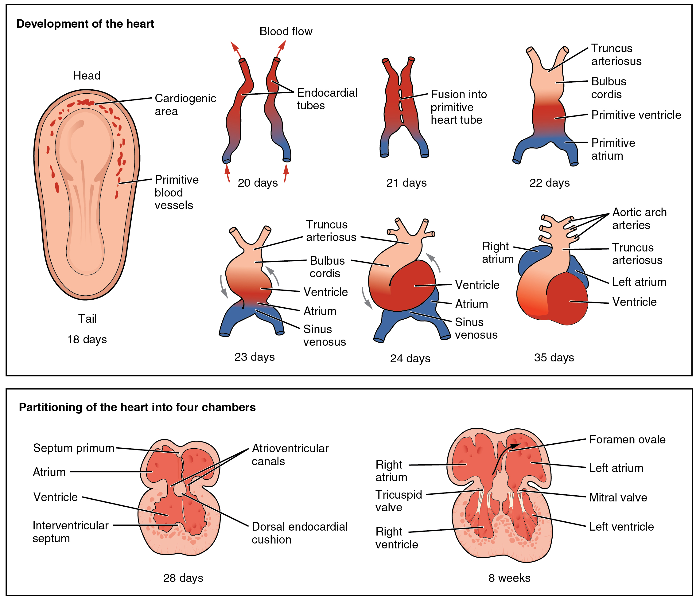
A ventricle is one of two large chambers toward the bottom of the heart that collect and expel blood towards the peripheral beds within the body and lungs. The blood pumped by a ventricle is supplied by an atrium, an adjacent chamber in the upper heart that is smaller than a ventricle. Interventricular means between the ventricles (for example the interventricular septum), while intraventricular means within one ventricle (for example an intraventricular block). In a four-chambered heart, such as that in humans, there are two ventricles that operate in a double circulatory system: the right ventricle pumps blood into the pulmonary circulation to the lungs, and the left ventricle pumps blood into the systemic circulation through the aorta. Structure Ventricles have thicker walls than atria and generate higher blood pressures. The physiological load on the ventricles requiring pumping of blood throughout the body and lungs is much greater than the pressure generated by the atria ... [...More Info...] [...Related Items...] OR: [Wikipedia] [Google] [Baidu] |
Electrical Conduction System Of The Heart
The cardiac conduction system (CCS) (also called the electrical conduction system of the heart) transmits the signals generated by the sinoatrial node – the heart's pacemaker, to cause the heart muscle to contract, and pump blood through the body's circulatory system. The pacemaking signal travels through the right atrium to the atrioventricular node, along the bundle of His, and through the bundle branches to Purkinje fibers in the walls of the ventricles. The Purkinje fibers transmit the signals more rapidly to stimulate contraction of the ventricles. The conduction system consists of specialized heart muscle cells, situated within the myocardium. There is a skeleton of fibrous tissue that surrounds the conduction system which can be seen on an ECG. Dysfunction of the conduction system can cause irregular heart rhythms including rhythms that are too fast or too slow. Structure Electrical signals arising in the SA node (located in the right atrium) stimulate the atr ... [...More Info...] [...Related Items...] OR: [Wikipedia] [Google] [Baidu] |
Cardiac Action Potential
The cardiac action potential is a brief change in voltage ( membrane potential) across the cell membrane of heart cells. This is caused by the movement of charged atoms (called ions) between the inside and outside of the cell, through proteins called ion channels. The cardiac action potential differs from action potentials found in other types of electrically excitable cells, such as nerves. Action potentials also vary within the heart; this is due to the presence of different ion channels in different cells. Unlike the action potential in skeletal muscle cells, the cardiac action potential is not initiated by nervous activity. Instead, it arises from a group of specialized cells known as pacemaker cells, that have automatic action potential generation capability. In healthy hearts, these cells form the cardiac pacemaker and are found in the sinoatrial node in the right atrium. They produce roughly 60–100 action potentials every minute. The action potential passes along t ... [...More Info...] [...Related Items...] OR: [Wikipedia] [Google] [Baidu] |
Purkinje Fibers
The Purkinje fibers (; often incorrectly ; Purkinje tissue or subendocardial branches) are located in the inner ventricular walls of the heart, just beneath the endocardium in a space called the subendocardium. The Purkinje fibers are specialized conducting fibers composed of electrically excitable cells. They are larger than cardiomyocytes with fewer myofibrils and many mitochondria. They conduct cardiac action potentials more quickly and efficiently than any of the other cells in the heart's electrical conduction system. Purkinje fibers allow the heart's conduction system to create synchronized contractions of its ventricles, and are essential for maintaining a consistent heart rhythm. Histology Purkinje fibers are a unique cardiac end-organ. Further histologic examination reveals that these fibers are split in ventricles walls. The electrical origin of atrial Purkinje fibers arrives from the sinoatrial node. Given no aberrant channels, the Purkinje fibers are distinct ... [...More Info...] [...Related Items...] OR: [Wikipedia] [Google] [Baidu] |
Interventricular Septum
The interventricular septum (IVS, or ventricular septum, or during development septum inferius) is the stout wall separating the ventricles, the lower chambers of the heart, from one another. The ventricular septum is directed obliquely backward to the right and curved with the convexity toward the right ventricle; its margins correspond with the anterior and posterior interventricular sulci. The lower part of the septum, which is the major part, is thick and muscular, and its much smaller upper part is thin and membraneous. During each cardiac cycle the interventricular septum contracts by shortening longitudinally and becoming thicker. Structure The interventricular septum is the stout wall separating the ventricles, the lower chambers of the heart, from one another. The ventricular septum is directed obliquely backward to the right and curved with the convexity toward the right ventricle; its margins correspond with the anterior and posterior longitudinal sulci. The gr ... [...More Info...] [...Related Items...] OR: [Wikipedia] [Google] [Baidu] |
Bundle Branch Block
A bundle branch block is a defect in one the bundle branches in the electrical conduction system of the heart. Anatomy and physiology The heart's electrical activity begins in the sinoatrial node (the heart's natural pacemaker), which is situated on the upper right atrium. The impulse travels next through the left and right atria and summates at the atrioventricular node. From the AV node the electrical impulse travels down the bundle of His and divides into the right and left bundle branches. The right bundle branch contains one fascicle. The left bundle branch subdivides into two fascicles: the left anterior fascicle, and the left posterior fascicle. Other sources divide the left bundle branch into three fascicles: the left anterior, the left posterior, and the left septal fascicle. The thicker left posterior fascicle bifurcates, with one fascicle being in the septal aspect. Ultimately, the fascicles divide into millions of Purkinje fibres, which in turn interdigitate with ind ... [...More Info...] [...Related Items...] OR: [Wikipedia] [Google] [Baidu] |
Sunao Tawara
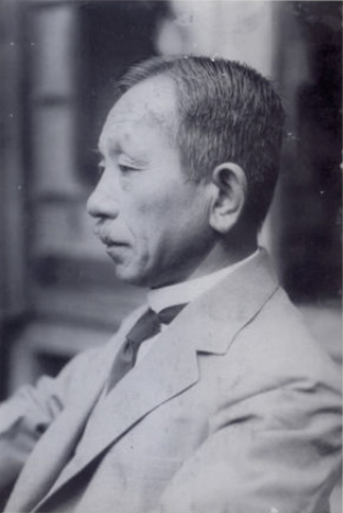
was a Japanese pathologist known for the discovery of the atrioventricular node. Tawara was born in Ōita Prefecture and studied at the Medical School, Imperial University of Tokyo in Tokyo, graduating in 1901 and receiving his Medical Doctor, Doctorate of Medical Science in 1908. Between 1903 and 1906 he spent in Philipps University of Marburg in Marburg, studying pathology and pathological anatomy with Ludwig Aschoff. It was here he undertook his important works on pathology and anatomy of heart. Upon returning to Japan he was appointed assistant professor of pathology at Kyushu University, Kyushu Imperial University in Fukuoka, obtaining full professorship in 1908. :''Node of Tawara'': a remnant of primitive fibers found in all mammalian hearts at the base of the interauricular septum, and forming the beginning of the auriculoventricular bundle or bundle of His, which is a muscular band, containing nerve fibers, connecting the auricles with the ventricles of the heart. The ' ... [...More Info...] [...Related Items...] OR: [Wikipedia] [Google] [Baidu] |
Das Reizleitungssystem Des Säugetierherzens
''Das Reizleitungssystem des Säugetierherzens'' (English: "''The Conduction System of the Mammalian Heart''") is a scientific monograph published in 1906 by Sunao Tawara. It has been recognized by cardiologists as a monumental discovery, and a milestone in cardiac electrophysiology". The monograph revealed the existence of the atrioventricular node and the function of Purkinje cells. It was used by Arthur Keith and Martin Flack as a detailed guide in their attempts to verify the existence of the Bundle of His, which subsequently led to their discovery of the sinoatrial node. Throughout the beginning of the 20th century, Tawara's monograph influenced the work of many cardiologists and it was later cited by Willem Einthoven in his anatomical interpretation of the electrocardiogram. Background Prior to Tawara's discoveries, it was assumed that electrical conduction through the Bundle of His was slow, because of the long interval between atrial and ventricular contractions. The ... [...More Info...] [...Related Items...] OR: [Wikipedia] [Google] [Baidu] |
Hans Eppinger
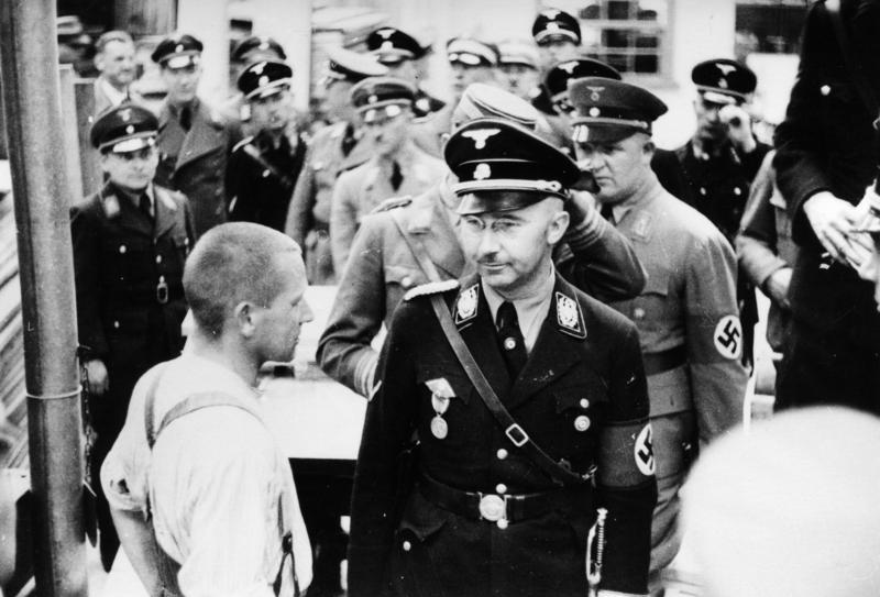
Hans Eppinger Jr. (5 January 1879, in Prague, Kingdom of Bohemia, Royal Bohemia, Austria-Hungary – 25 September 1946, in Vienna) was an Austrian physician of part-Jewish descent who performed experiments upon concentration camp prisoners. Early years Hans Eppinger was born in Prague, the son of the physician Professor Hans Eppinger [Sr] [1848-1916] a son of Heinrich Eppinger (1813–1868), notary and chancellery director in the monastery of Braunau (Broumov) in Bohemia and his wife Aloisia Salomon. Hans Eppinger Sr married Georgine Zetter in Klagenfurt and had two daughters and a son, Hans Eppinger junior. Hans Eppinger Jr received an education in Graz and Strasbourg. In 1903, he became a medical doctor in Graz, working at a medical clinic. He moved to Vienna in 1908, and in 1909 he specialized in internal medicine, particularly conditions of the liver. He became a professor in 1918, then taught in Freiburg in 1926 and in Cologne in 1930. In 1936 he is known to have travelle ... [...More Info...] [...Related Items...] OR: [Wikipedia] [Google] [Baidu] |
Carl Julius Rothberger
Carl may refer to: *Carl, Georgia, city in USA *Carl, West Virginia, an unincorporated community * Carl (name), includes info about the name, variations of the name, and a list of people with the name *Carl², a TV series * "Carl", an episode of television series ''Aqua Teen Hunger Force'' * An informal nickname for a student or alum of Carleton College CARL may refer to: *Canadian Association of Research Libraries *Colorado Alliance of Research Libraries See also * Carle (other) * Charles *Carle, a surname *Karl (other) *Karle (other) Karle may refer to: Places * Karle (Svitavy District), a municipality and village in the Czech Republic * Karli, India, a town in Maharashtra, India ** Karla Caves, a complex of Buddhist cave shrines * Karle, Belgaum, a settlement in Belgaum ... {{disambig ja:カール zh:卡尔 ... [...More Info...] [...Related Items...] OR: [Wikipedia] [Google] [Baidu] |
Heart Block
Heart block (HB) is a disorder in the heart's rhythm due to a fault in the natural pacemaker. This is caused by an obstruction – a block – in the electrical conduction system of the heart. Sometimes a disorder can be inherited. Despite the severe-sounding name, heart block may cause no symptoms at all in some cases, or occasional missed heartbeats in other cases (which can cause light-headedness, syncope (fainting), and palpitations), or may require the implantation of an artificial pacemaker, depending upon exactly where in the heart conduction is being impaired and how significantly it is affected. Heart block should not be confused with other conditions, which may or may not be co-occurring, relating to the heart and/or other nearby organs that are or can be serious, including angina (heart-related chest pain), heart attack (myocardial infarction), any type of heart failure, cardiogenic shock or other types of shock, different types of abnormal heart rhythms (arrhythmias ... [...More Info...] [...Related Items...] OR: [Wikipedia] [Google] [Baidu] |


