|
RYR2
Ryanodine receptor 2 (RYR2) is one of a class of ryanodine receptors and a protein found primarily in cardiac muscle. In humans, it is encoded by the ''RYR2'' gene. In the process of cardiac calcium-induced calcium release, RYR2 is the major mediator for sarcoplasmic release of stored calcium ions. Structure The channel is composed of RYR2 homotetramers and FK506-binding proteins found in a 1:4 stoichiometric ratio. Calcium channel function is affected by the specific type of FK506 isomer interacting with the RYR2 protein, due to binding differences and other factors. Function The RYR2 protein functions as the major component of a calcium channel located in the sarcoplasmic reticulum that supplies ions to the cardiac muscle during systole. To enable cardiac muscle contraction, calcium influx through voltage-gated L-type calcium channels in the plasma membrane allows calcium ions to bind to RYR2 located on the sarcoplasmic reticulum. This bindin ... [...More Info...] [...Related Items...] OR: [Wikipedia] [Google] [Baidu] |
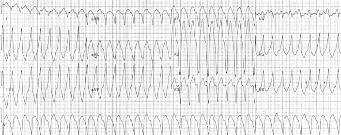
Catecholaminergic Polymorphic Ventricular Tachycardia
Catecholaminergic polymorphic ventricular tachycardia (CPVT) is an inherited genetic disorder that predisposes those affected to potentially life-threatening abnormal heart rhythms or arrhythmias. The arrhythmias seen in CPVT typically occur during exercise or at times of emotional stress, and classically take the form of bidirectional ventricular tachycardia or ventricular fibrillation. Those affected may be asymptomatic, but they may also experience blackouts or even sudden cardiac death. CPVT is caused by genetic mutations affecting proteins that regulate the concentrations of calcium within cardiac muscle cells. The most commonly identified gene is RYR2, which encodes a protein included in an ion channel known as the ryanodine receptor; this channel releases calcium from a cell's internal calcium store, the sarcoplasmic reticulum, during every heartbeat. CPVT is often diagnosed from an ECG recorded during an exercise tolerance test, but it may also be diagnosed with a g ... [...More Info...] [...Related Items...] OR: [Wikipedia] [Google] [Baidu] |
Ryanodine Receptor
Ryanodine receptors (RyR for short) form a class of intracellular calcium channels in various forms of excitable animal tissue like muscles and neurons. There are three major isoforms of the ryanodine receptor, which are found in different tissues and participate in different signaling pathways involving calcium release from intracellular organelles. The RYR2 ryanodine receptor isoform is the major cellular mediator of calcium-induced calcium release (CICR) in animal cells. Etymology The ryanodine receptors are named after the plant alkaloid ryanodine which shows a high affinity to them. Isoforms There are multiple isoforms of ryanodine receptors: * RyR1 is primarily expressed in skeletal muscle * RyR2 is primarily expressed in myocardium (heart muscle) * RyR3 is expressed more widely, but especially in the brain. * Non-mammalian vertebrates typically express two RyR isoforms, referred to as RyR-alpha and RyR-beta. * Many invertebrates, including the model organisms Dros ... [...More Info...] [...Related Items...] OR: [Wikipedia] [Google] [Baidu] |
Arrhythmogenic Right Ventricular Dysplasia
Arrhythmogenic cardiomyopathy (ACM), arrhythmogenic right ventricular dysplasia (ARVD), or arrhythmogenic right ventricular cardiomyopathy (ARVC), most commonly is an inherited heart disease. ACM is caused by genetic defects of the parts of heart muscle (also called ''myocardium'' or ''cardiac muscle'') known as desmosomes, areas on the surface of heart muscle cells which link the cells together. The desmosomes are composed of several proteins, and many of those proteins can have harmful mutations. ARVC can also develop in intense endurance athletes in the absence of desmosomal abnormalities. Exercise-induced ARVC cause possibly is a result of excessive right ventricular wall stress during high intensity exercise. The disease is a type of non-ischemic cardiomyopathy that primarily involves the right ventricle, though cases of exclusive left ventricular disease have been reported. It is characterized by hypokinetic areas involving the free wall of the ventricle, with fibrofatt ... [...More Info...] [...Related Items...] OR: [Wikipedia] [Google] [Baidu] |
Actin
Actin is a family of globular multi-functional proteins that form microfilaments in the cytoskeleton, and the thin filaments in muscle fibrils. It is found in essentially all eukaryotic cells, where it may be present at a concentration of over 100 μM; its mass is roughly 42 kDa, with a diameter of 4 to 7 nm. An actin protein is the monomeric subunit of two types of filaments in cells: microfilaments, one of the three major components of the cytoskeleton, and thin filaments, part of the contractile apparatus in muscle cells. It can be present as either a free monomer called G-actin (globular) or as part of a linear polymer microfilament called F-actin (filamentous), both of which are essential for such important cellular functions as the mobility and contraction of cells during cell division. Actin participates in many important cellular processes, including muscle contraction, cell motility, cell division and cytokinesis, vesicle and organelle movement, cell sign ... [...More Info...] [...Related Items...] OR: [Wikipedia] [Google] [Baidu] |
SRI (gene)
Sorcin is a protein that in humans is encoded by the ''SRI'' gene. Interactions SRI (gene) has been shown to interact with Ryanodine receptor 2, ANXA7 Annexin A7 is a protein that in humans is encoded by the ''ANXA7'' gene. Annexin VII is a member of the annexin family of calcium-dependent phospholipid binding proteins. The Annexin VII gene contains 14 exons and spans approximately 34 kb of DNA ... and GCA. References Further reading * * * * * * * * * * * * * * * * * {{gene-7-stub Penta-EF-hand proteins ... [...More Info...] [...Related Items...] OR: [Wikipedia] [Google] [Baidu] |
PRKACG
cAMP-dependent protein kinase catalytic subunit gamma is an enzyme that in humans is encoded by the ''PRKACG'' gene. Cyclic AMP-dependent protein kinase (PKA) consists of two catalytic subunits and a regulatory subunit dimer. This gene encodes the gamma form of its catalytic subunit. The gene is intronless and is thought to be a retrotransposon derived from the gene for the alpha form of the PKA catalytic subunit. Interactions PRKACG has been shown to interact with Ryanodine receptor 2 Ryanodine receptor 2 (RYR2) is one of a class of ryanodine receptors and a protein found primarily in cardiac muscle. In humans, it is encoded by the ''RYR2'' gene. In the process of cardiac calcium-induced calcium release, RYR2 is the major medi .... References Further reading * * * * * * * * * * * * * * * * EC 2.7.11 {{gene-9-stub ... [...More Info...] [...Related Items...] OR: [Wikipedia] [Google] [Baidu] |
PRKACB
cAMP-dependent protein kinase catalytic subunit beta is an enzyme that in humans is encoded by the ''PRKACB'' gene. cAMP is a signaling molecule important for a variety of cellular functions. cAMP exerts its effects by activating the protein kinase A (PKA), which transduces the signal through phosphorylation of different target proteins. The inactive holoenzyme of PKA is a tetramer composed of two regulatory and two catalytic subunits. cAMP causes the dissociation of the inactive holoenzyme into a dimer of regulatory subunits bound to four cAMP and two free monomeric catalytic subunits. Four different regulatory subunits and three catalytic subunits of PKA have been identified in humans. The protein encoded by this gene is a member of the serine/threonine protein kinase family and is a catalytic subunit of PKA. Three alternatively spliced transcript variants encoding distinct isoforms have been observed. Interactions PRKACB has been shown to interact with Ryanodine receptor 2 and ... [...More Info...] [...Related Items...] OR: [Wikipedia] [Google] [Baidu] |
PRKACA
The catalytic subunit α of protein kinase A is a key regulatory enzyme that in humans is encoded by the ''PRKACA'' gene. This enzyme is responsible for phosphorylating other proteins and substrates, changing their activity. Protein kinase A catalytic subunit (PKA Cα) is a member of the AGC kinase family (protein kinases A, G, and C), and contributes to the control of cellular processes that include glucose metabolism, cell division, and contextual memory. PKA Cα is part of a larger protein complex that is responsible for controlling when and where proteins are phosphorylated. Defective regulation of PKA holoenzyme activity has been linked to the progression of cardiovascular disease, certain endocrine disorders and cancers. Discovery Edmond H. Fischer and Edwin G. Krebs at the University of Washington discovered PKA in the late 1950s while working through the mechanisms that govern glycogen phosphorylase. They realized that a key metabolic enzyme called phosphorylase kina ... [...More Info...] [...Related Items...] OR: [Wikipedia] [Google] [Baidu] |
AKAP6
A-kinase anchor protein 6 is an enzyme that in humans is encoded by the ''AKAP6'' gene. The A-kinase anchor proteins (AKAPs) are a group of structurally diverse proteins, which have the common function of binding to the regulatory subunit of protein kinase A (PKA) and confining the holoenzyme to discrete locations within the cell. This gene encodes a member of the AKAP family. The encoded protein is highly expressed in various brain regions and cardiac and skeletal muscle. It is specifically localized to the sarcoplasmic reticulum and nuclear membrane, and is involved in anchoring PKA to the nuclear membrane or sarcoplasmic reticulum. Interactions AKAP6 has been shown to interact with Ryanodine receptor 2 Ryanodine receptor 2 (RYR2) is one of a class of ryanodine receptors and a protein found primarily in cardiac muscle. In humans, it is encoded by the ''RYR2'' gene. In the process of cardiac calcium-induced calcium release, RYR2 is the major medi ... and PDE4D3. References ... [...More Info...] [...Related Items...] OR: [Wikipedia] [Google] [Baidu] |
ATPase
ATPases (, Adenosine 5'-TriPhosphatase, adenylpyrophosphatase, ATP monophosphatase, triphosphatase, SV40 T-antigen, ATP hydrolase, complex V (mitochondrial electron transport), (Ca2+ + Mg2+)-ATPase, HCO3−-ATPase, adenosine triphosphatase) are a class of enzymes that catalyze the decomposition of ATP into ADP and a free phosphate ion or the inverse reaction. This dephosphorylation reaction releases energy, which the enzyme (in most cases) harnesses to drive other chemical reactions that would not otherwise occur. This process is widely used in all known forms of life. Some such enzymes are integral membrane proteins (anchored within biological membranes), and move solutes across the membrane, typically against their concentration gradient. These are called transmembrane ATPases. Functions Transmembrane ATPases import metabolites necessary for cell metabolism and export toxins, wastes, and solutes that can hinder cellular processes. An important example is the sodium-potass ... [...More Info...] [...Related Items...] OR: [Wikipedia] [Google] [Baidu] |
.png)


