|
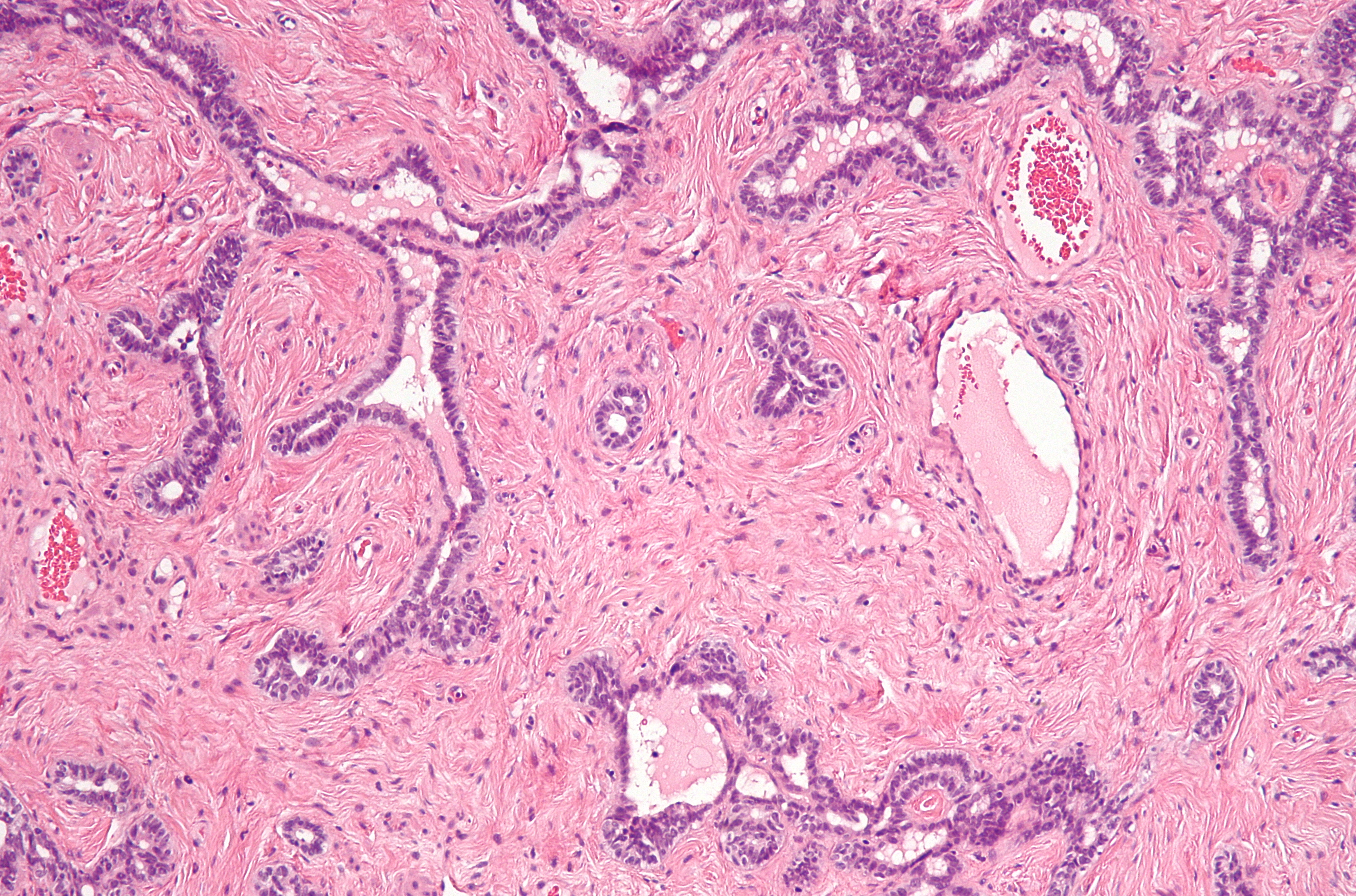
Rete Testis
The rete testis ( ; : retia testes) is an anastomosis, anastomosing network of delicate tubules located in the hilum of the testicle (mediastinum testis) that carries spermatozoon, sperm from the seminiferous tubules to the efferent ducts. It is the Homology (biology), homologue of the rete ovarii in females. Its function is to provide a site for fluid reabsorption. Structure The rete testis is the network of interconnecting tubules where the Tubuli seminiferi recti, straight seminiferous tubules (the terminal part of the seminiferous tubules) empty. It is located within a highly vascular connective tissue in the mediastinum testis. The epithelium, epithelial cells form a single layer that lines the inner surface of the tubules. These cells are Simple cuboidal epithelium, cuboidal, with microvillus, microvilli and a single cilium on their surface. Development In the development of the urinary and reproductive organs, the testis is developed in much the same way as the ovary, orig ... [...More Info...] [...Related Items...] OR: [Wikipedia] [Google] [Baidu] |
Testicular Septa
The septa testis are fibrous partitions of the testis dividing the testis into compartments - the Lobules of testis, lobules of the testis. The septa are formed by extensions of the Tunica albuginea of testis, tunica albuginea - the dense fibrous connective tissue surface covering of the testis - into the substance of the testis. The septa converge towards the mediastinum testis. Additional images File:Gray1145.png, Transverse section through the left side of the scrotum and the left testis. File:Gray1149.png, Vertical section of the testis, to show the arrangement of the ducts. References External links * - "Inguinal Region, Scrotum and Testes: The Cross-Section of the Testis" Testicle {{genitourinary-stub ... [...More Info...] [...Related Items...] OR: [Wikipedia] [Google] [Baidu] |
Connective Tissue
Connective tissue is one of the four primary types of animal tissue, a group of cells that are similar in structure, along with epithelial tissue, muscle tissue, and nervous tissue. It develops mostly from the mesenchyme, derived from the mesoderm, the middle embryonic germ layer. Connective tissue is found in between other tissues everywhere in the body, including the nervous system. The three meninges, membranes that envelop the brain and spinal cord, are composed of connective tissue. Most types of connective tissue consists of three main components: elastic and collagen fibers, ground substance, and cells. Blood and lymph are classed as specialized fluid connective tissues that do not contain fiber. All are immersed in the body water. The cells of connective tissue include fibroblasts, adipocytes, macrophages, mast cells and leukocytes. The term "connective tissue" (in German, ) was introduced in 1830 by Johannes Peter Müller. The tissue was already recognized as ... [...More Info...] [...Related Items...] OR: [Wikipedia] [Google] [Baidu] |
Neo-Latin
Neo-LatinSidwell, Keith ''Classical Latin-Medieval Latin-Neo Latin'' in ; others, throughout. (also known as New Latin and Modern Latin) is the style of written Latin used in original literary, scholarly, and scientific works, first in Italy during the Italian Renaissance of the fourteenth and fifteenth centuries, and then across northern Europe after about 1500, as a key feature of the humanist movement. Through comparison with Classical Latin, Latin of the Classical period, scholars from Petrarch onwards promoted a standard of Latin closer to that of the ancient Romans, especially in grammar, style, and spelling. The term ''Neo-Latin'' was however coined much later, probably in Germany in the late eighteenth century, as ''Neulatein'', spreading to French and other languages in the nineteenth century. Medieval Latin had diverged quite substantially from the classical standard and saw notable regional variation and influence from vernacular languages. Neo-Latin attempts to retur ... [...More Info...] [...Related Items...] OR: [Wikipedia] [Google] [Baidu] |
Cysts
A cyst is a closed Wikt:sac, sac, having a distinct Cell envelope, envelope and cell division, division compared with the nearby Biological tissue, tissue. Hence, it is a cluster of Cell (biology), cells that have grouped together to form a sac (like the manner in which water molecules group together to form a bubble); however, the distinguishing aspect of a cyst is that the cells forming the "shell" of such a sac are distinctly abnormal (in both appearance and behaviour) when compared with all surrounding cells for that given location. A cyst may contain air, fluids, or semi-solid material. A collection of pus is called an abscess, not a cyst. Once formed, a cyst may resolve on its own. When a cyst fails to resolve, it may need to be removed surgically, but that would depend upon its type and location. Cancer-related cysts are formed as a defense mechanism for the body following the development of mutations that lead to an uncontrolled cellular division. Once that mutation has o ... [...More Info...] [...Related Items...] OR: [Wikipedia] [Google] [Baidu] |
Rete Tubular Ectasia
Rete tubular ectasia, also known as cystic transformation of rete testis is a benign condition, usually found in older men, involving numerous small, tubular cystic structures within the rete testis. Presentation It is usually found in men older than 55 years and is frequently found on bilateral testes but is often asymmetrical. Mechanism The formation of cysts in the rete testis is associated with the obstruction of the efferent ducts, which connect the rete testis with the head of the epididymis. They are often bilateral. Diagnosis The condition can be detected with ultrasonography. Cystic lesions us usually found at the mediastinum testis with elongated shaped lesions displacing the mediastinum. It is commonly associated with epididymal abnormalities, such as spermatocele, epididymal cyst, and epididymitis. The condition shares a common location with cystic dysplasia of the testis and intratesticular cysts. Unlike cystic neoplasms, they don't present specific tumor mark ... [...More Info...] [...Related Items...] OR: [Wikipedia] [Google] [Baidu] |
Hilum (anatomy)
 In human anatomy, the hilum (; : hila), sometimes formerly called a hilus (; : hili), is a depression or fissure where structures such as blood vessels and nerves enter an Organ (anatomy), organ. Examples include:
* Hilum of kidney, admits the renal artery, renal vein, vein, ureter, and nerves
* Splenic hilum, on the surface of the spleen, admits the splenic artery, splenic vein, vein, lymph vessels, and nerves
* Hilum of lung, a triangular depression where the structures which form the root of the lung enter and leave the Organ (biology)#Viscera, viscus
* Hilum of lymph node, the portion of a lymph node where the Efferent lymphatic vessel, efferent vessels exit
* Hippocampus proper#CA4, Hilus of dentate gyrus, part of hippocampus that contains the Mossy fiber (hippocampus), mossy cells.
...
[...More Info...] [...Related Items...] OR: [Wikipedia] [Google] [Baidu] |
Mesonephros
The mesonephros () is one of three excretory system, excretory organs that develop in vertebrates. It serves as the main excretory organ of aquatic vertebrates and as a temporary kidney in reptiles, birds, and mammals. The mesonephros is included in the Wolffian body after Caspar Friedrich Wolff who described it in 1759. (The Wolffian body is composed of: mesonephros + paramesonephrotic blastema) Structure The mesonephros acts as a structure similar to the human kidney, kidney that, in humans, functions between the sixth and tenth weeks of embryological life. Despite the similarity in structure, function, and terminology, however, the mesonephric nephrons do not form any part of the mature kidney or nephrons. In humans, the mesonephros consists of units which are similar in structure and function to nephrons of the adult kidney. Each of these consists of a Glomerulus (kidney), glomerulus, a tuft of capillary, capillaries which arises from lateral branches of dorsal aorta and drain ... [...More Info...] [...Related Items...] OR: [Wikipedia] [Google] [Baidu] |
Mesothelium
The mesothelium is a membrane composed of simple squamous epithelium, simple squamous epithelial cells of mesodermal origin, which forms the lining of several body cavities: the pleura (pleural cavity around the lungs), peritoneum (abdominopelvic cavity including the mesentery, omentum (other), omenta, falciform ligament and the perimetrium) and pericardium (around the heart). Mesothelial tissue also surrounds the male testis (as the tunica vaginalis) and occasionally the spermatic cord (in a patent processus vaginalis). Mesothelium that tunica (biology), covers the internal organs is called visceral mesothelium, while one that covers the surrounding body walls is called the :wikt:parietal, parietal mesothelium. The mesothelium that secretes serous fluid as a main function is also known as a serosa. Origin Mesothelium derives from the embryonic mesoderm cell layer, that lines the body cavity, coelom (body cavity) in the embryo. It develops into the layer of cells that c ... [...More Info...] [...Related Items...] OR: [Wikipedia] [Google] [Baidu] |
Ovary
The ovary () is a gonad in the female reproductive system that produces ova; when released, an ovum travels through the fallopian tube/ oviduct into the uterus. There is an ovary on the left and the right side of the body. The ovaries are endocrine glands, secreting various hormones that play a role in the menstrual cycle and fertility. The ovary progresses through many stages beginning in the prenatal period through menopause. Structure Each ovary is whitish in color and located alongside the lateral wall of the uterus in a region called the ovarian fossa. The ovarian fossa is the region that is bounded by the external iliac artery and in front of the ureter and the internal iliac artery. This area is about 4 cm x 3 cm x 2 cm in size.Daftary, Shirish; Chakravarti, Sudip (2011). Manual of Obstetrics, 3rd Edition. Elsevier. pp. 1-16. . The ovaries are surrounded by a capsule, and have an outer cortex and an inner medulla. The capsule is of dense connect ... [...More Info...] [...Related Items...] OR: [Wikipedia] [Google] [Baidu] |
Testis
A testicle or testis ( testes) is the gonad in all male bilaterians, including humans, and is Homology (biology), homologous to the ovary in females. Its primary functions are the production of sperm and the secretion of Androgen, androgens, primarily testosterone. The release of testosterone is regulated by luteinizing hormone (LH) from the anterior pituitary gland. Sperm production is controlled by follicle-stimulating hormone (FSH) from the anterior pituitary gland and by testosterone produced within the gonads. Structure Appearance Males have two testicles of similar size contained within the scrotum, which is an extension of the abdominal wall. Scrotal asymmetry, in which one testicle extends farther down into the scrotum than the other, is common. This is because of the differences in the vasculature's anatomy. For 85% of men, the right testis hangs lower than the left one. Measurement and volume The volume of the testicle can be estimated by palpating it and compari ... [...More Info...] [...Related Items...] OR: [Wikipedia] [Google] [Baidu] |
Development Of The Urinary And Reproductive Organs
The development of the urinary system begins during prenatal development, and relates to the development of the urogenital system – both the organs of the urinary system and the sex organs of the reproductive system. The development continues as a part of sexual differentiation. The urinary and reproductive organs are developed from the intermediate mesoderm. The permanent organs of the adult are preceded by a set of structures which are purely embryonic, and which with the exception of the ducts disappear almost entirely before birth. These embryonic structures are on either side; the pronephros, the mesonephros and the metanephros of the kidney, and the Wolffian duct, Wolffian and Müllerian ducts of the sex organ. The pronephros disappears very early; the structural elements of the mesonephros mostly degenerate, but the gonad is developed in their place, with which the Wolffian duct remains as the duct in males, and the Müllerian as that of the female. Some of the tubules of ... [...More Info...] [...Related Items...] OR: [Wikipedia] [Google] [Baidu] |
Cilium
The cilium (: cilia; ; in Medieval Latin and in anatomy, ''cilium'') is a short hair-like membrane protrusion from many types of eukaryotic cell. (Cilia are absent in bacteria and archaea.) The cilium has the shape of a slender threadlike projection that extends from the surface of the much larger cell body. Eukaryotic flagella found on sperm cells and many protozoans have a similar structure to motile cilia that enables swimming through liquids; they are longer than cilia and have a different undulating motion. There are two major classes of cilia: ''motile'' and ''non-motile'' cilia, each with two subtypes, giving four types in all. A cell will typically have one primary cilium or many motile cilia. The structure of the cilium core, called the axoneme, determines the cilium class. Most motile cilia have a central pair of single microtubules surrounded by nine pairs of double microtubules called a 9+2 axoneme. Most non-motile cilia have a 9+0 axoneme that lacks the centr ... [...More Info...] [...Related Items...] OR: [Wikipedia] [Google] [Baidu] |


