|
Papilledema
Papilledema or papilloedema is optic disc swelling that is caused by increased intracranial pressure due to any cause. The swelling is usually bilateral and can occur over a period of hours to weeks. Unilateral presentation is extremely rare. In intracranial hypertension, the optic disc swelling most commonly occurs bilaterally. When papilledema is found on fundoscopy, further evaluation is warranted because vision loss can result if the underlying condition is not treated. Further evaluation with a CT or MRI of the brain and/or spine is usually performed. Recent research has shown that point-of-care ultrasound can be used to measure optic nerve sheath diameter for detection of increased intracranial pressure and shows good diagnostic test accuracy compared to CT. Thus, if there is a question of papilledema on fundoscopic examination or if the optic disc cannot be adequately visualized, ultrasound can be used to rapidly assess for increased intracranial pressure and help direct ... [...More Info...] [...Related Items...] OR: [Wikipedia] [Google] [Baidu] |
Papilledema Revealed By Scanning Laser Ophthalmoscopy And Laser Doppler Holography
Papilledema or papilloedema is optic disc swelling that is caused by increased intracranial pressure due to any cause. The swelling is usually bilateral and can occur over a period of hours to weeks. Unilateral presentation is extremely rare. In intracranial hypertension, the optic disc swelling most commonly occurs bilaterally. When papilledema is found on fundoscopy, further evaluation is warranted because vision loss can result if the underlying condition is not treated. Further evaluation with a CT or MRI of the brain and/or spine is usually performed. Recent research has shown that point-of-care ultrasound can be used to measure optic nerve sheath diameter for detection of increased intracranial pressure and shows good diagnostic test accuracy compared to CT. Thus, if there is a question of papilledema on fundoscopic examination or if the optic disc cannot be adequately visualized, ultrasound can be used to rapidly assess for increased intracranial pressure and help direct ... [...More Info...] [...Related Items...] OR: [Wikipedia] [Google] [Baidu] |
Intracranial Pressure
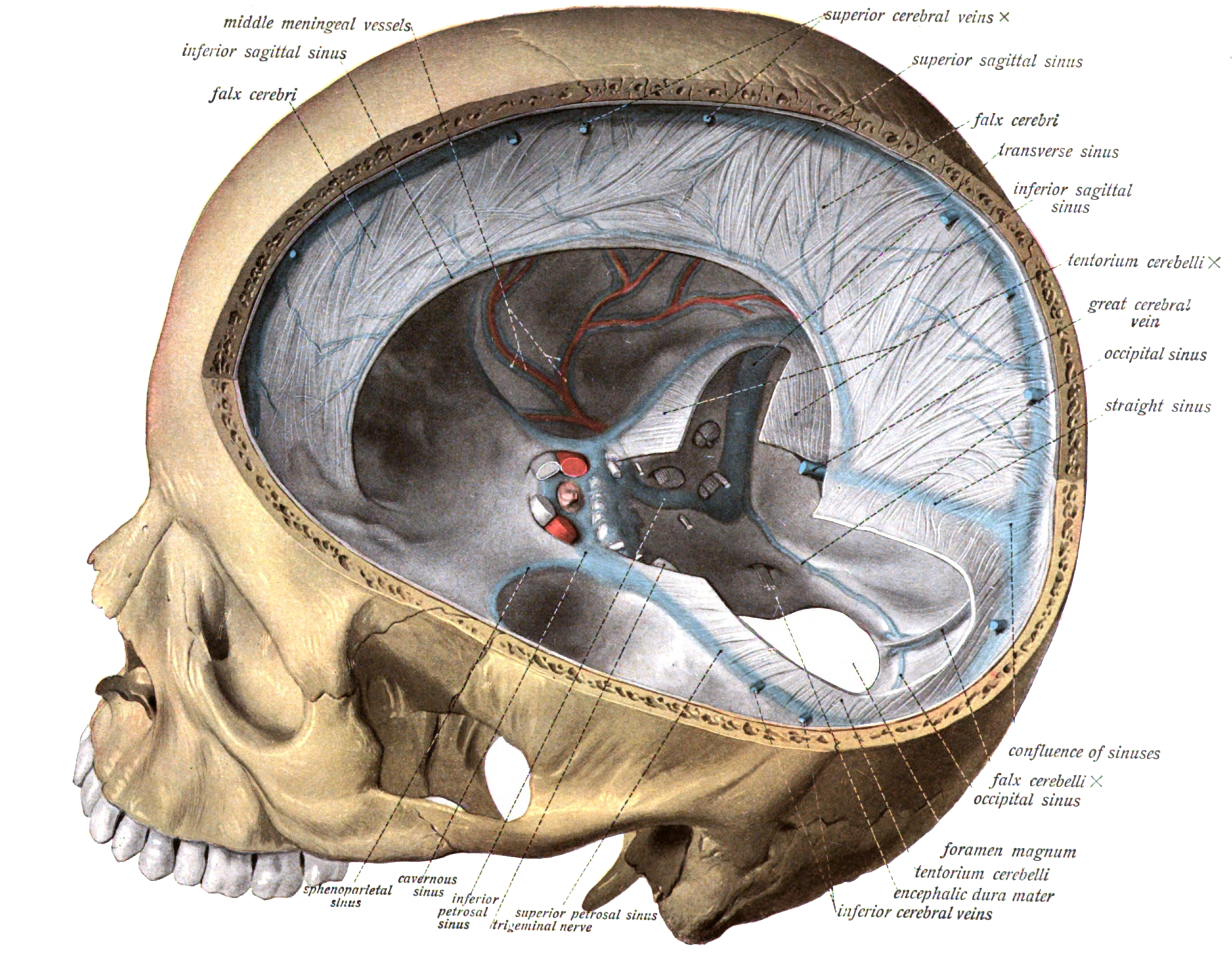
Intracranial pressure (ICP) is the pressure exerted by fluids such as cerebrospinal fluid (CSF) inside the skull and on the brain tissue. ICP is measured in millimeters of mercury ( mmHg) and at rest, is normally 7–15 mmHg for a supine adult. The body has various mechanisms by which it keeps the ICP stable, with CSF pressures varying by about 1 mmHg in normal adults through shifts in production and absorption of CSF. Changes in ICP are attributed to volume changes in one or more of the constituents contained in the cranium. CSF pressure has been shown to be influenced by abrupt changes in intrathoracic pressure during coughing (which is induced by contraction of the diaphragm and abdominal wall muscles, the latter of which also increases intra-abdominal pressure), the valsalva maneuver, and communication with the vasculature ( venous and arterial systems). Intracranial hypertension (IH), also called increased ICP (IICP) or raised intracranial pressure (RICP), is elevation ... [...More Info...] [...Related Items...] OR: [Wikipedia] [Google] [Baidu] |
Foster Kennedy Syndrome
Foster Kennedy syndrome is a constellation of findings associated with tumors of the frontal lobe. Although Foster Kennedy syndrome is sometimes called "Kennedy syndrome", it should not be confused with Kennedy disease, or spinal and bulbar muscular atrophy, which is named after William R. Kennedy. Pseudo-Foster Kennedy syndrome is defined as one-sided optic atrophy with papilledema in the other eye but with the absence of a mass. Presentation The syndrome is defined as the following changes: * optic atrophy in the ipsilateral eye * disc edema in the contralateral eye * central scotoma (loss of vision in the middle of the visual fields) in the ipsilateral eye * anosmia (loss of smell) ipsilaterally This syndrome is due to optic nerve compression, olfactory nerve compression, and increased intracranial pressure (ICP) secondary to a mass (such as meningioma or plasmacytoma, usually an olfactory groove meningioma). There are other symptoms present in some cases such as nause ... [...More Info...] [...Related Items...] OR: [Wikipedia] [Google] [Baidu] |
POEMS Syndrome
POEMS syndrome (also termed osteosclerotic myeloma, Crow–Fukase syndrome, Takatsuki disease, or PEP syndrome) is a rare paraneoplastic syndrome caused by a clone of aberrant plasma cells. The name POEMS is an acronym for some of the disease's major signs and symptoms ( polyneuropathy, organomegaly, endocrinopathy, myeloma protein, and skin changes), as is PEP ( polyneuropathy, endocrinopathy, plasma cell dyscrasia). The signs and symptoms of most neoplasms are due to their mass effects caused by the invasion and destruction of tissues by the neoplasms' cells. Signs and symptoms of a cancer causing a paraneoplastic syndrome result from the release of humoral factors such as hormones, cytokines, or immunoglobulins by the syndrome's neoplastic cells and/or the response of the immune system to the neoplasm. Many of the signs and symptoms in POEMS syndrome are due at least in part to the release of an aberrant immunoglobulin, i.e. a myeloma protein, as well as certain c ... [...More Info...] [...Related Items...] OR: [Wikipedia] [Google] [Baidu] |
Ophthalmoscope
Ophthalmoscopy, also called funduscopy, is a test that allows a health professional to see inside the fundus of the eye and other structures using an ophthalmoscope (or funduscope). It is done as part of an eye examination and may be done as part of a routine physical examination. It is crucial in determining the health of the retina, optic disc, and vitreous humor. The pupil is a hole through which the eye's interior will be viewed. Opening the pupil wider (dilating it) is a simple and effective way to better see the structures behind it. Therefore, dilation of the pupil (mydriasis) is often accomplished with medicated eye drops before funduscopy. However, although dilated fundus examination is ideal, undilated examination is more convenient and is also helpful (albeit not as comprehensive), and it is the most common type in primary care. An alternative or complement to ophthalmoscopy is to perform a fundus photography, where the image can be analysed later by a professional. ... [...More Info...] [...Related Items...] OR: [Wikipedia] [Google] [Baidu] |
Hypervitaminosis A
Hypervitaminosis A refers to the toxic effects of ingesting too much preformed vitamin A (retinyl esters, retinol, and retinal). Symptoms arise as a result of altered bone metabolism and altered metabolism of other fat-soluble vitamins. Hypervitaminosis A is believed to have occurred in early humans, and the problem has persisted throughout human history. Toxicity results from ingesting too much preformed vitamin A from foods (such as fish liver or animal liver), supplements, or prescription medications and can be prevented by ingesting no more than the recommended daily amount. Diagnosis can be difficult, as serum retinol is not sensitive to toxic levels of vitamin A, but there are effective tests available. Hypervitaminosis A is usually treated by stopping intake of the offending food(s), supplement(s), or medication. Most people make a full recovery. High intake of provitamin carotenoids (such as beta-carotene) from vegetables and fruits does not cause hypervitaminosis A. ... [...More Info...] [...Related Items...] OR: [Wikipedia] [Google] [Baidu] |
Cerebral Venous Sinus Thrombosis
Cerebral venous sinus thrombosis (CVST), cerebral venous and sinus thrombosis or cerebral venous thrombosis (CVT), is the presence of a blood clot in the dural venous sinuses (which drain blood from the brain), the cerebral veins, or both. Symptoms may include severe headache, visual symptoms, any of the symptoms of stroke such as weakness of the face and limbs on one side of the body, and seizures, which occur in around 40% of patients. The diagnosis is usually by computed tomography (CT scan) or magnetic resonance imaging (MRI) to demonstrate obstruction of the venous sinuses. After confirmation of the diagnosis, investigations may be performed to determine the underlying cause, especially if one is not readily apparent. Treatment is typically with anticoagulants (medications that suppress blood clotting) such as low molecular weight heparin. Rarely, thrombolysis (enzymatic destruction of the blood clot) or mechanical thrombectomy is used, although evidence for this the ... [...More Info...] [...Related Items...] OR: [Wikipedia] [Google] [Baidu] |
Fundoscopy
Ophthalmoscopy, also called funduscopy, is a test that allows a health professional to see inside the fundus of the eye and other structures using an ophthalmoscope (or funduscope). It is done as part of an eye examination and may be done as part of a routine physical examination. It is crucial in determining the health of the retina, optic disc, and vitreous humor. The pupil is a hole through which the eye's interior will be viewed. Opening the pupil wider (dilating it) is a simple and effective way to better see the structures behind it. Therefore, dilation of the pupil (mydriasis) is often accomplished with medicated eye drops before funduscopy. However, although dilated fundus examination is ideal, undilated examination is more convenient and is also helpful (albeit not as comprehensive), and it is the most common type in primary care. An alternative or complement to ophthalmoscopy is to perform a fundus photography, where the image can be analysed later by a professional. ... [...More Info...] [...Related Items...] OR: [Wikipedia] [Google] [Baidu] |
Optic Disc
The optic disc or optic nerve head is the point of exit for ganglion cell axons leaving the eye. Because there are no rods or cones overlying the optic disc, it corresponds to a small blind spot in each eye. The ganglion cell axons form the optic nerve after they leave the eye. The optic disc represents the beginning of the optic nerve and is the point where the axons of retinal ganglion cells come together. The optic disc is also the entry point for the major blood vessels that supply the retina. The optic disc in a normal human eye carries 1–1.2 million afferent nerve fibers from the eye towards the brain. Structure The optic disc is placed 3 to 4 mm to the nasal side of the fovea. It is a vertical oval, with average dimensions of 1.76mm horizontally by 1.92mm vertically. There is a central depression, of variable size, called the optic cup. This depression can be a variety of shapes from a shallow indentation to a bean pot—this shape can be significant for ... [...More Info...] [...Related Items...] OR: [Wikipedia] [Google] [Baidu] |
Neuro-ophthalmology
Neuro-ophthalmology is an academically-oriented subspecialty that merges the fields of neurology and ophthalmology, often dealing with complex systemic diseases that have manifestations in the visual system. Neuro-ophthalmologists initially complete a residency in either neurology or ophthalmology, then do a fellowship in the complementary field. Since diagnostic studies can be normal in patients with significant neuro-ophthalmic disease, a detailed medical history and physical exam is essential, and neuro-ophthalmologists often spend a significant amount of time with their patients. Common pathology referred to a neuro-ophthalmologist includes afferent visual system disorders (e.g. optic neuritis, optic neuropathy, papilledema, brain tumors or strokes) and efferent visual system disorders (e.g. anisocoria, diplopia, ophthalmoplegia, ptosis, nystagmus, blepharospasm, seizures of the eye or eye muscles, and hemifacial spasm). The largest international society of neuro-ophthalmol ... [...More Info...] [...Related Items...] OR: [Wikipedia] [Google] [Baidu] |
Protein
Proteins are large biomolecules and macromolecules that comprise one or more long chains of amino acid residues. Proteins perform a vast array of functions within organisms, including catalysing metabolic reactions, DNA replication, responding to stimuli, providing structure to cells and organisms, and transporting molecules from one location to another. Proteins differ from one another primarily in their sequence of amino acids, which is dictated by the nucleotide sequence of their genes, and which usually results in protein folding into a specific 3D structure that determines its activity. A linear chain of amino acid residues is called a polypeptide. A protein contains at least one long polypeptide. Short polypeptides, containing less than 20–30 residues, are rarely considered to be proteins and are commonly called peptides. The individual amino acid residues are bonded together by peptide bonds and adjacent amino acid residues. The sequence of amino acid ... [...More Info...] [...Related Items...] OR: [Wikipedia] [Google] [Baidu] |
Respiratory Failure
Respiratory failure results from inadequate gas exchange by the respiratory system, meaning that the arterial oxygen, carbon dioxide, or both cannot be kept at normal levels. A drop in the oxygen carried in the blood is known as hypoxemia; a rise in arterial carbon dioxide levels is called hypercapnia. Respiratory failure is classified as either Type 1 or Type 2, based on whether there is a high carbon dioxide level, and can be acute or chronic. In clinical trials, the definition of respiratory failure usually includes increased respiratory rate, abnormal blood gases (hypoxemia, hypercapnia, or both), and evidence of increased work of breathing. Respiratory failure causes an altered mental status due to ischemia in the brain. The typical partial pressure reference values are oxygen Pa more than 80 mmHg (11 kPa) and carbon dioxide Pa less than 45 mmHg (6.0 kPa). Cause Several types of conditions can potentially result in respiratory failure: * Conditions that reduce the f ... [...More Info...] [...Related Items...] OR: [Wikipedia] [Google] [Baidu] |


