|
Pneumoperitoneum
Pneumoperitoneum is pneumatosis (abnormal presence of air or other gas) in the peritoneal cavity, a potential space within the abdominal cavity. The most common cause is a perforated abdominal organ, generally from a perforated peptic ulcer, although any part of the bowel may perforate from a benign ulcer, tumor or abdominal trauma. A perforated appendix rarely causes a pneumoperitoneum. Spontaneous pneumoperitoneum is a rare case that is not caused by an abdominal organ rupture. This is also called an idiopathic spontaneous pneumoperitoneum when the cause is not known. In the mid-twentieth century, an "artificial" pneumoperitoneum was sometimes intentionally administered as a treatment for a hiatal hernia. This was achieved by insufflating the abdomen with carbon dioxide. The practice is currently used by surgical teams in order to aid in performing laparoscopic surgery. Causes * Perforated duodenal ulcer – The most common cause of rupture in the abdomen. Especially of the ... [...More Info...] [...Related Items...] OR: [Wikipedia] [Google] [Baidu] |
Abdominal Trauma
Abdominal trauma is an injury to the abdomen. Signs and symptoms include abdominal pain, tenderness (medicine), tenderness, rigidity, and bruise, bruising of the external abdomen. Complications may include blood loss and infection. Diagnosis may involve ultrasonography, computed tomography, and peritoneal lavage, and treatment may involve surgery. It is divided into two types blunt trauma, blunt or penetrating trauma, penetrating and may involve damage to the abdominal Organ (anatomy), organs. Injury to the lower chest may cause splenic or liver injuries. Signs and symptoms Signs and symptoms are not seen in early days and after some days initial pain is seen. People injured in motor vehicle collisions may present with a "seat belt sign", bruising on the abdomen along the site of the lap portion of the safety belt; this sign is associated with a high rate of injury to the abdominal organs. Seatbelts may also cause abrasions and hematomas; up to 30 percent of people with such ... [...More Info...] [...Related Items...] OR: [Wikipedia] [Google] [Baidu] |
Pneumatosis
Pneumatosis is the abnormal presence of air or other gas within tissues. In the lungs, emphysema involves enlargement of the distal airspaces,page 64 in: and is a major feature of (COPD). Other pneumatoses in the lungs are focal (localized) blebs and bullae, pulmonary cysts and cavities. Pneumoperitoneum (or per ... [...More Info...] [...Related Items...] OR: [Wikipedia] [Google] [Baidu] |
Chest X-ray
A chest radiograph, chest X-ray (CXR), or chest film is a Projectional radiography, projection radiograph of the chest used to diagnose conditions affecting the chest, its contents, and nearby structures. Chest radiographs are the most common film taken in medicine. Like all methods of radiography, chest radiography employs ionizing radiation in the form of X-rays to generate images of the chest. The mean radiation dose to an adult from a chest radiograph is around 0.02 Sievert, mSv (2 Roentgen equivalent man, mrem) for a front view (PA, or posteroanterior) and 0.08 mSv (8 mrem) for a side view (LL, or latero-lateral). Together, this corresponds to a background radiation equivalent time of about 10 days. Medical uses Conditions commonly identified by chest radiography * Pneumonia * Pneumothorax * Interstitial lung disease * Heart failure * Fracture (bone), Bone fracture * Hiatal hernia * Pulmonary tuberculosis Chest radiographs are used to diagnose many conditions involving th ... [...More Info...] [...Related Items...] OR: [Wikipedia] [Google] [Baidu] |
Penetrating Trauma
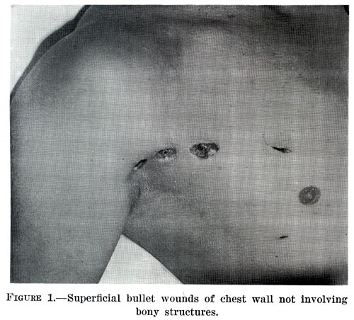
Penetrating trauma is an open wound injury that occurs when an object pierces the Human skin, skin and enters a tissue (biology), tissue of the body, creating a deep but relatively narrow entry wound. In contrast, a blunt trauma, blunt or ''non-penetrating'' trauma may have some deep damage, but the overlying skin is not necessarily broken and the wound is still closed to the outside environment. The penetrating object may foreign body, remain in the tissues, come back out the path it entered, or pass through the full thickness of the tissues and exit from another area. A penetrating injury in which an object enters the body or a structure and passes all the way through an exit wound is called a perforating trauma, while the term ''penetrating trauma'' implies that the object does not perforate wholly through. In gunshot wounds, perforating trauma is associated with an entrance wound and an often larger exit wound. Penetrating trauma can be caused by a foreign object or by fragm ... [...More Info...] [...Related Items...] OR: [Wikipedia] [Google] [Baidu] |
United Kingdom
The United Kingdom of Great Britain and Northern Ireland, commonly known as the United Kingdom (UK) or Britain, is a country in Northwestern Europe, off the coast of European mainland, the continental mainland. It comprises England, Scotland, Wales and Northern Ireland. The UK includes the island of Great Britain, the north-eastern part of the island of Ireland, and most of List of islands of the United Kingdom, the smaller islands within the British Isles, covering . Northern Ireland shares Republic of Ireland–United Kingdom border, a land border with the Republic of Ireland; otherwise, the UK is surrounded by the Atlantic Ocean, the North Sea, the English Channel, the Celtic Sea and the Irish Sea. It maintains sovereignty over the British Overseas Territories, which are located across various oceans and seas globally. The UK had an estimated population of over 68.2 million people in 2023. The capital and largest city of both England and the UK is London. The cities o ... [...More Info...] [...Related Items...] OR: [Wikipedia] [Google] [Baidu] |
Peritoneal Dialysis
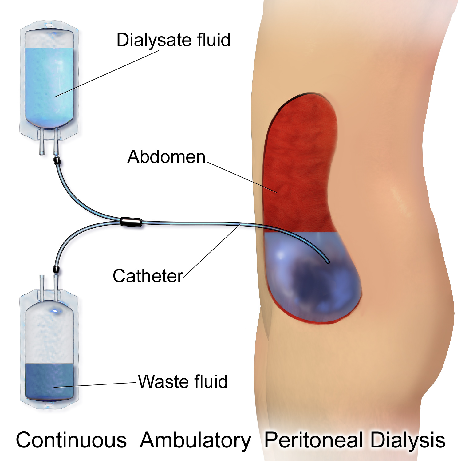
Peritoneal dialysis (PD) is a type of kidney dialysis, dialysis that uses the peritoneum in a person's abdomen as the membrane through which fluid and dissolved substances are exchanged with the blood. It is used to remove excess fluid, correct electrolyte problems, and remove toxins in those with kidney failure. Peritoneal dialysis has better outcomes than hemodialysis during the first two years. Other benefits include greater flexibility and better tolerability in those with significant heart disease. Side effects Complications may include peritonitis, infections within the abdomen, hernias, high blood sugar, bleeding in the abdomen, and blockage of the catheter. Peritoneal dialysis is not possible in those with significant prior abdominal surgery or inflammatory bowel disease. It requires some degree of technical skill to be done properly. Mechanism In peritoneal dialysis, a specific solution is introduced and then removed through a permanent tube in the lower abdomen. Th ... [...More Info...] [...Related Items...] OR: [Wikipedia] [Google] [Baidu] |
Endoscopy
An endoscopy is a procedure used in medicine to look inside the body. The endoscopy procedure uses an endoscope to examine the interior of a hollow organ or cavity of the body. Unlike many other medical imaging techniques, endoscopes are inserted directly into the organ. There are many types of endoscopies. Depending on the site in the body and type of procedure, an endoscopy may be performed by a doctor or a surgeon. During the procedure, a patient may be fully conscious or anaesthesia, anaesthetised. Most often, the term ''endoscopy'' is used to refer to an examination of the upper part of the human gastrointestinal tract, gastrointestinal tract, known as an esophagogastroduodenoscopy. Similar instruments are called borescopes for nonmedical use. History Adolf Kussmaul was fascinated by sword swallowers who would insert a sword down their throat without gagging. This drew inspiration to insert a hollow tube for observation; the next problem to solve was how to shine light th ... [...More Info...] [...Related Items...] OR: [Wikipedia] [Google] [Baidu] |
Surgical Anastomosis
A surgical anastomosis is a surgical technique used to make a new connection between two body structures that carry fluid, such as blood vessels or bowel. For example, an Artery, arterial anastomosis is used in vascular bypass and a Colon (anatomy), colonic anastomosis is used to restore colonic continuity after the resection of Colorectal cancer#surgery, colon cancer. A surgical anastomosis can be created using suture sewn by hand, mechanical staplers and biological glues, depending on the circumstances. While an anastomosis may be end-to-end, equally it could be performed side-to-side or end-to-side depending on the circumstances of the required reconstruction or Coronary artery bypass surgery, bypass. The term reanastomosis is also used to describe a surgical reconnection usually reversing a prior surgery to disconnect an anatomical anastomosis, e.g. tubal reversal after tubal ligation. __TOC__ Medical uses * Blood vessels: Arteries and veins. Most vascular procedures, includi ... [...More Info...] [...Related Items...] OR: [Wikipedia] [Google] [Baidu] |
Laparoscopy
Laparoscopy () is an operation performed in the abdomen or pelvis using small incisions (usually 0.5–1.5 cm) with the aid of a camera. The laparoscope aids diagnosis or therapeutic interventions with a few small cuts in the abdomen.MedlinePlus > Laparoscopy Update Date: 21 August 2009. Updated by: James Lee, MD // No longer valid Laparoscopic surgery, also called minimally invasive procedure, bandaid surgery, or keyhole surgery, is a modern surgical technique. There are a number of advantages to the patient with laparoscopic surgery versus an exploratory laparotomy. These include reduced pain due to smaller incisions, reduced hemorrhaging, and shorter recovery time. The key element is the use of a laparoscope, a long fiber optic cable system that allows viewing of the affected area by snaking the cable from a more distant, but more easily accessible location. Laparoscopic surgery includes operations within the abdominal or pelvic cavities, whereas keyhole surgery per ... [...More Info...] [...Related Items...] OR: [Wikipedia] [Google] [Baidu] |
Laparotomy
A laparotomy is a surgical procedure involving a surgical incision through the abdominal wall to gain access into the abdominal cavity. It is also known as a celiotomy. Origins and history The first successful laparotomy was performed without anesthesia by Ephraim McDowell in 1809 in Danville, Kentucky. On July 13, 1881, George E. Goodfellow treated a miner outside Tombstone, Arizona Territory, who had been shot in the abdomen with a .32-caliber Colt revolver. Goodfellow was able to operate on the man nine days after he was shot, when he performed the first laparotomy to treat a bullet wound. Terminology The term comes from the Greek word λᾰπάρᾱ (lapara) 'the soft part of the body between the ribs and hip, flank' and the suffix ''-tomy'', from the Greek word τομή (tome) '(surgical) cut'. In diagnostic laparotomy (most often referred to as an exploratory laparotomy and abbreviated ex-lap), the nature of the disease is unknown, and laparotomy is deemed the bes ... [...More Info...] [...Related Items...] OR: [Wikipedia] [Google] [Baidu] |
Steroids
A steroid is an organic compound with four fused rings (designated A, B, C, and D) arranged in a specific molecular configuration. Steroids have two principal biological functions: as important components of cell membranes that alter membrane fluidity; and as signaling molecules. Examples include the lipid cholesterol, sex hormones estradiol and testosterone, anabolic steroids, and the anti-inflammatory corticosteroid drug dexamethasone. Hundreds of steroids are found in fungi, plants, and animals. All steroids are manufactured in cells from a sterol: cholesterol (animals), lanosterol ( opisthokonts), or cycloartenol (plants). All three of these molecules are produced via cyclization of the triterpene squalene. Structure The steroid nucleus ( core structure) is called gonane (cyclopentanoperhydrophenanthrene). It is typically composed of seventeen carbon atoms, bonded in four fused rings: three six-member cyclohexane rings (rings A, B and C in the first illus ... [...More Info...] [...Related Items...] OR: [Wikipedia] [Google] [Baidu] |
Ischemic Colitis
Ischemic colitis (also spelled ischaemic colitis) is a medical condition in which inflammation and injury of the large intestine result from inadequate blood supply (ischemia). Although uncommon in the general population, ischemic colitis occurs with greater frequency in the elderly, and is the most common form of bowel ischemia. http://www.guideline.gov/summary/summary.aspx?ss=15&doc_id=3069&nbr=2295 Causes of the reduced blood flow can include changes in the systemic circulation (e.g. low blood pressure) or local factors such as constriction of blood vessels or a blood clot. In most cases, no specific cause can be identified. Ischemic colitis is usually suspected on the basis of the clinical setting, physical examination, and laboratory test results; the diagnosis can be confirmed by endoscopy or by using sigmoid or endoscopic placement of a visible light spectroscopic catheter (see Diagnosis). Ischemic colitis can span a wide spectrum of severity; most patients are treated ... [...More Info...] [...Related Items...] OR: [Wikipedia] [Google] [Baidu] |


