|
Plank (exercise)
The plank (also called a front hold, hover, or abdominal bridge) is an isometric exercise, isometric core (anatomy), core strength exercise that involves maintaining a position similar to a push-up. Form The most common plank is the forearm plank which is held in a push-up-like position, with the body's weight borne on forearms, elbows, and toes. Many variations exist such as the side plank and the reverse plank. The plank is commonly practiced in Pilates and yoga as exercise where it is called Chaturanga Dandasana, and by those training for boxing and other sports. The "extended plank" adds substantial difficulty to the standard plank exercise. To perform the extended plank, a person begins in the push-up position and then extends the arms or hands as far forward as possible. Effect The plank strengthens the abdominals, back, and shoulders. Muscles involved in the front plank include: * Primary muscles: erector spinae muscles, erector spinae, rectus abdominis muscle, rec ... [...More Info...] [...Related Items...] OR: [Wikipedia] [Google] [Baidu] [Amazon] |
Isometric Exercise
An isometric exercise is an exercise involving the static contraction of a muscle without any visible movement in the angle of the joint. The term "isometric" combines the Greek words ''isos'' (equal) and ''-metria'' (measuring), meaning that in these exercises the length of the muscle and the angle of the joint do not change, though contraction strength may be varied. This is in contrast to ''isotonic contractions'', in which the contraction strength does not change, though the muscle length and joint angle do. The three main types of isometric exercise are isometric presses, pulls, and holds. They may be included in a strength training regime in order to improve the body's ability to apply power from a static position or, in the case of isometric holds, improve the body's ability to maintain a position for a period of time. Considered as an action, isometric presses are also of fundamental importance to the body's ability to prepare itself to perform immediately subsequent pow ... [...More Info...] [...Related Items...] OR: [Wikipedia] [Google] [Baidu] [Amazon] |
Blood Pressure
Blood pressure (BP) is the pressure of Circulatory system, circulating blood against the walls of blood vessels. Most of this pressure results from the heart pumping blood through the circulatory system. When used without qualification, the term "blood pressure" refers to the pressure in a brachial artery, where it is most commonly measured. Blood pressure is usually expressed in terms of the systolic pressure (maximum pressure during one Cardiac cycle, heartbeat) over diastolic pressure (minimum pressure between two heartbeats) in the cardiac cycle. It is measured in Millimetre of mercury, millimetres of mercury (mmHg) above the surrounding atmospheric pressure, or in Pascal (unit), kilopascals (kPa). The difference between the systolic and diastolic pressures is known as pulse pressure, while the average pressure during a cardiac cycle is known as mean arterial pressure. Blood pressure is one of the vital signs—together with respiratory rate, heart rate, Oxygen saturation (me ... [...More Info...] [...Related Items...] OR: [Wikipedia] [Google] [Baidu] [Amazon] |
Wall Sit
The imaginary chair or wall sit is a means of exercise or punishment, where one positions themselves against a wall as if seated. A wall sit specifically refers to an exercise done to strengthen the quadriceps muscles. The exercise is characterized by the two right angles formed by the body, one at the hips and one at the knees. The person wall sitting places their back against a wall with their feet shoulder-width apart and a little ways out from the wall. Then, keeping their back against the wall, they lower their hips until their knees form right angles. This is a very intense workout for the quadriceps muscles, and it can be very painful to hold this position for extended periods. Wall sits are used as a primary strengthening exercise in many sports requiring strong quadriceps including fencing, ice hockey, sailing (mostly small boat racing), skiing and track and field. Benefits Wall sitting primarily builds isometric strength and endurance in glutes, calves, quadriceps, ... [...More Info...] [...Related Items...] OR: [Wikipedia] [Google] [Baidu] [Amazon] |
British Journal Of Sports Medicine
The ''British Journal of Sports Medicine'' is a twice-monthly peer-reviewed medical journal covering sports science and sports medicine including sport physiotherapy. It is published by the BMJ Group. It was established in 1964 and the editor-in-chief from 2008 to 2020 was Karim M. Khan (University of British Columbia). Jonathan Drezner (University of Washington) has been editor-in-chief since January 1, 2021. Abstracting and indexing According to the ''Journal Citation Reports'', the journal has a 2023 impact factor of 11.8. International Olympic Committee consensus statements Since 2009, the journal has partnered with the International Olympic Committee to produce regular consensus statements regarding important issues in sports injury prevention and elite sport. Some of the recent examples include Consensus Statements on concussions in sport (the "Berlin guidelines"), relative energy deficiency in sport, the relationship between training load and injury, mental health issues ... [...More Info...] [...Related Items...] OR: [Wikipedia] [Google] [Baidu] [Amazon] |
Hamstring
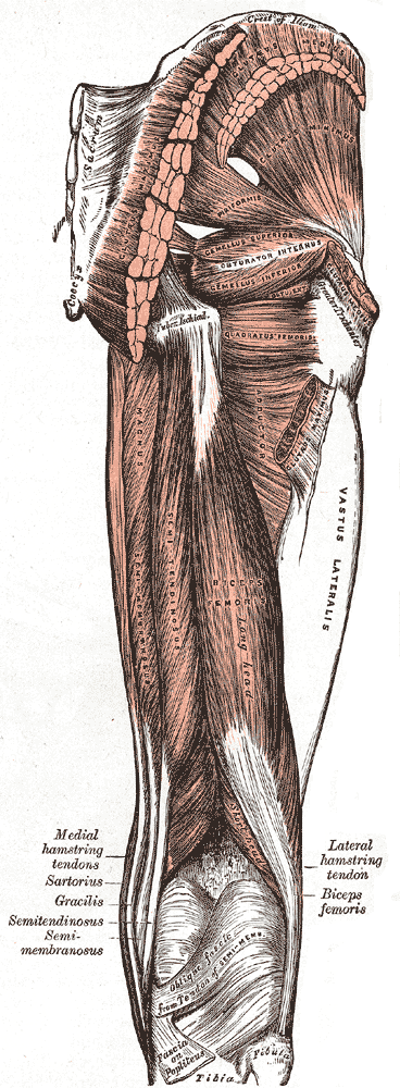
A hamstring () is any one of the three posterior thigh muscles in human anatomy between the hip and the knee: from medial to lateral, the semimembranosus, semitendinosus and biceps femoris. Etymology The word " ham" is derived from the Old English “ham” or “hom” meaning the hollow or bend of the knee, from a Germanic base where it meant "crooked". It gained the meaning of the leg of an animal around the 15th century. ''String'' refers to tendons, and thus the hamstrings' string-like tendons felt on either side of the back of the knee. Criteria The common criteria of any hamstring muscles are: # Muscles should originate from ischial tuberosity. # Muscles should be inserted over the knee joint, in the tibia or in the fibula. # Muscles will be innervated by the tibial branch of the sciatic nerve. # Muscle will participate in flexion of the knee joint and extension of the hip joint. Those muscles which fulfill all of the four criteria are called true hamstrings. ... [...More Info...] [...Related Items...] OR: [Wikipedia] [Google] [Baidu] [Amazon] |
Abdominal Internal Oblique Muscle
The abdominal internal oblique muscle, also internal oblique muscle or interior oblique, is an abdominal muscle in the abdominal wall that lies below the external oblique muscle and just above the transverse abdominal muscle. Structure Its fibers run perpendicular to the external oblique muscle, beginning in the thoracolumbar fascia of the lower back, the anterior 2/3 of the iliac crest (upper part of hip bone) and the lateral half of the inguinal ligament. The muscle fibers run from these points superomedially (up and towards midline) to the muscle's insertions on the inferior borders of the 10th through 12th ribs and the linea alba. In males, the cremaster muscle is also attached to the internal oblique. Nerve supply The internal oblique is supplied by the lower intercostal nerves, as well as the iliohypogastric nerve and the ilioinguinal nerve. Function The internal oblique performs two major functions. Firstly as an accessory muscle of respiration, it acts as an ... [...More Info...] [...Related Items...] OR: [Wikipedia] [Google] [Baidu] [Amazon] |
Abdominal External Oblique Muscle
The abdominal external oblique muscle (also external oblique muscle or exterior oblique) is the largest and outermost of the three flat Abdomen#Muscles, abdominal muscles of the lateral anterior abdomen. Structure The external oblique is situated on the lateral and anterior parts of the abdomen. It is broad, thin, and irregularly quadrilateral, its muscular portion occupying the side, its aponeurosis the anterior wall of the abdomen. In most humans, the oblique is not visible, due to subcutaneous adipose, fat deposits and the small size of the muscle. It arises from eight fleshy digitations, each from the external surfaces and inferior borders of the fifth to twelfth ribs (lower eight ribs). These digitations are arranged in an oblique line which runs inferiorly and anteriorly, with the upper digitations being attached close to the cartilages of the corresponding ribs, the lowest to the apex of the cartilage of the last rib, the intermediate ones to the ribs at some distance fr ... [...More Info...] [...Related Items...] OR: [Wikipedia] [Google] [Baidu] [Amazon] |
Adductor Muscles Of The Hip
The adductor muscles of the hip are a group of muscles in the medial compartment of the thigh mostly used for bringing the thighs together (called adduction). Structure The adductor group is made up of: * Adductor brevis * Adductor longus * Adductor magnus * Adductor minimus This is often considered to be a part of adductor magnus. * pectineus * gracilis * Obturator externusPlatzer, Werner (2004), Color Atlas of Human Anatomy, Vol. 1, Locomotor System', Thieme, 5th ed, p 240 is also part of the medial compartment of thigh The adductors originate on the pubis and ischium bones and insert mainly on the medial posterior surface of the femur. Nerve supply The pectineus is the only adductor muscle that is innervated by the femoral nerve. The other adductor muscles are innervated by the obturator nerve with the exception of a small part of the adductor magnus which is innervated by the tibial nerve. Variation In 33% of people a supernumerary muscle is found between ... [...More Info...] [...Related Items...] OR: [Wikipedia] [Google] [Baidu] [Amazon] |
Gluteus Minimus Muscle
The gluteus minimus, or glutæus minimus, the smallest of the three gluteal muscles, is situated immediately beneath the gluteus medius. Structure It is fan-shaped, arising from the outer surface of the ilium, between the anterior and inferior gluteal lines, and behind, from the margin of the greater sciatic notch. The fibers converge to the deep surface of a radiated aponeurosis, and this ends in a tendon which is inserted into an impression on the anterior border of the greater trochanter, and gives an expansion to the capsule of the hip joint. Relations A bursa is interposed between the tendon and the greater trochanter. Between the gluteus medius and gluteus minimus are the deep branches of the superior gluteal vessels and the superior gluteal nerve. The deep surface of the gluteus minimus is in relation with the reflected tendon of the rectus femoris and the capsule of the hip joint. Variations The muscle may be divided into an anterior and a posterior part, or ... [...More Info...] [...Related Items...] OR: [Wikipedia] [Google] [Baidu] [Amazon] |
Gluteus Medius Muscle
The gluteus medius, one of the three gluteal muscles, is a broad, thick, radiating muscle. It is situated on the outer surface of the pelvis. Its posterior third is covered by the gluteus maximus, its anterior two-thirds by the gluteal aponeurosis, which separates it from the superficial fascia and integument. Structure The gluteus medius muscle starts, or "originates", on the outer surface of the ilium between the iliac crest and the posterior gluteal line above, and the anterior gluteal line below; the gluteus medius also originates from its own fascia, the gluteal aponeurosis, that covers its outer surface. The fibers of the muscle converge into a strong flattened tendon that inserts on the lateral surface of the greater trochanter. More specifically, the muscle's tendon inserts into an oblique ridge that runs downward and forward on the lateral surface of the greater trochanter. Before the insertion the fibers cross from anterior to posterior and vice versa. Relations ... [...More Info...] [...Related Items...] OR: [Wikipedia] [Google] [Baidu] [Amazon] |
Gastrocnemius Muscle
The gastrocnemius muscle (plural ''gastrocnemii'') is a superficial two-headed muscle that is in the back part of the lower leg of humans. It is located superficial to the soleus in the posterior (back) compartment of the leg. It runs from its two heads just above the knee to the heel, extending across a total of three joints (knee, ankle and subtalar joints). The muscle is named via Latin, from Greek γαστήρ (''gaster'') 'belly' or 'stomach' and κνήμη (''knḗmē'') 'leg', meaning 'stomach of the leg' (referring to the bulging shape of the calf). Structure Origin/proximal attachment The lateral head originates from the lateral condyle of the femur, while the medial head originates from the medial condyle of the femur. Insertion/distal attachment Its other end forms a common tendon with the soleus muscle; this tendon is known as the calcaneal tendon or Achilles tendon and inserts onto the posterior surface of the calcaneus, or heel bone. Relations The ga ... [...More Info...] [...Related Items...] OR: [Wikipedia] [Google] [Baidu] [Amazon] |


