|
Peritoneal
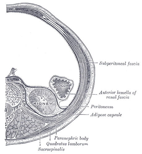
The peritoneum is the serous membrane forming the lining of the abdominal cavity or coelom in amniotes and some invertebrates, such as annelids. It covers most of the intra-abdominal (or coelomic) organs, and is composed of a layer of mesothelium supported by a thin layer of connective tissue. This peritoneal lining of the cavity supports many of the abdominal organs and serves as a conduit for their blood vessels, lymphatic vessels, and nerves. The abdominal cavity (the space bounded by the vertebrae, abdominal muscles, diaphragm, and pelvic floor) is different from the intraperitoneal space (located within the abdominal cavity but wrapped in peritoneum). The structures within the intraperitoneal space are called "intraperitoneal" (e.g., the stomach and intestines), the structures in the abdominal cavity that are located behind the intraperitoneal space are called "retroperitoneal" (e.g., the kidneys), and those structures below the intraperitoneal space are called "subperit ... [...More Info...] [...Related Items...] OR: [Wikipedia] [Google] [Baidu] |
Intraperitoneal Injection
Intraperitoneal injection or IP injection is the injection of a substance into the peritoneum (body cavity). It is more often applied to non-human animals than to humans. In general, it is preferred when large amounts of blood replacement fluids are needed or when low blood pressure or other problems prevent the use of a suitable blood vessel for intravenous injection. In humans, the method is widely used to administer chemotherapy drugs to treat some cancers, particularly ovarian cancer. Although controversial, intraperitoneal use in ovarian cancer has been recommended as a standard of care. Fluids are injected intraperitoneally in infants, also used for peritoneal dialysis. Intraperitoneal injections are a way to administer therapeutics and drugs through a peritoneal route (body cavity). They are one of the few ways drugs can be administered through injection, and have uses in research involving animals, drug administration to treat ovarian cancers, and much more. Understandin ... [...More Info...] [...Related Items...] OR: [Wikipedia] [Google] [Baidu] |
Retroperitoneal
The retroperitoneal space (retroperitoneum) is the anatomical space (sometimes a potential space) behind (''retro'') the peritoneum. It has no specific delineating anatomical structures. Organs are retroperitoneal if they have peritoneum on their anterior side only. Structures that are not suspended by mesentery in the abdominal cavity and that lie between the parietal peritoneum and abdominal wall are classified as retroperitoneal. This is different from organs that are not retroperitoneal, which have peritoneum on their posterior side and are suspended by mesentery in the abdominal cavity. The retroperitoneum can be further subdivided into the following: *Perirenal (or perinephric) space *Anterior pararenal (or paranephric) space *Posterior pararenal (or paranephric) space Retroperitoneal structures Structures that lie behind the peritoneum are termed "retroperitoneal". Organs that were once suspended within the abdominal cavity by mesentery but migrated posterior to the pe ... [...More Info...] [...Related Items...] OR: [Wikipedia] [Google] [Baidu] |
Peritoneal Cavity
The peritoneal cavity is a potential space located between the two layers of the peritoneum—the parietal peritoneum, the serous membrane that lines the abdominal wall, and visceral peritoneum, which surrounds the internal organs. While situated within the abdominal cavity, the term ''peritoneal cavity'' specifically refers to the potential space enclosed by these peritoneal membranes. The cavity contains a thin layer of lubricating serous fluid that enables the organs to move smoothly against each other, facilitating the movement and expansion of internal organs during digestion. The parietal and visceral peritonea are named according to their location and function. The peritoneal cavity, derived from the coelomic cavity in the embryo, is one of several body cavities, including the pleural cavities surrounding the lungs and the pericardial cavity around the heart. The peritoneal cavity is the largest serosal sac and fluid-filled cavity in the body, it secretes approximately ... [...More Info...] [...Related Items...] OR: [Wikipedia] [Google] [Baidu] |
Mesothelium
The mesothelium is a membrane composed of simple squamous epithelium, simple squamous epithelial cells of mesodermal origin, which forms the lining of several body cavities: the pleura (pleural cavity around the lungs), peritoneum (abdominopelvic cavity including the mesentery, omentum (other), omenta, falciform ligament and the perimetrium) and pericardium (around the heart). Mesothelial tissue also surrounds the male testis (as the tunica vaginalis) and occasionally the spermatic cord (in a patent processus vaginalis). Mesothelium that tunica (biology), covers the internal organs is called visceral mesothelium, while one that covers the surrounding body walls is called the :wikt:parietal, parietal mesothelium. The mesothelium that secretes serous fluid as a main function is also known as a serosa. Origin Mesothelium derives from the embryonic mesoderm cell layer, that lines the body cavity, coelom (body cavity) in the embryo. It develops into the layer of cells that c ... [...More Info...] [...Related Items...] OR: [Wikipedia] [Google] [Baidu] |
Kidney
In humans, the kidneys are two reddish-brown bean-shaped blood-filtering organ (anatomy), organs that are a multilobar, multipapillary form of mammalian kidneys, usually without signs of external lobulation. They are located on the left and right in the retroperitoneal space, and in adult humans are about in length. They receive blood from the paired renal artery, renal arteries; blood exits into the paired renal veins. Each kidney is attached to a ureter, a tube that carries excreted urine to the urinary bladder, bladder. The kidney participates in the control of the volume of various body fluids, fluid osmolality, Acid-base homeostasis, acid-base balance, various electrolyte concentrations, and removal of toxins. Filtration occurs in the glomerulus (kidney), glomerulus: one-fifth of the blood volume that enters the kidneys is filtered. Examples of substances reabsorbed are solute-free water, sodium, bicarbonate, glucose, and amino acids. Examples of substances secreted are hy ... [...More Info...] [...Related Items...] OR: [Wikipedia] [Google] [Baidu] |
Thoracic Diaphragm
The thoracic diaphragm, or simply the diaphragm (; ), is a sheet of internal Skeletal striated muscle, skeletal muscle in humans and other mammals that extends across the bottom of the thoracic cavity. The diaphragm is the most important Muscles of respiration, muscle of respiration, and separates the thoracic cavity, containing the heart and lungs, from the abdominal cavity: as the diaphragm contracts, the volume of the thoracic cavity increases, creating a negative pressure there, which draws air into the lungs. Its high oxygen consumption is noted by the many mitochondria and capillaries present; more than in any other skeletal muscle. The term ''diaphragm'' in anatomy, created by Gerard of Cremona, can refer to other flat structures such as the urogenital diaphragm or Pelvic floor, pelvic diaphragm, but "the diaphragm" generally refers to the thoracic diaphragm. In humans, the diaphragm is slightly asymmetric—its right half is higher up (superior) to the left half, since th ... [...More Info...] [...Related Items...] OR: [Wikipedia] [Google] [Baidu] |
Intestine
The gastrointestinal tract (GI tract, digestive tract, alimentary canal) is the tract or passageway of the digestive system that leads from the mouth to the anus. The tract is the largest of the body's systems, after the cardiovascular system. The GI tract contains all the major organs of the digestive system, in humans and other animals, including the esophagus, stomach, and intestines. Food taken in through the mouth is digested to extract nutrients and absorb energy, and the waste expelled at the anus as feces. ''Gastrointestinal'' is an adjective meaning of or pertaining to the stomach and intestines. Most animals have a "through-gut" or complete digestive tract. Exceptions are more primitive ones: sponges have small pores ( ostia) throughout their body for digestion and a larger dorsal pore ( osculum) for excretion, comb jellies have both a ventral mouth and dorsal anal pores, while cnidarians and acoels have a single pore for both digestion and excretion. The human gas ... [...More Info...] [...Related Items...] OR: [Wikipedia] [Google] [Baidu] |
Abdominal Cavity
The abdominal cavity is a large body cavity in humans and many other animals that contain Organ (anatomy), organs. It is a part of the abdominopelvic cavity. It is located below the thoracic cavity, and above the pelvic cavity. Its dome-shaped roof is the thoracic diaphragm, a thin sheet of muscle under the lungs, and its floor is the pelvic inlet, opening into the pelvis. Structure Organs Organs of the abdominal cavity include the stomach, liver, gallbladder, spleen, pancreas, small intestine, kidneys, large intestine, and adrenal glands. Peritoneum The abdominal cavity is lined with a protective membrane termed the peritoneum. The inside wall is covered by the parietal peritoneum. The kidneys are located behind the peritoneum, in the retroperitoneum, outside the abdominal cavity. The viscera are also covered by visceral peritoneum. Between the visceral and parietal peritoneum is the peritoneal cavity, which is a potential space. It contains a serous fluid called peritoneal ... [...More Info...] [...Related Items...] OR: [Wikipedia] [Google] [Baidu] |
Abdominal Wall
In anatomy, the abdominal wall represents the boundaries of the abdominal cavity. The abdominal wall is split into the anterolateral and posterior walls. There is a common set of layers covering and forming all the walls: the deepest being the visceral peritoneum, which covers many of the abdominal organs (most of the large and small intestines, for example), and the parietal peritoneum—which covers the visceral peritoneum below it, the extraperitoneal fat, the transversalis fascia, the internal and external oblique and transversus abdominis aponeurosis, and a layer of fascia, which has different names according to what it covers (e.g., transversalis, psoas fascia). In medical vernacular, the term 'abdominal wall' most commonly refers to the layers composing the anterior abdominal wall which, in addition to the layers mentioned above, includes the three layers of muscle: the transversus abdominis (transverse abdominal muscle), the internal oblique, internal (obliquus internus) a ... [...More Info...] [...Related Items...] OR: [Wikipedia] [Google] [Baidu] |
Abdomen
The abdomen (colloquially called the gut, belly, tummy, midriff, tucky, or stomach) is the front part of the torso between the thorax (chest) and pelvis in humans and in other vertebrates. The area occupied by the abdomen is called the abdominal cavity. In arthropods, it is the posterior (anatomy), posterior tagma (biology), tagma of the body; it follows the thorax or cephalothorax. In humans, the abdomen stretches from the thorax at the thoracic diaphragm to the pelvis at the pelvic brim. The pelvic brim stretches from the lumbosacral joint (the intervertebral disc between Lumbar vertebrae, L5 and Vertebra#Sacrum, S1) to the pubic symphysis and is the edge of the pelvic inlet. The space above this inlet and under the thoracic diaphragm is termed the abdominal cavity. The boundary of the abdominal cavity is the abdominal wall in the front and the peritoneal surface at the rear. In vertebrates, the abdomen is a large body cavity enclosed by the abdominal muscles, at the front an ... [...More Info...] [...Related Items...] OR: [Wikipedia] [Google] [Baidu] |
Bladder
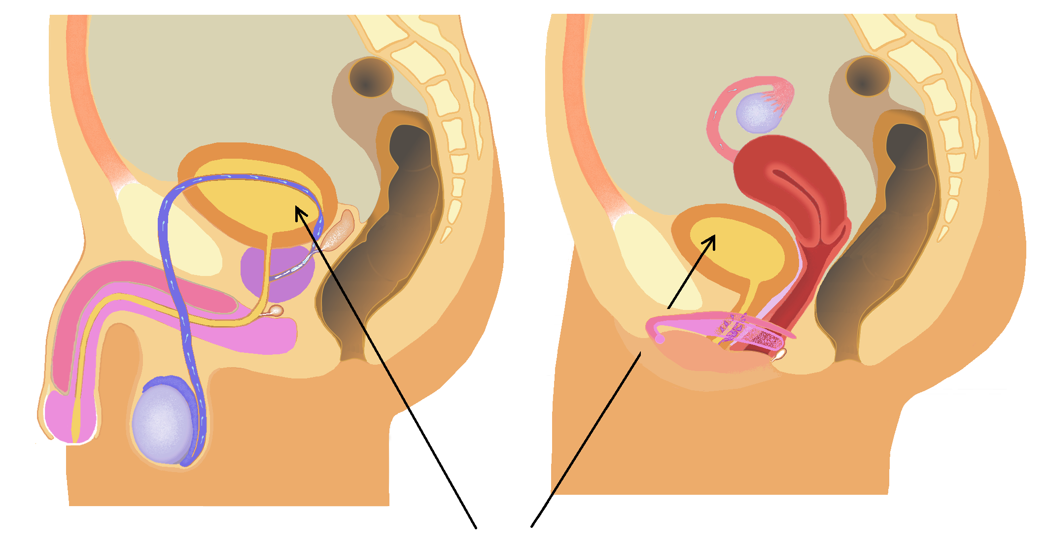
The bladder () is a hollow organ in humans and other vertebrates that stores urine from the kidneys. In placental mammals, urine enters the bladder via the ureters and exits via the urethra during urination. In humans, the bladder is a distensible organ that sits on the pelvic floor. The typical adult human bladder will hold between 300 and (10 and ) before the urge to empty occurs, but can hold considerably more. The Latin phrase for "urinary bladder" is ''vesica urinaria'', and the term ''vesical'' or prefix ''vesico-'' appear in connection with associated structures such as vesical veins. The modern Latin word for "bladder" – ''cystis'' – appears in associated terms such as cystitis (inflammation of the bladder). Structure In humans, the bladder is a hollow muscular organ situated at the base of the pelvis. In gross anatomy, the bladder can be divided into a broad (base), a body, an apex, and a neck. The apex (also called the vertex) is directed forward toward th ... [...More Info...] [...Related Items...] OR: [Wikipedia] [Google] [Baidu] |


