|
Perineal Branch
The perineal branches of the posterior femoral cutaneous nerve are distributed to the skin at the upper and medial side of the thigh. One long perineal branch, inferior pudendal (long scrotal nerve), curves forward below and in front of the ischial tuberosity, pierces the fascia lata, and runs forward beneath the superficial fascia of the perineum to the skin of the scrotum in the male, and of the labium majus in the female. It communicates with the inferior anal nerves and the posterior scrotal nerves. See also * Posterior cutaneous nerve of thigh The posterior cutaneous nerve of the thigh (also called the posterior femoral cutaneous nerve) is a sensory nerve of the thigh. It is a branch of the sacral plexus. It supplies the skin of the posterior surface of the thigh, leg, buttock, and als ... References External links * () {{Authority control Nerves of the lower limb and lower torso Scrotum ... [...More Info...] [...Related Items...] OR: [Wikipedia] [Google] [Baidu] |
Posterior Femoral Cutaneous Nerve
The posterior cutaneous nerve of the thigh (also called the posterior femoral cutaneous nerve) is a sensory nerve of the thigh. It is a branch of the sacral plexus. It supplies the skin of the posterior surface of the thigh, leg, buttock, and also the perineum. Unlike most nerves termed "cutaneous" which are subcutaneous, only the terminal branches of this nerve pass into subcutaneous tissue before being distributed to the skin, with most of the nerve itself situated deep to the deep fascia. Structure Origin The posterior cutaneous nerve of the thigh is a branch of the sacral plexus. It arises from the posterior divisions of the S1- S2, and the anterior divisions of S2- S3 sacral spinal nerves. Course It leaves the pelvis through the greater sciatic foramen inferior to the piriformis muscle. It then descends deep to the gluteus maximus muscle, medial or posterior to the sciatic nerve, and alongside the inferior gluteal artery. It descends within the posterior thigh deep ... [...More Info...] [...Related Items...] OR: [Wikipedia] [Google] [Baidu] |
Thigh
In anatomy, the thigh is the area between the hip (pelvis) and the knee. Anatomically, it is part of the lower limb. The single bone in the thigh is called the femur. This bone is very thick and strong (due to the high proportion of bone tissue), and forms a ball and socket joint at the hip, and a modified hinge joint at the knee. Structure Bones The femur is the only bone in the thigh and serves as an attachment site for all thigh muscles. The head of the femur articulates with the acetabulum in the pelvic bone forming the hip joint, while the distal part of the femur articulates with the tibia and patella forming the knee. By most measures, the femur is the strongest and longest bone in the body. The femur is categorised as a long bone and comprises a diaphysis, the shaft (or body) and two epiphyses, the lower extremity and the upper extremity of femur, that articulate with adjacent bones in the hip and knee. Muscular compartments In cross-section, the thigh is d ... [...More Info...] [...Related Items...] OR: [Wikipedia] [Google] [Baidu] |
Ischial Tuberosity
The ischial tuberosity (or tuberosity of the ischium, tuber ischiadicum), also known colloquially as the sit bones or sitz bones, or as a pair the sitting bones, is a large posterior bony protuberance on the superior ramus of the ischium. It marks the lateral boundary of the pelvic outlet. When sitting, the weight is frequently placed upon the ischial tuberosity. The gluteus maximus provides cover in the upright posture, but leaves it free in the seated position.Platzer (2004), p 236 The distance between a cyclist's ischial tuberosities is one of the factors in the choice of a bicycle saddle. Divisions The tuberosity is divided into two portions: a lower, rough, somewhat triangular part, and an upper, smooth, quadrilateral portion. * The ''lower portion'' is subdivided by a prominent longitudinal ridge, passing from base to apex, into two parts: ** The outer gives attachment to the adductor magnus ** The inner to the sacrotuberous ligament * The ''upper portion'' is subdiv ... [...More Info...] [...Related Items...] OR: [Wikipedia] [Google] [Baidu] |
Fascia Lata
The fascia lata is the deep fascia of the thigh. It encloses the thigh muscles and forms the outer limit of the fascial compartments of thigh, which are internally separated by the medial intermuscular septum and the lateral intermuscular septum. The fascia lata is thickened at its lateral side where it forms the iliotibial tract, a structure that runs to the tibia and serves as a site of muscle attachment. Structure The fascia lata is an investment for the whole of the thigh, but varies in thickness in different parts. It is thicker in the upper and lateral part of the thigh, where it receives a fibrous expansion from the gluteus maximus, and where the tensor fasciae latae is inserted between its layers; it is very thin behind and at the upper and medial part, where it covers the adductor muscles, and again becomes stronger around the knee, receiving fibrous expansions from the tendon of the biceps femoris laterally, from the sartorius medially, and from the quadriceps fem ... [...More Info...] [...Related Items...] OR: [Wikipedia] [Google] [Baidu] |
Superficial Fascia Of The Perineum
The subcutaneous tissue of perineum (or superficial perineal fascia) is a layer of subcutaneous tissue surrounding the region of the perineal body. The superficial fascia of this region consists of two layers, superficial and deep. * The superficial layer is thick, loose, areolar in texture, and contains in its meshes much adipose tissue, the amount of which varies in different subjects. In front, it is continuous with the dartos tunic of the scrotum; behind, with the subcutaneous areolar tissue surrounding the anus; and, on either side, with the same fascia on the inner sides of the thighs. In the middle line, it is adherent to the skin on the raphe and to the deep layer of the superficial fascia. * The deep layer of superficial fascia (fascia of Colles The membranous layer of the superficial fascia of the perineum (Colles' fascia) is the deeper layer (membranous layer) of the superficial perineal fascia. It is thin, aponeurotic in structure, and of considerable strength, ser ... [...More Info...] [...Related Items...] OR: [Wikipedia] [Google] [Baidu] |
Scrotum
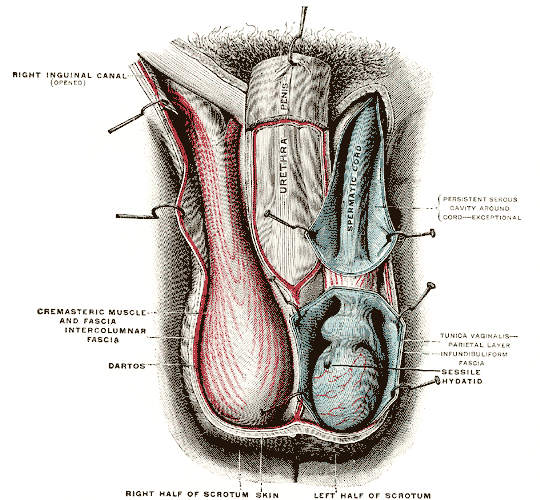
In most terrestrial mammals, the scrotum (: scrotums or scrota; possibly from Latin ''scortum'', meaning "hide" or "skin") or scrotal sac is a part of the external male genitalia located at the base of the penis. It consists of a sac of skin containing the external spermatic fascia, testicles, epididymides, and vasa deferentia. The scrotum will usually tighten when exposed to cold temperatures. The scrotum is homologous to the labia majora in females. Structure In regards to humans, the scrotum is a suspended two-chambered sac of skin and muscular tissue containing the testicles and the lower part of the spermatic cords. It is located behind the penis and above the perineum. The perineal raphe is a small, vertical ridge of skin that expands from the anus and runs through the middle of the scrotum front to back. The scrotum is also a distention of the perineum and carries some abdominal tissues into its cavity including the testicular artery, testicular vein, and ... [...More Info...] [...Related Items...] OR: [Wikipedia] [Google] [Baidu] |
Labium Majus
In primates, and specifically in humans, the labia majora (: labium majus), also known as the outer lips or outer labia, are two prominent longitudinal skin folds that extend downward and backward from the mons pubis to the perineum. Together with the labia minora, they form the labia of the vulva. The labia majora are homologous to the male scrotum. Etymology ''Labia majora'' is the Latin plural for big ("major") lips. The Latin term ''labium/labia'' is used in anatomy for a number of usually paired parallel structures, but in English, it is mostly applied to two pairs of parts of the vulva—labia majora and labia minora. Traditionally, to avoid confusion with other lip-like structures of the body, the vulvar labia were termed by anatomists in Latin as ''labia majora (''or ''minora) pudendi.'' Embryology Embryologically, they develop from labioscrotal folds. The labia majora after puberty may become of a darker color than the skin outside them and grow pubic hair on their ex ... [...More Info...] [...Related Items...] OR: [Wikipedia] [Google] [Baidu] |
Inferior Anal Nerves
The inferior rectal nerves (inferior anal nerves, inferior hemorrhoidal nerve) usually branch from the pudendal nerve but occasionally arises directly from the sacral plexus; they cross the ischiorectal fossa along with the inferior rectal artery and veins, toward the anal canal and the lower end of the rectum, and is distributed to the sphincter ani externus (external anal sphincter, EAS) and to the integument (skin) around the anus. Branches of this nerve communicate with the perineal branch of the posterior femoral cutaneous and with the posterior scrotal nerves at the forepart of the perineum. Supplies Cutaneous innervation below the pectinate line and external anal sphincter. See also * Inferior rectal artery Additional images File:Gray405.png, The perineum. The integument and superficial layer of superficial fascia reflected. File:Gray837.png, Sacral plexus of the right side. (Hemorrhoidal branch of pudic labeled at bottom right.) References External links Detai ... [...More Info...] [...Related Items...] OR: [Wikipedia] [Google] [Baidu] |
Posterior Scrotal Nerves
The posterior scrotal branches are two in number, medial and lateral. They are branches of the perineal nerve, which is itself a branch of the pudendal nerve. The pudendal nerve arises from spinal roots S2 through S4, travels through the pudendal canal on the fascia of the obturator internus muscle, and gives off the perineal nerve in the perineum. The major branch of the perineal nerve is the posterior scrotal/posterior labial. They pierce the fascia of the urogenital diaphragm, and run forward along the lateral part of the urethral triangle in company with the posterior scrotal branches of the perineal artery; they are distributed to the skin of the scrotum In most terrestrial mammals, the scrotum (: scrotums or scrota; possibly from Latin ''scortum'', meaning "hide" or "skin") or scrotal sac is a part of the external male genitalia located at the base of the penis. It consists of a sac of skin ... or labia and communicate with the perineal branch of the posterior femo ... [...More Info...] [...Related Items...] OR: [Wikipedia] [Google] [Baidu] |
Posterior Cutaneous Nerve Of Thigh
The posterior cutaneous nerve of the thigh (also called the posterior femoral cutaneous nerve) is a sensory nerve of the thigh. It is a branch of the sacral plexus. It supplies the skin of the posterior surface of the thigh, leg, buttock, and also the perineum. Unlike most nerves termed "cutaneous" which are subcutaneous, only the terminal branches of this nerve pass into subcutaneous tissue before being distributed to the skin, with most of the nerve itself situated deep to the deep fascia. Structure Origin The posterior cutaneous nerve of the thigh is a branch of the sacral plexus. It arises from the posterior divisions of the S1- S2, and the anterior divisions of S2- S3 sacral spinal nerves. Course It leaves the pelvis through the greater sciatic foramen inferior to the piriformis muscle. It then descends deep to the gluteus maximus muscle, medial or posterior to the sciatic nerve, and alongside the inferior gluteal artery. It descends within the posterior thigh dee ... [...More Info...] [...Related Items...] OR: [Wikipedia] [Google] [Baidu] |
Nerves Of The Lower Limb And Lower Torso
A nerve is an enclosed, cable-like bundle of nerve fibers (called axons). Nerves have historically been considered the basic units of the peripheral nervous system. A nerve provides a common pathway for the electrochemical nerve impulses called action potentials that are transmitted along each of the axons to peripheral organs or, in the case of sensory nerves, from the periphery back to the central nervous system. Each axon is an extension of an individual neuron, along with other supportive cells such as some Schwann cells that coat the axons in myelin. Each axon is surrounded by a layer of connective tissue called the endoneurium. The axons are bundled together into groups called fascicles, and each fascicle is wrapped in a layer of connective tissue called the perineurium. The entire nerve is wrapped in a layer of connective tissue called the epineurium. Nerve cells (often called neurons) are further classified as either sensory or motor. In the central nervous system, t ... [...More Info...] [...Related Items...] OR: [Wikipedia] [Google] [Baidu] |


