|
Pennate Muscle
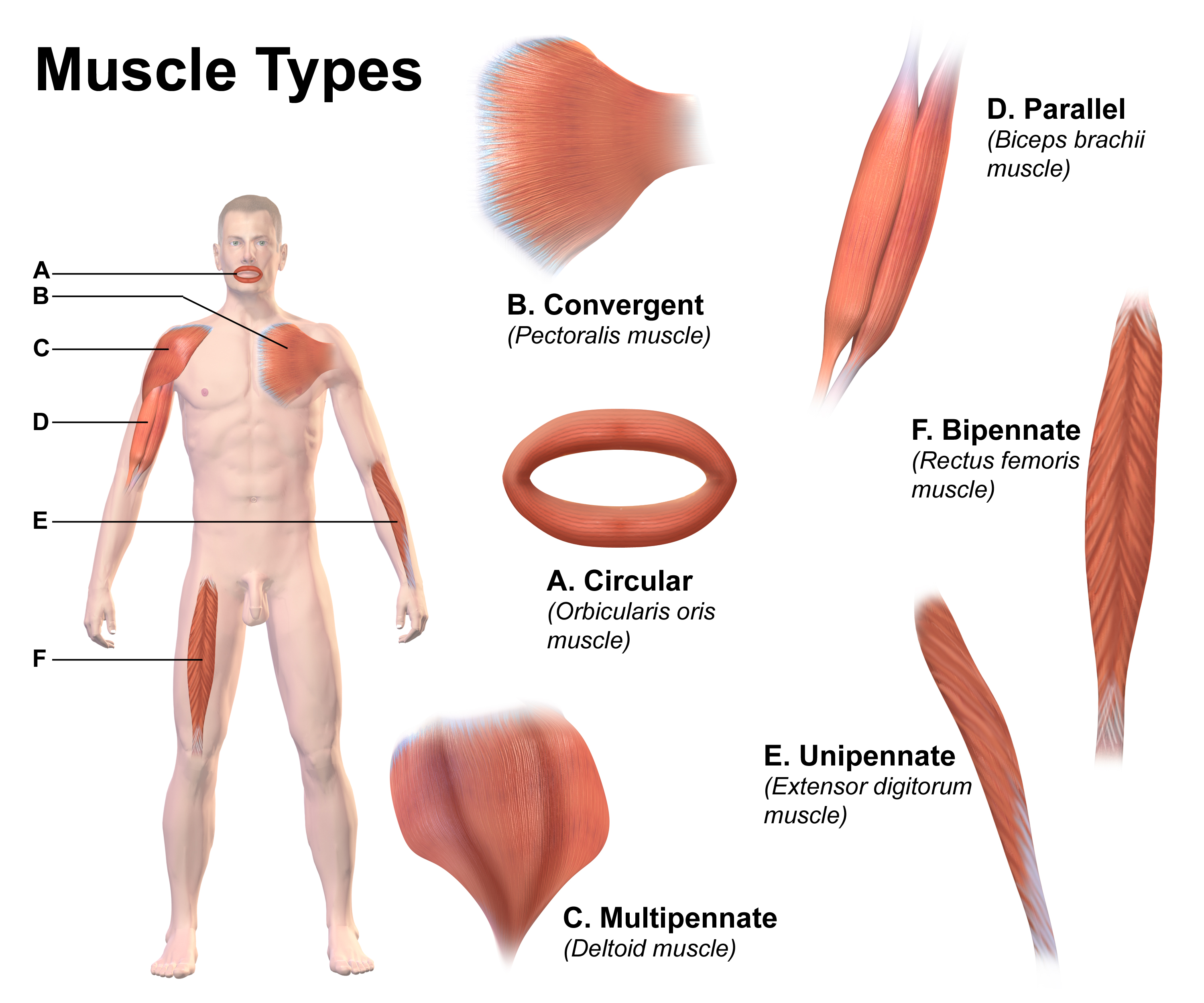
A pennate or pinnate muscle (also called a penniform muscle) is a type of skeletal muscle with fascicles that attach obliquely (in a slanting position) to its tendon. This type of muscle generally allows higher force production but a smaller range of motion. When a muscle contracts and shortens, the pennation angle increases. Etymology The term "pennate" comes from the Latin ''pinnātus'' ("feathered, winged"), from ''pinna'' ("feather, wing"). Types of pennate muscle In skeletal muscle tissue, 10-100 endomysium-sheathed muscle fibers are organized into perimysium-wrapped bundles known as fascicles. Each muscle is composed of a number of fascicles grouped together by a sleeve of connective tissue, known as an epimysium. In a pennate muscle, aponeuroses run along each side of the muscle and attach to the tendon. The fascicles attach to the aponeuroses and form an angle (the pennation angle) to the load axis of the muscle. If all the fascicles are on the same side of the t ... [...More Info...] [...Related Items...] OR: [Wikipedia] [Google] [Baidu] |
Anterior Inferior Iliac Spine
The anterior inferior iliac spine (AIIS) is a bony eminence on the anterior border of the hip bone, or, more precisely, the wing of the ilium. Structure The AIIS is a bony eminence on the anterior border of the ilium. It is below the anterior superior iliac spine. Development The AIIS is formed from a separate ossification centre to the rest of the ilium. Function The upper portion of the spine gives origin to the straight head of the rectus femoris muscle. A teardrop-shaped lower portion gives origin to the iliofemoral ligament of the hip joint and borders the rim of the acetabulum. Anteromedially and inferiorly to the AIIS is the iliopsoas groove, the passage for the iliopsoas muscle as it passes down to the lesser trochanter of the femur. A vague line, the inferior gluteal line, might run from the AIIS to the greater sciatic notch which delineates the inferior extent of the origin of gluteus minimus muscle. Clinical significance Rectus femoris muscle may avu ... [...More Info...] [...Related Items...] OR: [Wikipedia] [Google] [Baidu] |
Epimysium
Epimysium (plural ''epimysia'') (Greek ''epi-'' for on, upon, or above + Greek ''mys'' for muscle) is the fibrous tissue envelope that surrounds muscle. It is a layer of dense irregular connective tissue which ensheaths the entire muscle and protects muscles from friction against other muscles and bones. It also allows a muscle to contract and move powerfully while maintaining its structural integrity. It is continuous with fascia and other connective tissue wrappings of muscle including the endomysium and perimysium. It is also continuous with tendons, where it becomes thicker and collagenous. While the epimysium is irregular on muscles, it is regular on tendons. See also *Endomysium * Perimysium *Mucous membrane A mucous membrane or mucosa is a membrane that lines various cavities in the body of an organism and covers the surface of internal organs. It consists of one or more layers of epithelial cells overlying a layer of loose connective tissue. It ... References ... [...More Info...] [...Related Items...] OR: [Wikipedia] [Google] [Baidu] |
Sarcomere
A sarcomere (Greek σάρξ ''sarx'' "flesh", μέρος ''meros'' "part") is the smallest functional unit of striated muscle tissue. It is the repeating unit between two Z-lines. Skeletal striated muscle, Skeletal muscles are composed of tubular muscle cells (called muscle fibers or myofibers) which are formed during embryonic development, embryonic myogenesis. Muscle fibers contain numerous tubular myofibrils. Myofibrils are composed of repeating sections of sarcomeres, which appear under the microscope as alternating dark and light bands. Sarcomeres are composed of long, fibrous proteins as filaments that slide past each other when a muscle contracts or relaxes. The costamere is a different component that connects the sarcomere to the sarcolemma. Two of the important proteins are myosin, which forms the thick filament, and actin, which forms the thin filament. Myosin has a long fibrous tail and a globular head that binds to actin. The myosin head also binds to Adenosine triphos ... [...More Info...] [...Related Items...] OR: [Wikipedia] [Google] [Baidu] |
Human Development (biology)
Development of the human body is the process of growth to maturity. The process begins with fertilization, where an egg released from the ovary of a female is penetrated by a sperm cell from a male. The resulting zygote develops through mitosis and cell differentiation, and the resulting embryo then implants in the uterus, where the embryo continues development through a fetal stage until birth. Further growth and development continues after birth, and includes both physical and psychological development that is influenced by genetic, hormonal, environmental and other factors. This continues throughout life: through childhood and adolescence into adulthood. Before birth Development before birth, or prenatal development () is the process in which a zygote, and later an embryo, and then a fetus develops during gestation. Prenatal development starts with fertilization and the formation of the zygote, the first stage in embryonic development which continues in fetal de ... [...More Info...] [...Related Items...] OR: [Wikipedia] [Google] [Baidu] |
Tetanic Contraction
A tetanic contraction (also called tetanized state, tetanus, or physiologic tetanus, the latter to differentiate from the disease called tetanus) is a sustained muscle contraction evoked when the motor nerve that innervates a skeletal muscle emits action potentials at a very high rate. During this state, a motor unit has been maximally stimulated by its motor neuron and remains that way for some time. This occurs when a muscle's motor unit is stimulated by multiple impulses at a sufficiently high frequency. Each stimulus causes a twitch. If stimuli are delivered slowly enough, the tension in the muscle will relax between successive twitches. If stimuli are delivered at high frequency, the twitches will overlap, resulting in tetanic contraction. A tetanic contraction can be either ''unfused (incomplete) or fused (complete)''. An unfused tetanus is when the muscle fibers do not completely relax before the next stimulus because they are being stimulated at a fast rate; however there is ... [...More Info...] [...Related Items...] OR: [Wikipedia] [Google] [Baidu] |
Physiological Cross Sectional Area
In muscle physiology, physiological cross-sectional area (PCSA) is the area of the cross section of a muscle perpendicular to its fibers, generally at its largest point. It is typically used to describe the contraction properties of pennate muscles. It is not the same as the anatomical cross-sectional area (ACSA), which is the area of the crossection of a muscle perpendicular to its longitudinal axis. In a non-pennate muscle the fibers are parallel to the longitudinal axis, and therefore PCSA and ACSA coincide. Definition One advantage of pennate muscles is that more muscle fibers can be packed in parallel, thus allowing the muscle to produce more force, although the fiber angle to the direction of action means that the maximum force in that direction is somewhat less than the maximum force in the fiber direction. C. Gans (1982). Fiber architecture and muscle function. Exercise & Sports Science Reviews. 10:160–207. E. Otten (1988). Concepts and models of functional architectu ... [...More Info...] [...Related Items...] OR: [Wikipedia] [Google] [Baidu] |
Pennation Angle Of Fibers In Pennate Muscle
Pinnation (also called pennation) is the arrangement of feather-like or multi-divided features arising from both sides of a common Anatomical terms of location#Axes, axis. Pinnation occurs in biological morphology (biology), morphology, in Crystal, crystals, such as some forms of Ice crystals, ice or Metallic crystal, metal crystals, and in patterns of erosion or Stream bed, stream beds. The term derives from the Latin word ''pinna'' meaning "feather", "wing", or "fin". A similar concept is "pectination", which is a comb-like arrangement of parts (arising from one side of an axis only). Pinnation is commonly referred to in contrast to "palmation", in which the parts or structures radiate out from a common point. The terms "pinnation" and "pennation" are cognate, and although they are sometimes used distinctly, there is no consistent difference in the meaning or usage of the two words.Jackson, Benjamin, Daydon; ''A Glossary of Botanic Terms with their Derivation and Accent''. Geral ... [...More Info...] [...Related Items...] OR: [Wikipedia] [Google] [Baidu] |
Shoulder
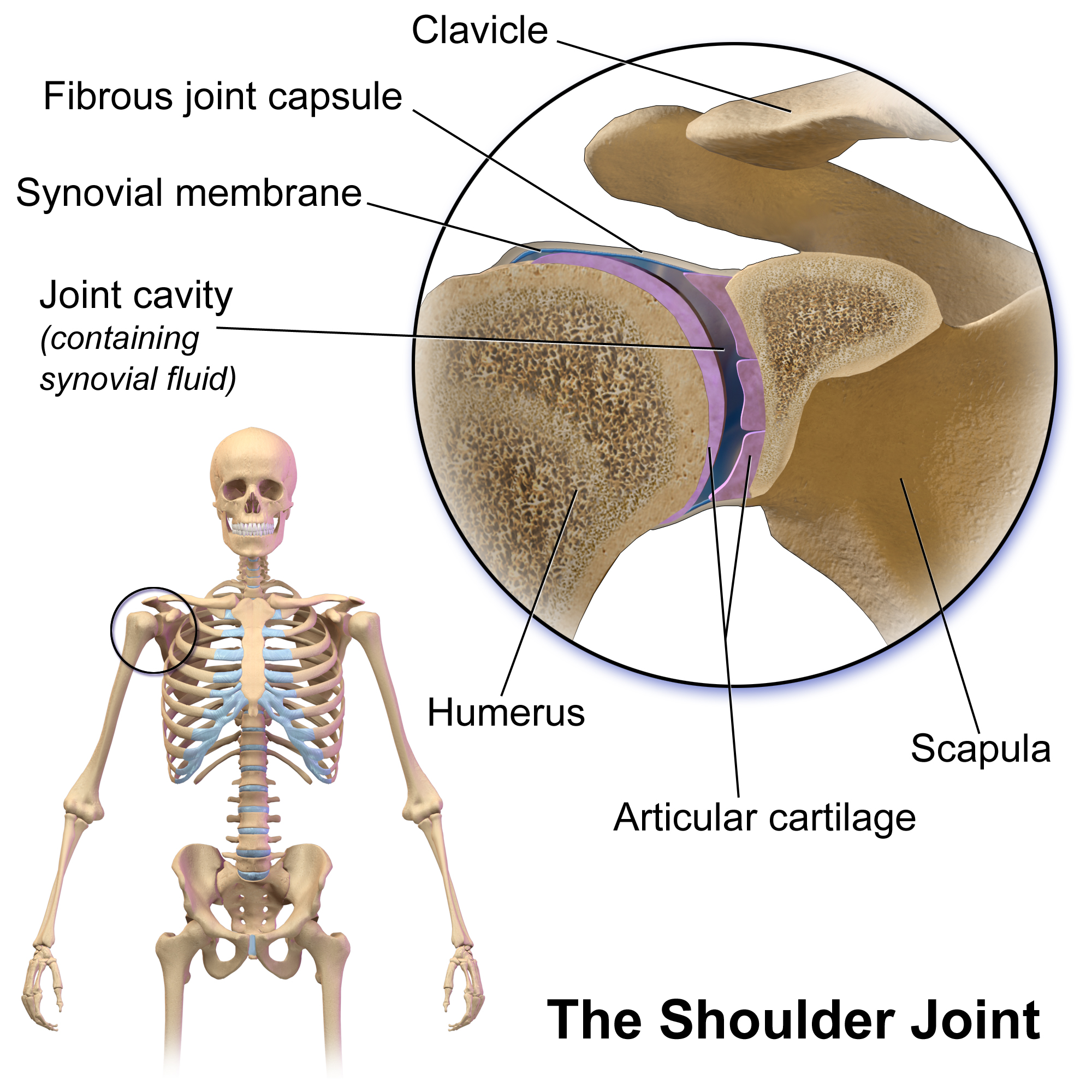
The human shoulder is made up of three bones: the clavicle (collarbone), the scapula (shoulder blade), and the humerus (upper arm bone) as well as associated muscles, ligaments and tendons. The articulations between the bones of the shoulder make up the shoulder joints. The shoulder joint, also known as the glenohumeral joint, is the major joint of the shoulder, but can more broadly include the acromioclavicular joint. In human anatomy, the shoulder joint comprises the part of the body where the humerus attaches to the scapula, and the head sits in the glenoid cavity. The shoulder is the group of structures in the region of the joint. The shoulder joint is the main joint of the shoulder. It is a ball and socket joint that allows the arm to rotate in a circular fashion or to hinge out and up away from the body. The joint capsule is a soft tissue envelope that encircles the glenohumeral joint and attaches to the scapula, humerus, and head of the biceps. It is lined by a ... [...More Info...] [...Related Items...] OR: [Wikipedia] [Google] [Baidu] |
Deltoid Muscle
The deltoid muscle is the muscle forming the rounded contour of the shoulder, human shoulder. It is also known as the 'common shoulder muscle', particularly in other animals such as the domestic cat. Anatomically, the deltoid muscle is made up of three distinct sets of muscle fibers, namely the # anterior or clavicular part (pars clavicularis) ( More commonly known as the front delt.) # posterior or scapular part (pars scapularis) ( More commonly known as the rear delt.) # intermediate or acromial part (pars acromialis) ( More commonly known as the side delt) The deltoid's fibres are pennate muscle. However, electromyography suggests that it consists of at least seven groups that can be independently coordinated by the nervous system. It was previously called the deltoideus (plural ''deltoidei'') and the name is still used by some anatomists. It is called so because it is in the shape of the Greek alphabet, Greek capital letter Delta (letter), delta (Δ). Deltoid is also further ... [...More Info...] [...Related Items...] OR: [Wikipedia] [Google] [Baidu] |
Quadriceps
The quadriceps femoris muscle (, also called the quadriceps extensor, quadriceps or quads) is a large muscle group that includes the four prevailing muscles on the front of the thigh. It is the sole extensor muscle of the knee, forming a large fleshy mass which covers the front and sides of the femur. The name derives . Structure Parts The quadriceps femoris muscle is subdivided into four separate muscles (the 'heads'), with the first superficial to the other three over the femur (from the trochanters to the condyles): * The rectus femoris muscle occupies the middle of the thigh, covering most of the other three quadriceps muscles. It originates on the ilium. It is named for its straight course. * The vastus lateralis muscle is on the ''lateral side'' of the femur (i.e. on the outer side of the thigh). * The vastus medialis muscle is on the ''medial side'' of the femur (i.e. on the inner part thigh). * The vastus intermedius muscle lies between vastus lateralis and vas ... [...More Info...] [...Related Items...] OR: [Wikipedia] [Google] [Baidu] |
Rectus Femoris
The rectus femoris muscle is one of the four quadriceps muscles of the human body. The others are the vastus medialis, the vastus intermedius (deep to the rectus femoris), and the vastus lateralis. All four parts of the quadriceps muscle attach to the patella (knee cap) by the quadriceps tendon. The rectus femoris is situated in the middle of the front of the thigh; it is fusiform in shape, and its superficial fibers are arranged in a bipenniform manner, the deep fibers running straight () down to the deep aponeurosis. Its functions are to flex the thigh at the hip joint and to extend the leg at the knee joint. Structure It arises by two tendons: one, the anterior or straight, from the anterior inferior iliac spine; the other, the posterior or reflected, from a groove above the rim of the acetabulum. The two unite at an acute angle and spread into an aponeurosis that is prolonged downward on the anterior surface of the muscle, and from this the muscular fibers arise. The ... [...More Info...] [...Related Items...] OR: [Wikipedia] [Google] [Baidu] |
Bipennate
Muscle architecture is the physical arrangement of muscle fibers at the macroscopic level that determines a muscle's mechanical function. There are several different muscle architecture types including: parallel, pennate and hydrostats. Force production and gearing vary depending on the different muscle parameters such as muscle length, fiber length, pennation angle, and the physiological cross-sectional area (PCSA). Architecture types Parallel and pennate (also known as pinnate) are two main types of muscle architecture. A third subcategory, muscular hydrostats, can also be considered. Architecture type is determined by the direction in which the muscle fibers are oriented relative to the force-generating axis. The force produced by a given muscle is proportional to the cross-sectional area, or the number of parallel sarcomeres present. Parallel The parallel muscle architecture is found in muscles where the fibers are parallel to the force-generating axis. These muscles are ... [...More Info...] [...Related Items...] OR: [Wikipedia] [Google] [Baidu] |


