|
PET–MRI
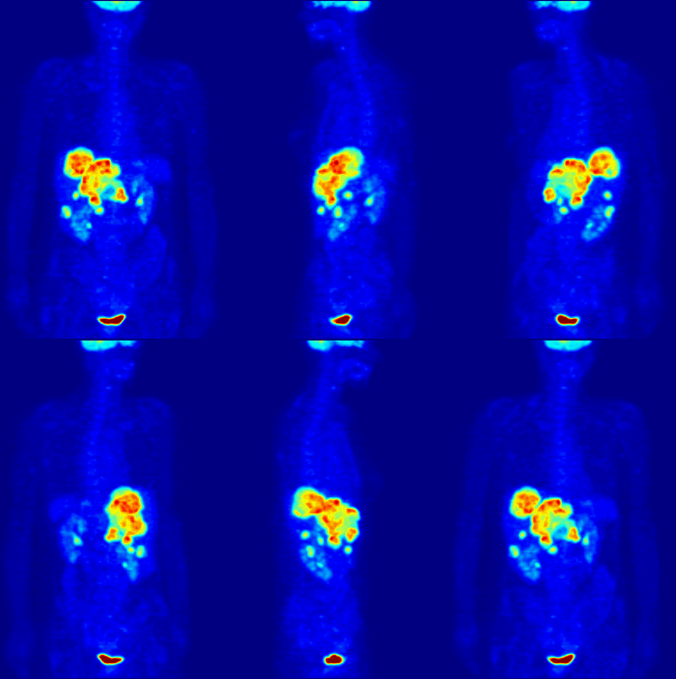
Positron emission tomography–magnetic resonance imaging (PET–MRI) is a hybrid imaging technology that incorporates magnetic resonance imaging (MRI) soft tissue morphological imaging and positron emission tomography (PET) functional imaging. The combination of PET and MRI was mentioned in a 1991 Phd thesis by R. Raylman. Simultaneous PET/MR detection was first demonstrated in 1997, however it took another 13 years, and new detector technologies, for clinical systems to become commercially available. Applications Presently, the main clinical fields of PET–MRI are oncology, cardiology, neurology, and neuroscience. Research studies are actively conducted at the moment to understand benefits of the new PET–MRI diagnostic method. The technology combines the exquisite structural and functional characterization of tissue provided by MRI with the extreme sensitivity of PET imaging of metabolism and tracking of uniquely labeled cell types or cell receptors. Manufacturers Several co ... [...More Info...] [...Related Items...] OR: [Wikipedia] [Google] [Baidu] |
Positron Emission Tomography
Positron emission tomography (PET) is a functional imaging technique that uses radioactive substances known as radiotracers to visualize and measure changes in metabolic processes, and in other physiological activities including blood flow, regional chemical composition, and absorption. Different tracers are used for various imaging purposes, depending on the target process within the body, such as: * Fluorodeoxyglucose ( 18F">sup>18FDG or FDG) is commonly used to detect cancer; * 18Fodium fluoride">sup>18Fodium fluoride (Na18F) is widely used for detecting bone formation; * Oxygen-15 (15O) is sometimes used to measure blood flow. PET is a common imaging technique, a medical scintillography technique used in nuclear medicine. A radiopharmaceutical—a radioisotope attached to a drug—is injected into the body as a tracer. When the radiopharmaceutical undergoes beta plus decay, a positron is emitted, and when the positron interacts with an ordinary electron, the tw ... [...More Info...] [...Related Items...] OR: [Wikipedia] [Google] [Baidu] |
Imaging Technology
Imaging is the representation or reproduction of an object's form; especially a visual representation (i.e., the formation of an image). Imaging technology is the application of materials and methods to create, preserve, or duplicate images. Imaging science is a multidisciplinary field concerned with the generation, collection, duplication, analysis, modification, and visualization of images,Joseph P. Hornak, ''Encyclopedia of Imaging Science and Technology'' (John Wiley & Sons, 2002) including imaging things that the human eye cannot detect. As an evolving field it includes research and researchers from physics, mathematics, electrical engineering, computer vision, computer science, and perceptual psychology. '' Imagers'' are imaging sensors. Imaging chain The foundation of imaging science as a discipline is the "imaging chain" – a conceptual model describing all of the factors which must be considered when developing a system for creating visual renderings (images). In ... [...More Info...] [...Related Items...] OR: [Wikipedia] [Google] [Baidu] |
Computed Tomography
A computed tomography scan (CT scan), formerly called computed axial tomography scan (CAT scan), is a medical imaging technique used to obtain detailed internal images of the body. The personnel that perform CT scans are called radiographers or radiology technologists. CT scanners use a rotating X-ray tube and a row of detectors placed in a gantry to measure X-ray attenuations by different tissues inside the body. The multiple X-ray measurements taken from different angles are then processed on a computer using tomographic reconstruction algorithms to produce tomographic (cross-sectional) images (virtual "slices") of a body. CT scans can be used in patients with metallic implants or pacemakers, for whom magnetic resonance imaging (MRI) is contraindicated. Since its development in the 1970s, CT scanning has proven to be a versatile imaging technique. While CT is most prominently used in medical diagnosis, it can also be used to form images of non-living objects. The 1979 N ... [...More Info...] [...Related Items...] OR: [Wikipedia] [Google] [Baidu] |
Neuroimaging
Neuroimaging is the use of quantitative (computational) techniques to study the neuroanatomy, structure and function of the central nervous system, developed as an objective way of scientifically studying the healthy human brain in a non-invasive manner. Increasingly it is also being used for quantitative research studies of brain disease and psychiatric illness. Neuroimaging is highly multidisciplinary involving neuroscience, computer science, psychology and statistics, and is not a medical specialty. Neuroimaging is sometimes confused with neuroradiology. Neuroradiology is a medical specialty that uses non-statistical brain imaging in a clinical setting, practiced by radiologists who are medical practitioners. Neuroradiology primarily focuses on recognizing brain lesions, such as vascular diseases, strokes, tumors, and inflammatory diseases. In contrast to neuroimaging, neuroradiology is qualitative (based on subjective impressions and extensive clinical training) but sometime ... [...More Info...] [...Related Items...] OR: [Wikipedia] [Google] [Baidu] |
MRI Artifact
An MRI artifact is a visual artifact (an anomaly seen during visual representation) in magnetic resonance imaging (MRI). It is a feature appearing in an image that is not present in the original object. Many different artifacts can occur during MRI, some affecting the diagnostic quality, while others may be confused with pathology. Artifacts can be classified as patient-related, signal processing-dependent and hardware (machine)-related. Patient-related MR artifacts Motion artifacts A motion artifact is one of the most common artifacts in MR imaging. Motion can cause either ghost images or diffuse image noise in the phase-encoding direction. The reason for mainly affecting data sampling in the phase-encoding direction is the significant difference in the time of acquisition in the frequency- and phase-encoding directions. Frequency-encoding sampling in all the rows of the matrix (128, 256 or 512) takes place during a single echo (milliseconds). Phase-encoded sampling takes severa ... [...More Info...] [...Related Items...] OR: [Wikipedia] [Google] [Baidu] |
Voxel
In computing, a voxel is a representation of a value on a three-dimensional regular grid, akin to the two-dimensional pixel. Voxels are frequently used in the Data visualization, visualization and analysis of medical imaging, medical and scientific data (e.g. geographic information systems (GIS)). Voxels also have technical and artistic applications in video games, largely originating with surface rendering in ''Outcast (video game), Outcast'' (1999). ''Minecraft'' (2011) makes use of an entirely voxelated world to allow for a fully destructable and constructable environment. Voxel art, of the sort used in ''Minecraft'' and elsewhere, is a style and format of 3D art analogous to pixel art. As with pixels in a 2D bitmap, voxels themselves do not typically have their position (i.e. coordinates) explicitly encoded with their values. Instead, Rendering (computer graphics), rendering systems infer the position of a voxel based upon its position relative to other voxels (i.e., its pos ... [...More Info...] [...Related Items...] OR: [Wikipedia] [Google] [Baidu] |
Radiation Treatment Planning
In radiotherapy, radiation treatment planning (RTP) is the process in which a team consisting of radiation oncologists, radiation therapist, medical physicists and medical dosimetrists plan the appropriate external beam radiotherapy or internal brachytherapy treatment technique for a patient with cancer. History In the early days of radiotherapy planning was performed on 2D x-ray images, often by hand and with manual calculations. Computerised treatment planning systems began to be used in the 1970s to improve the accuracy and speed of dose calculations. By the 1990s CT scans, more powerful computers, improved dose calculation algorithms and Multileaf collimators (MLCs) lead to 3D conformal planning (3DCRT), categorised as a Level 2 technique by the European Dynarad consortium. 3DCRT uses MLCs to shape the radiotherapy beam to closely match the shape of a target tumour, reducing the dose to healthy surrounding tissue. Level 3 techniques such as IMRT and VMAT utilise inverse pl ... [...More Info...] [...Related Items...] OR: [Wikipedia] [Google] [Baidu] |
Image Segmentation
In digital image processing and computer vision, image segmentation is the process of partitioning a digital image into multiple image segments, also known as image regions or image objects (Set (mathematics), sets of pixels). The goal of segmentation is to simplify and/or change the representation of an image into something that is more meaningful and easier to analyze.Linda Shapiro, Linda G. Shapiro and George C. Stockman (2001): "Computer Vision", pp 279–325, New Jersey, Prentice-Hall, Image segmentation is typically used to locate objects and Boundary tracing, boundaries (lines, curves, etc.) in images. More precisely, image segmentation is the process of assigning a label to every pixel in an image such that pixels with the same label share certain characteristics. The result of image segmentation is a set of segments that collectively cover the entire image, or a set of Contour line, contours extracted from the image (see edge detection). Each of the pixels in a region ... [...More Info...] [...Related Items...] OR: [Wikipedia] [Google] [Baidu] |
MRI Sequence
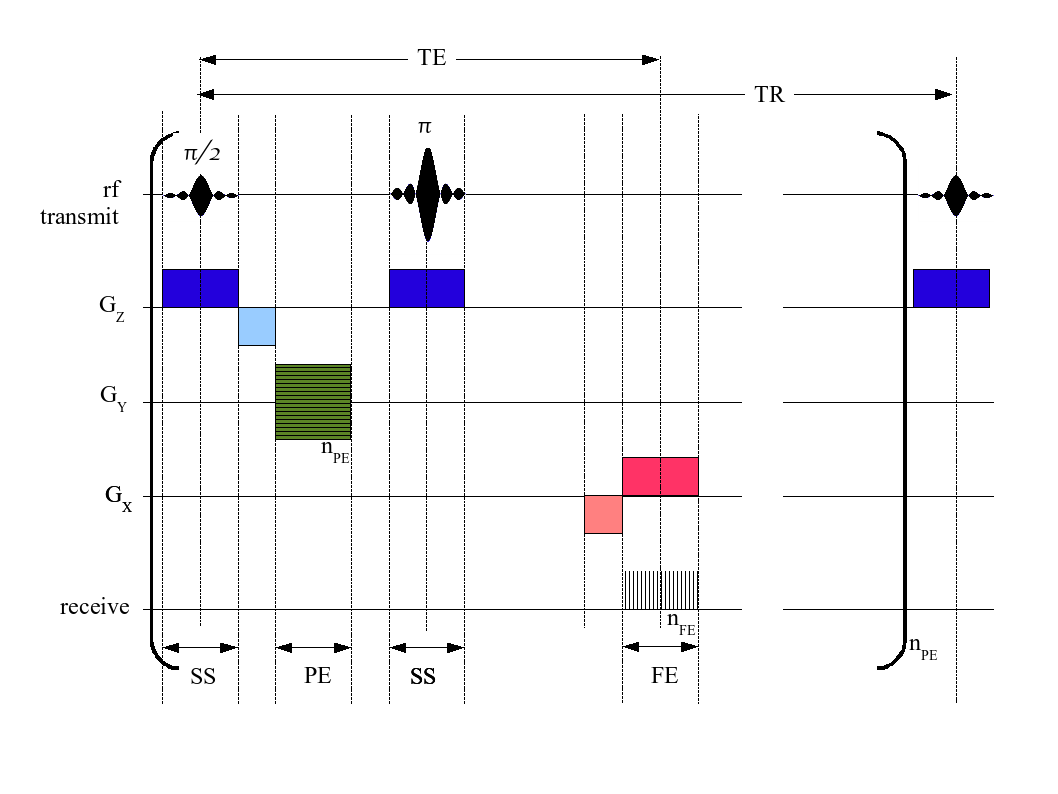
An MRI pulse sequence in magnetic resonance imaging (MRI) is a particular setting of pulse sequences and pulsed field gradients, resulting in a particular image appearance. A multiparametric MRI is a combination of two or more sequences, and/or including Magnetic resonance imaging#Other specialized configurations, other specialized MRI configurations such as In vivo magnetic resonance spectroscopy, spectroscopy. Spin echo T1 and T2 Each tissue returns to its equilibrium state after excitation by the independent relaxation processes of T1 (Spin–lattice relaxation, spin-lattice; that is, magnetization in the same direction as the static magnetic field) and T2 (Spin-spin relaxation time, spin-spin; transverse to the static magnetic field). To create a T1-weighted image, magnetization is allowed to recover before measuring the MR signal by changing the repetition time (TR). This image weighting is useful for assessing the cerebral cortex, identifying fatty tissue, characteriz ... [...More Info...] [...Related Items...] OR: [Wikipedia] [Google] [Baidu] |
Hounsfield Unit
The Hounsfield scale ( ), named after Sir Godfrey Hounsfield, is a quantitative scale for describing radiodensity. It is frequently used in CT scans, where its value is also termed CT number. Definition The Hounsfield unit (HU) scale is a linear transformation of the original linear attenuation coefficient measurement into one in which the radiodensity of distilled water at standard pressure and temperature (STP) is defined as 0 Hounsfield units (HU), while the radiodensity of air at STP is defined as −1000 HU. In a voxel with average linear attenuation coefficient \mu, the corresponding HU value is therefore given by: HU = 1000\times\frac where \mu_ and \mu_ are respectively the linear attenuation coefficients of water and air. Thus, a change of one Hounsfield unit (HU) represents a change of 0.1% of the attenuation coefficient of water since the attenuation coefficient of air is nearly zero. Calibration tests of HU with reference to water and other materials may be done to ... [...More Info...] [...Related Items...] OR: [Wikipedia] [Google] [Baidu] |
Germanium-68
Germanium (32Ge) has five naturally occurring isotopes, 70Ge, 72Ge, 73Ge, 74Ge, and 76Ge. Of these, 76Ge is very slightly radioactive, decaying by double beta decay with a half-life of 1.78 × 10²¹ years (130 billion times the age of the universe). Stable 74Ge is the most common isotope, having a natural abundance of approximately 36%. 76Ge is the least common with a natural abundance of approximately 7%. At least 27 radioisotopes have also been synthesized ranging in atomic mass from 58 to 89. The most stable of these is 68Ge, decaying by electron capture with a half-life of 270.95 d. It decays to the medically useful positron-emitting isotope 68Ga. (See gallium-68 generator for notes on the source of this isotope, and its medical use.) The least stable known germanium isotope is 59Ge with a half-life of 13.3 ms. While most of germanium's radioisotopes decay by beta decay, 61Ge and 65Ge can also decay by β+-delayed proton emission. 84Ge through 87Ge also ha ... [...More Info...] [...Related Items...] OR: [Wikipedia] [Google] [Baidu] |
Attenuation
In physics, attenuation (in some contexts, extinction) is the gradual loss of flux intensity through a Transmission medium, medium. For instance, dark glasses attenuate sunlight, lead attenuates X-rays, and water and air attenuate both light and sound at variable attenuation rates. Hearing protection device, Hearing protectors help reduce Sound power, acoustic flux from flowing into the ears. This phenomenon is called acoustic attenuation and is measured in decibels (dBs). In electrical engineering and telecommunications, attenuation affects the Wave propagation, propagation of waves and signals in electrical circuits, in optical fibers, and in air. Attenuator (electronics), Electrical attenuators and optical attenuators are commonly manufactured components in this field. Background In many cases, attenuation is an exponential function of the path length through the medium. In optics and in chemical spectroscopy, this is known as the Beer–Lambert law. In engineering, attenu ... [...More Info...] [...Related Items...] OR: [Wikipedia] [Google] [Baidu] |


