|
Neurofibril
Neurofilaments (NF) are classed as type IV intermediate filaments found in the cytoplasm of neurons. They are protein polymers measuring 10 nm in diameter and many micrometers in length. Together with microtubules (~25 nm) and microfilaments (7 nm), they form the neuronal cytoskeleton. They are believed to function primarily to provide structural support for axons and to regulate axon diameter, which influences nerve conduction velocity. The proteins that form neurofilaments are members of the intermediate filament protein family, which is divided into six types based on their gene organization and protein structure. Types I and II are the keratins which are expressed in epithelia. Type III contains the proteins vimentin, desmin, peripherin and glial fibrillary acidic protein (GFAP). Type IV consists of the neurofilament proteins NF-L, NF-M, NF-H and α-internexin. Type V consists of the nuclear lamins, and type VI consists of the protein nestin. The ... [...More Info...] [...Related Items...] OR: [Wikipedia] [Google] [Baidu] |
Neurofibrillary Tangle
Neurofibrillary tangles (NFTs) are intracellular aggregates of hyperphosphorylated tau protein that are most commonly known as a primary Biomarker (medicine), biomarker of Alzheimer's disease. Their presence is also found in numerous other diseases known as Tauopathy, tauopathies. Little is known about their exact relationship to the different pathologies. Formation Neurofibrillary tangles are formed by phosphorylation, hyperphosphorylation of a microtubule-associated protein known as tau (protein), tau, causing it to aggregate, or group, in an insoluble form. (These aggregations of hyperphosphorylated tau protein are also referred to as PHF, or "paired helical filaments"). The precise mechanism of tangle formation is not completely understood, though it is typically recognized that tangles are a primary causative factor in neurodegenerative disease. Cytoskeletal changes Three different maturation states of NFT have been defined using anti-tau and anti-ubiquitin immunostaining. ... [...More Info...] [...Related Items...] OR: [Wikipedia] [Google] [Baidu] |
NEFL
Neurofilament light polypeptide is a protein that in humans is encoded by the NEFL gene. Structure Neurofilament light polypeptide is a member of the intermediate filament protein family. This protein family consists of over 50 human proteins divided into 5 major classes, the Class I and II keratins, Class III vimentin, Glial fibrillary acidic protein, GFAP, desmin and the others, the Class IV neurofilaments and the Class V nuclear lamins. There are four major neurofilament subunits, NF-L, NF-M, NF-H and α-internexin. These form heteropolymers which assemble to produce 10 nm neurofilaments which are only expressed in neurons where they are major structural proteins, particularly concentrated in large projection axons. The NF-L protein is encoded by the ''NEFL'' gene. Function These neurofilament heteropolymers assemble into the cytoskeleton of axons, where they provide structural support and help regulate axonal diameter and conduction velocity. Axons are particularly sens ... [...More Info...] [...Related Items...] OR: [Wikipedia] [Google] [Baidu] |
Internexin
Internexin, alpha-internexin, is a Class IV intermediate filament approximately 66 kDa. The protein was originally purified from rat optic nerve and spinal cord.Levavasseur F, Zhu Q, and JP Julien. No requirement of alpha-internexin for nervous system development and for radial growth of axons. Molecular Brain Research. 69:104-112. (1999). The protein copurifies with other neurofilament subunits, as it was originally discovered, however in some mature neurons it can be the only neurofilament expressed. The protein is present in developing neuroblasts and in the central nervous system of adults. The protein is a major component of the intermediate filament network in small interneurons and cerebellar granule cells, where it is present in the parallel fibers. Structure Alpha-internexin has a homologous central rod domain of approximately 310 amino acid residues that form a highly conserved alpha helical region. The central rod domain is responsible for coiled-coil structure an ... [...More Info...] [...Related Items...] OR: [Wikipedia] [Google] [Baidu] |
Lysine
Lysine (symbol Lys or K) is an α-amino acid that is a precursor to many proteins. Lysine contains an α-amino group (which is in the protonated form when the lysine is dissolved in water at physiological pH), an α-carboxylic acid group (which is in the deprotonated form when the lysine is dissolved in water at physiological pH), and a side chain (which is partially protonated when the lysine is dissolved in water at physiological pH), and so it is classified as a basic, charged (in water at physiological pH), aliphatic amino acid. It is encoded by the codons AAA and AAG. Like almost all other amino acids, the α-carbon is chiral and lysine may refer to either enantiomer or a racemic mixture of both. For the purpose of this article, lysine will refer to the biologically active enantiomer L-lysine, where the α-carbon is in the ''S'' configuration. The human body cannot synthesize lysine. It is essential in humans and must therefore be obtained from the diet. In orga ... [...More Info...] [...Related Items...] OR: [Wikipedia] [Google] [Baidu] |
Glutamic Acid
Glutamic acid (symbol Glu or E; known as glutamate in its anionic form) is an α- amino acid that is used by almost all living beings in the biosynthesis of proteins. It is a non-essential nutrient for humans, meaning that the human body can synthesize enough for its use. It is also the most abundant excitatory neurotransmitter in the vertebrate nervous system. It serves as the precursor for the synthesis of the inhibitory gamma-aminobutyric acid (GABA) in GABAergic neurons. Its molecular formula is . Glutamic acid exists in two optically isomeric forms; the dextrorotatory -form is usually obtained by hydrolysis of gluten or from the waste waters of beet-sugar manufacture or by fermentation.Webster's Third New International Dictionary of the English Language Unabridged, Third Edition, 1971. Its molecular structure could be idealized as HOOC−CH()−()2−COOH, with two carboxyl groups −COOH and one amino group −. However, in the solid state and mildly acidic water s ... [...More Info...] [...Related Items...] OR: [Wikipedia] [Google] [Baidu] |
SDS-PAGE
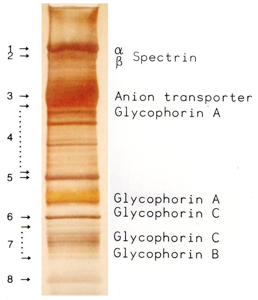
SDS-PAGE (sodium dodecyl sulfate–polyacrylamide gel electrophoresis) is a Discontinuous electrophoresis, discontinuous electrophoretic system developed by Ulrich K. Laemmli which is commonly used as a method to separate proteins with molecular masses between 5 and 250 Kilodalton, kDa. The combined use of sodium dodecyl sulfate (SDS, also known as sodium lauryl sulfate) and polyacrylamide gel eliminates the influence of structure and charge, and proteins are separated by differences in their size. At least up to 2012, the publication describing it was the most frequently cited paper by a single author, and the second most cited overall. Properties SDS-PAGE is an electrophoresis method that allows protein separation by mass. The medium (also referred to as ′matrix′) is a polyacrylamide-based discontinuous gel. The polyacrylamide-gel is typically sandwiched between two glass plates in a slab gel. Although tube gels (in glass cylinders) were used historically, they were rapid ... [...More Info...] [...Related Items...] OR: [Wikipedia] [Google] [Baidu] |
Molecular Mass
The molecular mass () is the mass of a given molecule, often expressed in units of daltons (Da). Different molecules of the same compound may have different molecular masses because they contain different isotopes of an element. The derived quantity relative molecular mass is the unitless ratio of the mass of a molecule to the atomic mass constant (which is equal to one dalton). The molecular mass and relative molecular mass are distinct from but related to the ''molar mass''. The molar mass is defined as the mass of a given substance divided by the amount of the substance, and is expressed in grams per mole (g/mol). That makes the molar mass an ''average'' of many particles or molecules (weighted by abundance of the isotopes), and the molecular mass the mass of one specific particle or molecule. The molar mass is usually the more appropriate quantity when dealing with macroscopic (weigh-able) quantities of a substance. The definition of molecular weight is most authoritat ... [...More Info...] [...Related Items...] OR: [Wikipedia] [Google] [Baidu] |
Horizontal Neurons
Horizontal cells are the laterally interconnecting neurons having cell bodies in the inner nuclear layer of the retina of vertebrate eyes. They help integrate and regulate the input from multiple photoreceptor cells. Among their functions, horizontal cells are believed to be responsible for increasing contrast via lateral inhibition and adapting both to bright and dim light conditions. Horizontal cells provide inhibitory feedback to rod and cone photoreceptors. They are thought to be important for the antagonistic center-surround property of the receptive fields of many types of retinal ganglion cells. Other retinal neurons include photoreceptor cells, bipolar cells, amacrine cells, and retinal ganglion cells. Structure Depending on the species, there are typically one or two classes of horizontal cells, with a third type sometimes proposed. Horizontal cells span across photoreceptors and summate inputs before synapsing onto photoreceptor cells. Horizontal cells may also sy ... [...More Info...] [...Related Items...] OR: [Wikipedia] [Google] [Baidu] |
Retina
The retina (; or retinas) is the innermost, photosensitivity, light-sensitive layer of tissue (biology), tissue of the eye of most vertebrates and some Mollusca, molluscs. The optics of the eye create a focus (optics), focused two-dimensional image of the visual world on the retina, which then processes that image within the retina and sends nerve impulses along the optic nerve to the visual cortex to create visual perception. The retina serves a function which is in many ways analogous to that of the photographic film, film or image sensor in a camera. The neural retina consists of several layers of neurons interconnected by Chemical synapse, synapses and is supported by an outer layer of pigmented epithelial cells. The primary light-sensing cells in the retina are the photoreceptor cells, which are of two types: rod cell, rods and cone cell, cones. Rods function mainly in dim light and provide monochromatic vision. Cones function in well-lit conditions and are responsible fo ... [...More Info...] [...Related Items...] OR: [Wikipedia] [Google] [Baidu] |
Peripherin 2
Peripherin-2 is a protein, that in humans is encoded by the ''PRPH2'' gene. Peripherin-2 is found in the rod and cone cells of the retina of the eye. Defects in this protein result in one form of retinitis pigmentosa, an incurable blindness. Mutations in the PRPH2 gene are associated with Vitelliform macular dystrophy. Function The protein encoded by this gene is a member of the transmembrane 4 superfamily, also known as the tetraspanin family. Most of these members are cell-surface proteins that are characterized by the presence of four transmembrane helices. Tetraspanins mediate signal transduction events that play a role in the regulation of cell development, activation, growth and motility. Peripherin 2 (sometimes referred to as peripherin/RDS or simply RDS) is a cell surface glycoprotein found in the outer segment of both rod and cone photoreceptor cells. It is located in the rim regions of the flattened disks that contain rhodopsin, which is the protein that is responsi ... [...More Info...] [...Related Items...] OR: [Wikipedia] [Google] [Baidu] |
Axonal Transport
Axonal transport, also called axoplasmic transport or axoplasmic flow, is a cellular process responsible for movement of mitochondria, lipids, synaptic vesicles, proteins, and other organelles to and from a neuron's cell body, through the cytoplasm of its axon called the axoplasm. Since some axons are on the order of meters long, neurons cannot rely on diffusion to carry products of the nucleus and organelles to the ends of their axons. Axonal transport is also responsible for moving molecules destined for degradation from the axon back to the cell body, where they are broken down by lysosomes. Movement toward the cell body is called retrograde transport and movement toward the synapse is called anterograde transport. Mechanism The vast majority of axonal proteins are synthesized in the neuronal cell body and transported along axons. Some mRNA translation has been demonstrated within axons.Si K, Giustetto Axonal transport occurs throughout the life of a neuron and is esse ... [...More Info...] [...Related Items...] OR: [Wikipedia] [Google] [Baidu] |
Neurodegenerative Disease
A neurodegenerative disease is caused by the progressive loss of neurons, in the process known as neurodegeneration. Neuronal damage may also ultimately result in their death. Neurodegenerative diseases include amyotrophic lateral sclerosis, multiple sclerosis, Parkinson's disease, Alzheimer's disease, Huntington's disease, multiple system atrophy, tauopathies, and prion diseases. Neurodegeneration can be found in the brain at many different levels of neuronal circuitry, ranging from molecular to systemic. Because there is no known way to reverse the progressive degeneration of neurons, these diseases are considered to be incurable; however research has shown that the two major contributing factors to neurodegeneration are oxidative stress and inflammation. Biomedical research has revealed many similarities between these diseases at the subcellular level, including atypical protein assemblies (like proteinopathy) and induced cell death. These similarities suggest that therap ... [...More Info...] [...Related Items...] OR: [Wikipedia] [Google] [Baidu] |


