|
Neonatal-onset Multisystem Inflammatory Disease
Neonatal-onset multisystem inflammatory disease is a rare genetic periodic fever syndrome which causes uncontrolled inflammation in multiple parts of the body starting in the newborn period. Symptoms include skin rashes, severe arthritis, and chronic meningitis leading to neurologic damage. It is one of the cryopyrin-associated periodic syndromes. NOMID can result from a mutation in the ''CIAS1'' gene (also known as ''NLRP3'' gene), which helps control inflammation. Mutations in this gene also cause familial cold urticaria and Muckle–Wells syndrome. NOMID has been successfully treated with the drug anakinra. This syndrome is also known as the Prieur–Griscelli syndrome as it was first described by these authors in 1981.Prieur AM, Griscelli C (1981) Arthropathy with rash, chronic meningitis, eye lesions, and mental retardation. J. Pediat 99:79-83 Signs and symptoms The age of onset is almost always before 3 months of age. Many infants are born preterm (1/3 cases) and dysmatur ... [...More Info...] [...Related Items...] OR: [Wikipedia] [Google] [Baidu] |
Rare Disease
A rare disease is any disease that affects a small percentage of the population. In some parts of the world, the term orphan disease describes a rare disease whose rarity results in little or no funding or research for treatments, without financial incentives from governments or other agencies. Orphan drugs are medications targeting orphan diseases. Most rare diseases are genetic in origin and thus are present throughout the person's entire life, even if symptoms do not immediately appear. Many rare diseases appear early in life, and about 30% of children with rare diseases will die before reaching their fifth birthdays. Fields condition is considered the rarest known disease, affecting three known individuals, two of whom are identical twins. With four diagnosed patients in 27 years, ribose-5-phosphate isomerase deficiency is considered the second rarest. While no single number has been agreed upon for which a disease is considered rare, several efforts have been undertaken to ... [...More Info...] [...Related Items...] OR: [Wikipedia] [Google] [Baidu] |
Central Nervous System
The central nervous system (CNS) is the part of the nervous system consisting primarily of the brain, spinal cord and retina. The CNS is so named because the brain integrates the received information and coordinates and influences the activity of all parts of the bodies of bilateria, bilaterally symmetric and triploblastic animals—that is, all multicellular animals except sponges and Coelenterata, diploblasts. It is a structure composed of nervous tissue positioned along the Anatomical_terms_of_location#Rostral,_cranial,_and_caudal, rostral (nose end) to caudal (tail end) axis of the body and may have an enlarged section at the rostral end which is a brain. Only arthropods, cephalopods and vertebrates have a true brain, though precursor structures exist in onychophorans, gastropods and lancelets. The rest of this article exclusively discusses the vertebrate central nervous system, which is radically distinct from all other animals. Overview In vertebrates, the brain and spinal ... [...More Info...] [...Related Items...] OR: [Wikipedia] [Google] [Baidu] |
Contractures
In pathology, a contracture is a shortening of muscles, tendons, skin, and nearby soft tissues that causes the joints to shorten and become very stiff, preventing normal movement. A contracture is usually permanent, but less commonly can be temporary (such as in McArdle disease), or resolve over time but reoccur later in life (such as in Bethlem myopathy 1). It is usually in response to prolonged hypertonic spasticity in a concentrated muscle area, such as is seen in the tightest muscles of people with conditions like spastic cerebral palsy, but can also be due to the congenital abnormal development of muscles and connective tissue in the womb. Contractures develop usually when normally elastic tissues such as muscles or tendons are replaced by inelastic tissues (fibrosis). This results in the shortening and hardening of these tissues, ultimately causing rigidity, joint deformities and a total loss of movement around the joint. Most of the physical therapy, occupational thera ... [...More Info...] [...Related Items...] OR: [Wikipedia] [Google] [Baidu] |
Patella
The patella (: patellae or patellas), also known as the kneecap, is a flat, rounded triangular bone which articulates with the femur (thigh bone) and covers and protects the anterior articular surface of the knee joint. The patella is found in many tetrapods, such as mice, cats, birds, and dogs, but not in whales, or most reptiles. In humans, the patella is the largest sesamoid bone (i.e., embedded within a tendon or a muscle) in the body. Babies are born with a patella of soft cartilage which begins to ossify into bone at about four years of age. Structure The patella is a sesamoid bone roughly triangular in shape, with the apex of the patella facing downwards. The apex is the most inferior (lowest) part of the patella. It is pointed in shape, and gives attachment to the patellar ligament. The front and back surfaces are joined by a thin margin and towards centre by a thicker margin. The tendon of the quadriceps femoris muscle attaches to the base of the patella., with th ... [...More Info...] [...Related Items...] OR: [Wikipedia] [Google] [Baidu] |
Wrist
In human anatomy, the wrist is variously defined as (1) the carpus or carpal bones, the complex of eight bones forming the proximal skeletal segment of the hand; "The wrist contains eight bones, roughly aligned in two rows, known as the carpal bones." (2) the wrist joint or radiocarpal joint, the joint between the radius and the carpus and; (3) the anatomical region surrounding the carpus including the distal parts of the bones of the forearm and the proximal parts of the metacarpus or five metacarpal bones and the series of joints between these bones, thus referred to as ''wrist joints''. "With the large number of bones composing the wrist (ulna, radius, eight carpas, and five metacarpals), it makes sense that there are many, many joints that make up the structure known as the wrist." This region also includes the carpal tunnel, the anatomical snuff box, bracelet lines, the flexor retinaculum, and the extensor retinaculum. As a consequence of these various definitions, f ... [...More Info...] [...Related Items...] OR: [Wikipedia] [Google] [Baidu] |
Elbow
The elbow is the region between the upper arm and the forearm that surrounds the elbow joint. The elbow includes prominent landmarks such as the olecranon, the cubital fossa (also called the chelidon, or the elbow pit), and the lateral and the medial epicondyles of the humerus. The elbow joint is a hinge joint between the arm and the forearm; more specifically between the humerus in the upper arm and the radius and ulna in the forearm which allows the forearm and hand to be moved towards and away from the body. The term ''elbow'' is specifically used for humans and other primates, and in other vertebrates it is not used. In those cases, forelimb plus joint is used. The name for the elbow in Latin is ''cubitus'', and so the word cubital is used in some elbow-related terms, as in ''cubital nodes'' for example. Structure Joint The elbow joint has three different portions surrounded by a common joint capsule. These are joints between the three bones of the elbow, the ... [...More Info...] [...Related Items...] OR: [Wikipedia] [Google] [Baidu] |
Ankle
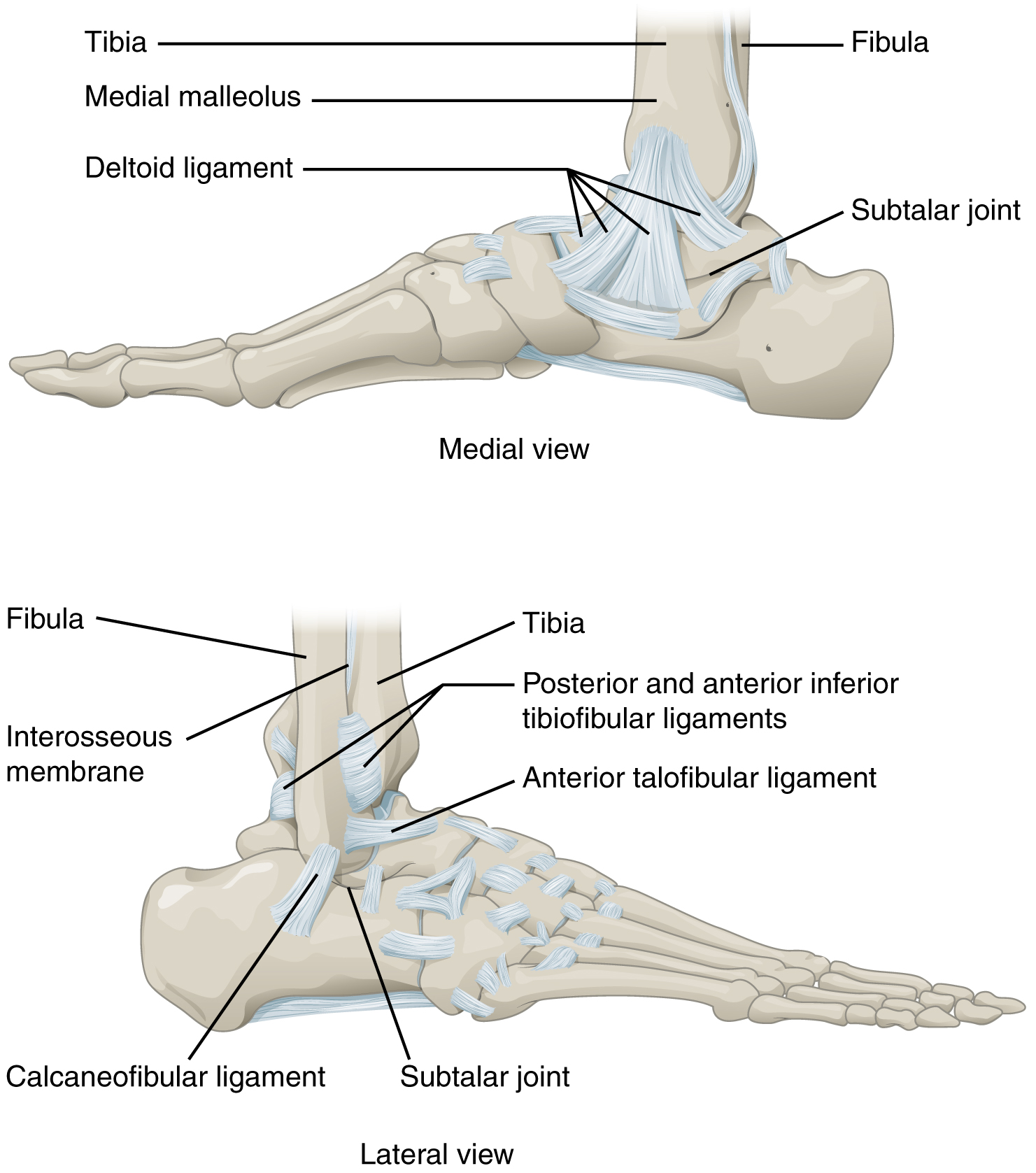
The ankle, the talocrural region or the jumping bone (informal) is the area where the foot and the leg meet. The ankle includes three joints: the ankle joint proper or talocrural joint, the subtalar joint, and the inferior tibiofibular joint. The movements produced at this joint are dorsiflexion and plantarflexion of the foot. In common usage, the term ankle refers exclusively to the ankle region. In medical terminology, "ankle" (without qualifiers) can refer broadly to the region or specifically to the talocrural joint. The main bones of the ankle region are the talus bone, talus (in the foot), the tibia, and fibula (both in the leg). The talocrural joint is a Synovial joint, synovial hinge joint that connects the distal ends of the tibia and fibula in the lower limb with the proximal end of the talus. The articulation between the tibia and the talus bears more weight than that between the smaller fibula and the talus. Structure Region The ankle region is found at the junction ... [...More Info...] [...Related Items...] OR: [Wikipedia] [Google] [Baidu] |
Knee
In humans and other primates, the knee joins the thigh with the leg and consists of two joints: one between the femur and tibia (tibiofemoral joint), and one between the femur and patella (patellofemoral joint). It is the largest joint in the human body. The knee is a modified hinge joint, which permits flexion and extension (kinesiology), extension as well as slight internal and external rotation. The knee is vulnerable to injury and to the development of osteoarthritis. It is often termed a ''compound joint'' having tibiofemoral and patellofemoral components. (The fibular collateral ligament is often considered with tibiofemoral components.) Structure The knee is a modified hinge joint, a type of synovial joint, which is composed of three functional compartments: the patellofemoral articulation, consisting of the patella, or "kneecap", and the patellar groove on the front of the femur through which it slides; and the medial and lateral tibiofemoral articulations linking the ... [...More Info...] [...Related Items...] OR: [Wikipedia] [Google] [Baidu] |
Hypertrophic
Hypertrophy is the increase in the volume of an organ or tissue due to the enlargement of its component cells. It is distinguished from hyperplasia, in which the cells remain approximately the same size but increase in number. Although hypertrophy and hyperplasia are two distinct processes, they frequently occur together, such as in the case of the hormonally induced proliferation and enlargement of the cells of the uterus during pregnancy. Eccentric hypertrophy is a type of hypertrophy where the walls and chamber of a hollow organ undergo growth in which the overall size and volume are enlarged. It is applied especially to the left ventricle of heart. Sarcomeres are added in series, as for example in dilated cardiomyopathy (in contrast to hypertrophic cardiomyopathy, a type of concentric hypertrophy, where sarcomeres are added in parallel). Gallery Gould Pyle 234.jpg, Breasts Hypertrophied clitoris.jpg, Clitoris Head of a boy with hypertrophy of the ear Wellcome L0062496.jpg, ... [...More Info...] [...Related Items...] OR: [Wikipedia] [Google] [Baidu] |
Epiphyses
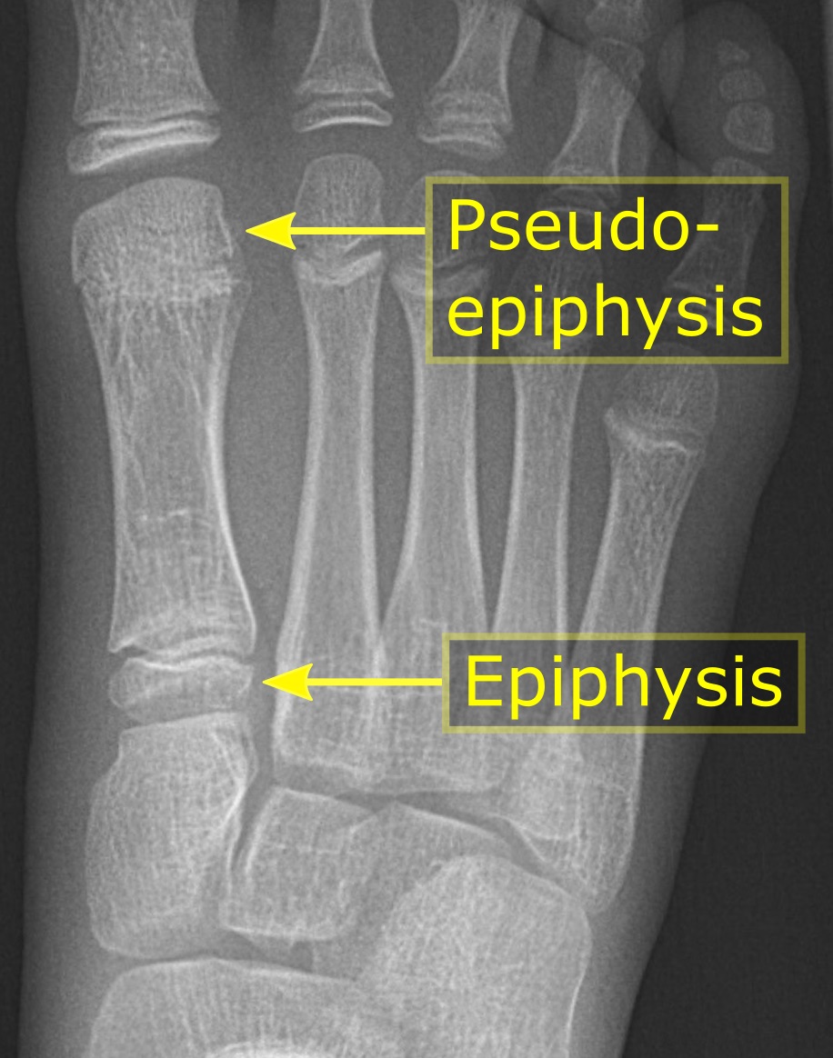
An epiphysis (; : epiphyses) is one of the rounded ends or tips of a long bone that ossify from one or more secondary centers of ossification. Between the epiphysis and diaphysis (the long midsection of the long bone) lies the metaphysis, including the epiphyseal plate (growth plate). During formation of the secondary ossification center, vascular canals (epiphysial canals) stemming from the perichondrium invade the epiphysis, supplying nutrients to the developing secondary centers of ossification. At the joint, the epiphysis is covered with articular cartilage; below that covering is a zone similar to the epiphyseal plate, known as subchondral bone. The epiphysis is mostly found in mammals but it is also present in some lizards. However, the secondary center of ossification may have evolved multiple times, having been found in the Jurassic sphenodont '' Sapheosaurus'' as well as in the therapsid '' Niassodon mfumukasi.'' The epiphysis is filled with red bone marrow, which ... [...More Info...] [...Related Items...] OR: [Wikipedia] [Google] [Baidu] |
Cartilage
Cartilage is a resilient and smooth type of connective tissue. Semi-transparent and non-porous, it is usually covered by a tough and fibrous membrane called perichondrium. In tetrapods, it covers and protects the ends of long bones at the joints as articular cartilage, and is a structural component of many body parts including the rib cage, the neck and the bronchial tubes, and the intervertebral discs. In other taxa, such as chondrichthyans and cyclostomes, it constitutes a much greater proportion of the skeleton. It is not as hard and rigid as bone, but it is much stiffer and much less flexible than muscle. The matrix of cartilage is made up of glycosaminoglycans, proteoglycans, collagen fibers and, sometimes, elastin. It usually grows quicker than bone. Because of its rigidity, cartilage often serves the purpose of holding tubes open in the body. Examples include the rings of the trachea, such as the cricoid cartilage and carina. Cartilage is composed of specialized c ... [...More Info...] [...Related Items...] OR: [Wikipedia] [Google] [Baidu] |
Sequelae
A sequela (, ; usually used in the plural, sequelae ) is a pathological condition resulting from a disease, injury, therapy, or other trauma. Derived from the Latin word meaning "sequel", it is used in the medical field to mean a complication or condition following a prior illness or disease. A typical sequela is a chronic complication of an acute condition—in other words, a long-term effect of a temporary disease or injury—which follows immediately from the condition. Sequelae differ from late effects, which can appear long after—even several decades after—the original condition has resolved. In general, non-medical usage, the terms ''sequela'' and ''sequelae'' mean consequence and consequences. Examples and uses Chronic kidney disease, for example, is sometimes a sequela of diabetes; "chronic constipation" or more accurately "obstipation" (that is, inability to pass stool or gas) is a sequela to an intestinal obstruction; and neck pain is a common sequela of whipl ... [...More Info...] [...Related Items...] OR: [Wikipedia] [Google] [Baidu] |


