|
Myocardial Infarction Complications
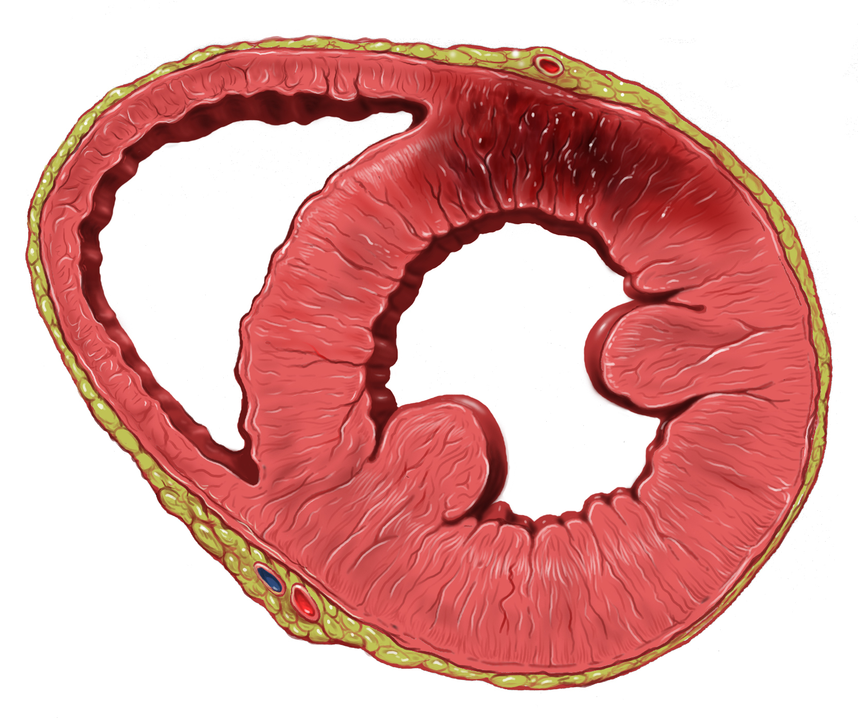
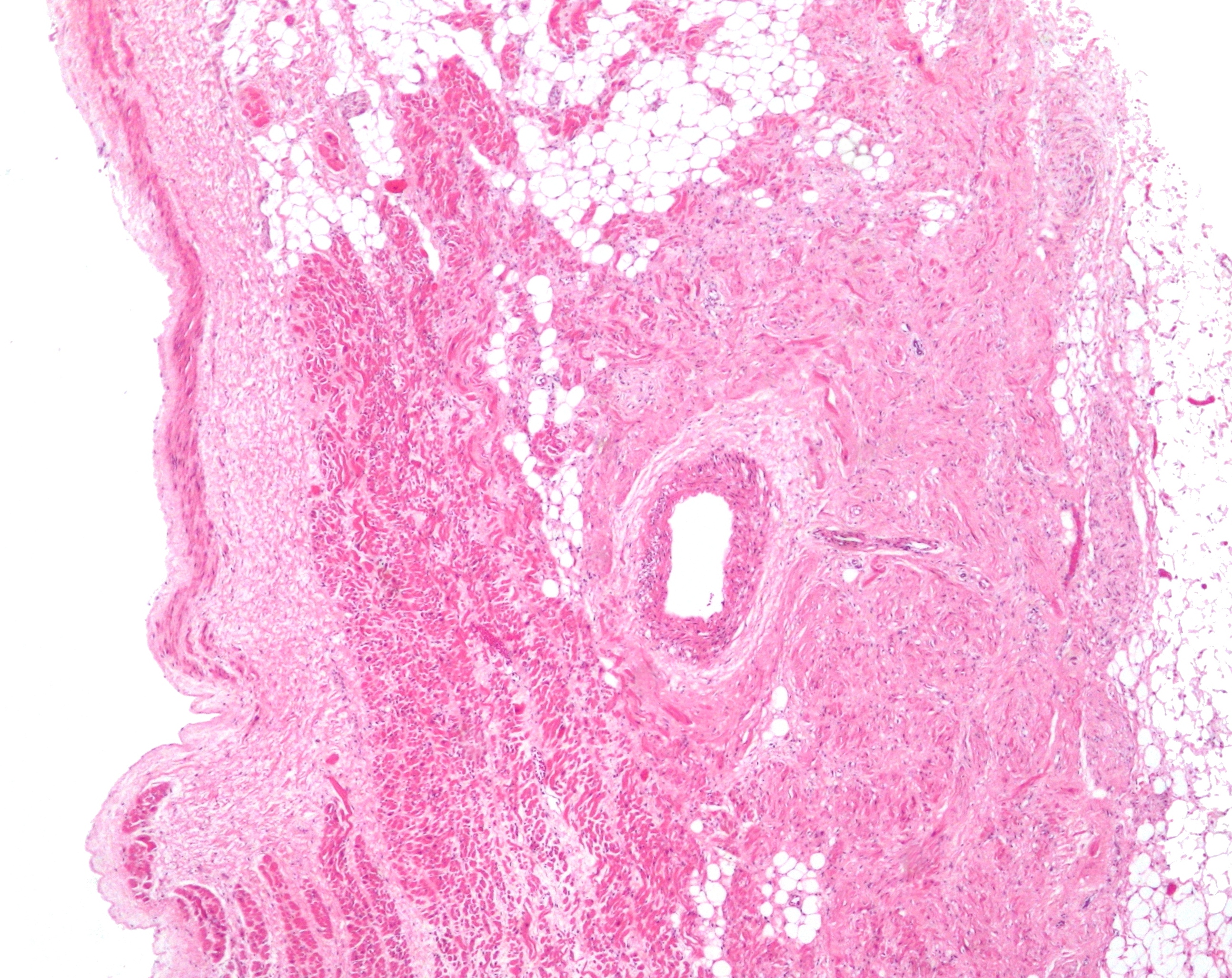
Myocardial infarction complications may occur immediately following a myocardial infarction (heart attack) (in the Acute (medical), acute phase), or may need time to develop (a Chronic (medicine), chronic problem). After an infarction, an obvious complication is a second infarction, which may occur in the domain of another atherosclerotic coronary artery, or in the same zone if there are any live cells left in the infarct. Post-myocardial complications occur after a period of ischemia, these changes can be seen in gross tissue changes and microscopic changes. Necrosis begins after 20 minutes of an infarction. Under 4 hours of ischemia, there are no gross or microscopic changes noted.Kumar, V., Abbas, A. K., & Aster, J. C. (2015). Robbins and Cotran pathologic basis of disease (Ninth edition.). Philadelphia, PA: Elsevier/Saunders. From 4-24 hours coagulative necrosis begins to be seen, which is characterized by the removal of dead Cardiac muscle cell, cardiomyocytes through heteroly ... [...More Info...] [...Related Items...] OR: [Wikipedia] [Google] [Baidu] |
Myocardial Infarction
A myocardial infarction (MI), commonly known as a heart attack, occurs when Ischemia, blood flow decreases or stops in one of the coronary arteries of the heart, causing infarction (tissue death) to the heart muscle. The most common symptom is retrosternal Angina, chest pain or discomfort that classically radiates to the left shoulder, arm, or jaw. The pain may occasionally feel like heartburn. This is the dangerous type of acute coronary syndrome. Other symptoms may include shortness of breath, nausea, presyncope, feeling faint, a diaphoresis, cold sweat, Fatigue, feeling tired, and decreased level of consciousness. About 30% of people have atypical symptoms. Women more often present without chest pain and instead have neck pain, arm pain or feel tired. Among those over 75 years old, about 5% have had an MI with little or no history of symptoms. An MI may cause heart failure, an Cardiac arrhythmia, irregular heartbeat, cardiogenic shock or cardiac arrest. Most MIs occur d ... [...More Info...] [...Related Items...] OR: [Wikipedia] [Google] [Baidu] |
Cardiac Tamponade
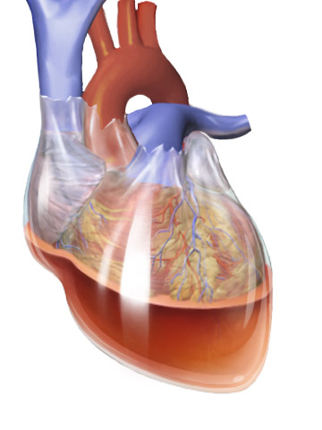
Cardiac tamponade, also known as pericardial tamponade (), is a compression of the heart due to pericardial effusion (the build-up of pericardial fluid in the pericardium, sac around the heart). Onset may be rapid or gradual. Symptoms typically include those of obstructive shock including shortness of breath, weakness, lightheadedness, and cough. Other symptoms may relate to the underlying cause. Common causes of cardiac tamponade include cancer, kidney failure, chest trauma, myocardial infarction, and pericarditis. Other causes include connective tissue disease, connective tissues diseases, hypothyroidism, aortic rupture, autoimmune disease, and complications of cardiac surgery. In Africa, tuberculosis is a relatively common cause. Diagnosis may be suspected based on hypotension, low blood pressure, jugular venous distension, or quiet heart sounds (together known as Beck's triad (cardiology), Beck's triad). A pericardial rub may be present in cases due to inflammation. The dia ... [...More Info...] [...Related Items...] OR: [Wikipedia] [Google] [Baidu] |
Sinoatrial Node
The sinoatrial node (also known as the sinuatrial node, SA node, sinus node or Keith–Flack node) is an ellipse, oval shaped region of special cardiac muscle in the upper back wall of the right atrium made up of Cell (biology), cells known as pacemaker cells. The sinus node is approximately 15 millimetre, mm long, 3 mm wide, and 1 mm thick, located directly below and to the side of the superior vena cava. These cells produce an Action potential, electrical impulse known as a cardiac action potential that travels through the electrical conduction system of the heart, causing it to muscle contraction, contract. In a healthy heart, the SA node continuously produces action potentials, setting the rhythm of the heart (sinus rhythm), and so is known as the heart's cardiac pacemaker, natural pacemaker. The rate of action potentials produced (and therefore the heart rate) is influenced by the nerves that supply it. Structure The sinoatrial node is an Ellipse, oval-shaped structure that ... [...More Info...] [...Related Items...] OR: [Wikipedia] [Google] [Baidu] |
Third Degree Heart Block
Third-degree atrioventricular block (AV block) is a medical condition in which the electrical impulse generated in the sinoatrial node (SA node) in the atrium of the heart can not propagate to the ventricles. Because the impulse is blocked, an accessory pacemaker in the lower chambers will typically activate the ventricles. This is known as an '' escape rhythm''. Since this accessory pacemaker also activates independently of the impulse generated at the SA node, two independent rhythms can be noted on the electrocardiogram (ECG). * The P waves with a regular P-to-P interval (in other words, a sinus rhythm) represent the first rhythm. * The QRS complexes with a regular R-to-R interval represent the second rhythm. The PR interval will be variable, as the hallmark of complete heart block is the lack of any apparent relationship between P waves and QRS complexes. Presentation People with third-degree AV block typically experience severe bradycardia (an abnormally low measured hea ... [...More Info...] [...Related Items...] OR: [Wikipedia] [Google] [Baidu] |
Electrical Conduction System Of The Heart
The cardiac conduction system (CCS, also called the electrical conduction system of the heart) transmits the Cardiac action potential, signals generated by the sinoatrial node – the heart's Cardiac pacemaker, pacemaker, to cause the heart muscle to Muscle contraction, contract, and pump blood through the body's circulatory system. The Cardiac pacemaker, pacemaking signal travels through the right atrium to the atrioventricular node, along the bundle of His, and through the bundle branches to Purkinje fibers in the Ventricle (heart), walls of the ventricles. The Purkinje fibers transmit the signals more rapidly to stimulate contraction of the ventricles. The conduction system consists of specialized Cardiomyocyte, heart muscle cells, situated within the myocardium. There is a cardiac skeleton, skeleton of fibrous tissue that surrounds the conduction system which can be seen on an ECG. Dysfunction of the conduction system can cause Heart arrhythmia, irregular heart rhythms includ ... [...More Info...] [...Related Items...] OR: [Wikipedia] [Google] [Baidu] |
Ventricular Fibrillation
Ventricular fibrillation (V-fib or VF) is an abnormal heart rhythm in which the Ventricle (heart), ventricles of the heart Fibrillation, quiver. It is due to disorganized electrical conduction system of the heart, electrical activity. Ventricular fibrillation results in cardiac arrest with loss of consciousness and no pulse. This is followed by sudden cardiac death in the absence of treatment. Ventricular fibrillation is initially found in about 10% of people with cardiac arrest. Ventricular fibrillation can occur due to coronary heart disease, valvular heart disease, cardiomyopathy, Brugada syndrome, long QT syndrome, electric shock, or intracranial hemorrhage. Diagnosis is by an electrocardiogram (ECG) showing irregular unformed QRS complexes without any clear P wave (electrocardiography), P waves. An important differential diagnosis is torsades de pointes. Treatment is with cardiopulmonary resuscitation (CPR) and defibrillation. Defibrillation#Change to a biphasic waveform, ... [...More Info...] [...Related Items...] OR: [Wikipedia] [Google] [Baidu] |
Ventricular Tachycardia
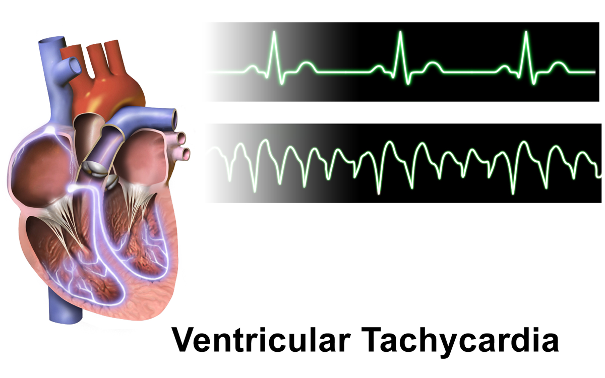
Ventricular tachycardia (V-tach or VT) is a cardiovascular disorder in which fast heart rate occurs in the ventricles of the heart. Although a few seconds of VT may not result in permanent problems, longer periods are dangerous; and multiple episodes over a short period of time are referred to as an electrical storm. Short periods may occur without symptoms, or present with lightheadedness, palpitations, shortness of breath, chest pain, and decreased level of consciousness. Ventricular tachycardia may lead to coma and persistent vegetative state due to lack of blood and oxygen to the brain. Ventricular tachycardia may result in ventricular fibrillation (VF) and turn into cardiac arrest. This conversion of the VT into VF is called the degeneration of the VT. It is found initially in about 7% of people in cardiac arrest. Ventricular tachycardia can occur due to coronary heart disease, aortic stenosis, cardiomyopathy, electrolyte imbalance, or a heart attack. Diagnosis is ... [...More Info...] [...Related Items...] OR: [Wikipedia] [Google] [Baidu] |
Arrhythmias
Arrhythmias, also known as cardiac arrhythmias, are irregularities in the heartbeat, including when it is too fast or too slow. Essentially, this is anything but normal sinus rhythm. A resting heart rate that is too fast – above 100 beats per minute in adults – is called tachycardia, and a resting heart rate that is too slow – below 60 beats per minute – is called bradycardia. Some types of arrhythmias have no symptoms. Symptoms, when present, may include palpitations or feeling a pause between heartbeats. In more serious cases, there may be lightheadedness, passing out, shortness of breath, chest pain, or decreased level of consciousness. While most cases of arrhythmia are not serious, some predispose a person to complications such as stroke or heart failure. Others may result in sudden death. Arrhythmias are often categorized into four groups: extra beats, supraventricular tachycardias, ventricular arrhythmias and bradyarrhythmias. Extra beats include prematu ... [...More Info...] [...Related Items...] OR: [Wikipedia] [Google] [Baidu] |
Myocardial Infarction
A myocardial infarction (MI), commonly known as a heart attack, occurs when Ischemia, blood flow decreases or stops in one of the coronary arteries of the heart, causing infarction (tissue death) to the heart muscle. The most common symptom is retrosternal Angina, chest pain or discomfort that classically radiates to the left shoulder, arm, or jaw. The pain may occasionally feel like heartburn. This is the dangerous type of acute coronary syndrome. Other symptoms may include shortness of breath, nausea, presyncope, feeling faint, a diaphoresis, cold sweat, Fatigue, feeling tired, and decreased level of consciousness. About 30% of people have atypical symptoms. Women more often present without chest pain and instead have neck pain, arm pain or feel tired. Among those over 75 years old, about 5% have had an MI with little or no history of symptoms. An MI may cause heart failure, an Cardiac arrhythmia, irregular heartbeat, cardiogenic shock or cardiac arrest. Most MIs occur d ... [...More Info...] [...Related Items...] OR: [Wikipedia] [Google] [Baidu] |
Cardiogenic Shock
Cardiogenic shock is a medical emergency resulting from inadequate blood flow to the body's organs due to the dysfunction of the heart. Signs of inadequate blood flow include low urine production (<30 mL/hour), cool arms and legs, and decreased level of consciousness. People may also have a severely low blood pressure and heart rate. Causes of cardiogenic shock include cardiomyopathic, arrhythmic, and mechanical. Cardiogenic shock is most commonly precipitated by a heart attack. Treatment of cardiogenic shock depends on the cause with the initial goals to improve blood flow to the body. If cardiogenic shock is due to a heart attack, attempts to open th ... [...More Info...] [...Related Items...] OR: [Wikipedia] [Google] [Baidu] |
Pulmonary Edema
Pulmonary edema (British English: oedema), also known as pulmonary congestion, is excessive fluid accumulation in the tissue or air spaces (usually alveoli) of the lungs. This leads to impaired gas exchange, most often leading to shortness of breath ( dyspnea) which can progress to hypoxemia and respiratory failure. Pulmonary edema has multiple causes and is traditionally classified as cardiogenic (caused by the heart) or noncardiogenic (all other types not caused by the heart). Various laboratory tests ( CBC, troponin, BNP, etc.) and imaging studies (chest x-ray, CT scan, ultrasound) are often used to diagnose and classify the cause of pulmonary edema. Treatment is focused on three aspects: * improving respiratory function, * treating the underlying cause, and * preventing further damage and allow full recovery to the lung. Pulmonary edema can cause permanent organ damage, and when sudden (acute), can lead to respiratory failure or cardiac arrest due to hypoxia ... [...More Info...] [...Related Items...] OR: [Wikipedia] [Google] [Baidu] |


