|
Mycobacterium Tusciae
''Mycobacterium tusciae'' is a slow-growing, scotochromogenic mycobacterium first isolated from a lymph node of an immunocompromised child and subsequently from tap water and from a respiratory specimen of a patient with chronic fibrosis. Etymology: tusciae referring to the Italian region of Tuscany, where the organisms were first isolated. Description Microscopy *Gram-positive, nonmotile and acid-fast rods. *Early microscopic morphology on Middlebrook 7H11 agar is characterized by a very elevated centre surrounded by an uneven flat fringe. Colony characteristics *Colonies are rough and strongly yellow-pigmented. Physiology *Slow growth on Löwenstein-Jensen medium at temperatures between 25 °C and 32 °C within 4 weeks. *Growth at 37 °C is inconsistent and requires longer incubation. *No growth at 42 °C and on MacConkey agar. *The type strain is susceptible ''in vitro'' to ciprofloxacin, clarithromycin, rifabutin, rifampicin, sparfloxacin and streptomyc ... [...More Info...] [...Related Items...] OR: [Wikipedia] [Google] [Baidu] |
Mycobacterium
''Mycobacterium'' is a genus of over 190 species in the phylum Actinomycetota, assigned its own family, Mycobacteriaceae. This genus includes pathogens known to cause serious diseases in mammals, including tuberculosis (''M. tuberculosis'') and leprosy ('' M. leprae'') in humans. The Greek prefix ''myco-'' means 'fungus', alluding to this genus' mold-like colony surfaces. Since this genus has cell walls with Gram-positive and Gram-negative features, acid-fast staining is used to emphasize their resistance to acids, compared to other cell types. Metabolism and Morphology Mycobacteria are aerobic with 0.2-0.6 µm wide and 1.0-10 µm long rod shapes. They are generally non-motile, except for the species ''Mycobacterium marinum'', which has been shown to be motile within macrophages. Mycobacteria possess capsules and most do not form endospores. ''M. marinum'' and perhaps ''M. bovis'' have been shown to sporulate; however, this has been contested by further research. The disting ... [...More Info...] [...Related Items...] OR: [Wikipedia] [Google] [Baidu] |
Etymology
Etymology () The New Oxford Dictionary of English (1998) – p. 633 "Etymology /ˌɛtɪˈmɒlədʒi/ the study of the class in words and the way their meanings have changed throughout time". is the study of the history of the form of words and, by extension, the origin and evolution of their semantic meaning across time. It is a subfield of historical linguistics, and draws upon comparative semantics, morphology, semiotics, and phonetics. For languages with a long written history, etymologists make use of texts, and texts about the language, to gather knowledge about how words were used during earlier periods, how they developed in meaning and form, or when and how they entered the language. Etymologists also apply the methods of comparative linguistics to reconstruct information about forms that are too old for any direct information to be available. By analyzing related languages with a technique known as the comparative method, linguists can make inferences about ... [...More Info...] [...Related Items...] OR: [Wikipedia] [Google] [Baidu] |
Microscopic
The microscopic scale () is the scale of objects and events smaller than those that can easily be seen by the naked eye, requiring a lens or microscope to see them clearly. In physics, the microscopic scale is sometimes regarded as the scale between the macroscopic scale and the quantum scale. Microscopic units and measurements are used to classify and describe very small objects. One common microscopic length scale unit is the micrometre (also called a ''micron'') (symbol: μm), which is one millionth of a metre. History Whilst compound microscopes were first developed in the 1590s, the significance of the microscopic scale was only truly established in the 1600s when Marcello Malphigi and Antonie van Leeuwenhoek microscopically observed frog lungs and microorganisms. As microbiology was established, the significance of making scientific observations at a microscopic level increased. Published in 1665, Robert Hooke’s book Micrographia details his microscopic observa ... [...More Info...] [...Related Items...] OR: [Wikipedia] [Google] [Baidu] |
Silica
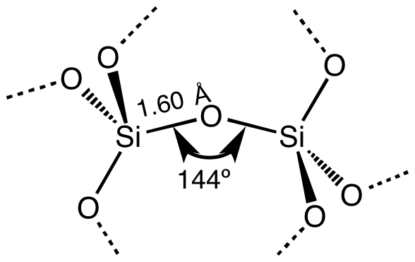
Silicon dioxide, also known as silica, is an oxide of silicon with the chemical formula , most commonly found in nature as quartz and in various living organisms. In many parts of the world, silica is the major constituent of sand. Silica is one of the most complex and most abundant families of materials, existing as a compound of several minerals and as a synthetic product. Notable examples include fused quartz, fumed silica, silica gel, opal and aerogels. It is used in structural materials, microelectronics (as an electrical insulator), and as components in the food and pharmaceutical industries. Structure In the majority of silicates, the silicon atom shows tetrahedral coordination, with four oxygen atoms surrounding a central Si atomsee 3-D Unit Cell. Thus, SiO2 forms 3-dimensional network solids in which each silicon atom is covalently bonded in a tetrahedral manner to 4 oxygen atoms. In contrast, CO2 is a linear molecule. The starkly different structures of th ... [...More Info...] [...Related Items...] OR: [Wikipedia] [Google] [Baidu] |
Dust Cell
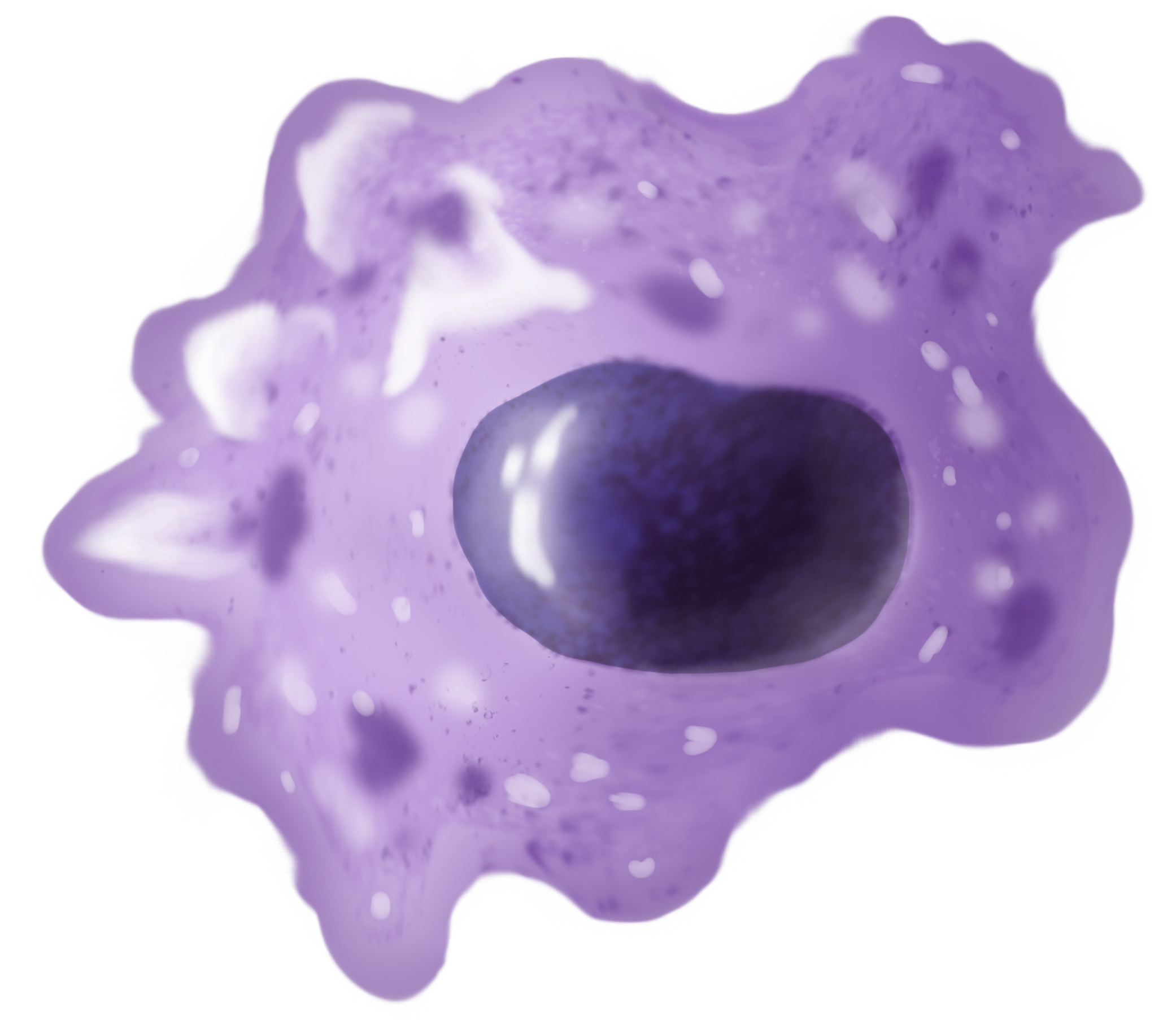
An alveolar macrophage, pulmonary macrophage, (or dust cell) is a type of macrophage, a professional phagocyte, found in the airways and at the level of the alveoli in the lungs, but separated from their walls. Activity of the alveolar macrophage is relatively high, because they are located at one of the major boundaries between the body and the outside world. They are responsible for removing particles such as dust or microorganisms from the respiratory surfaces. Alveolar macrophages are frequently seen to contain granules of exogenous material such as particulate carbon that they have picked up from respiratory surfaces. Such black granules may be especially common in smoker's lungs or long-term city dwellers. The alveolar macrophage is the third cell type in the alveolus, the others are the type I and type II pneumocytes. Comparison of pigmented pulmonary macrophages Function Alveolar macrophages are phagocytes that play a critical role in homeostasis, host defense, ... [...More Info...] [...Related Items...] OR: [Wikipedia] [Google] [Baidu] |
Macrophages
Macrophages (abbreviated as M φ, MΦ or MP) ( el, large eaters, from Greek ''μακρός'' (') = large, ''φαγεῖν'' (') = to eat) are a type of white blood cell of the immune system that engulfs and digests pathogens, such as cancer cells, microbes, cellular debris, and foreign substances, which do not have proteins that are specific to healthy body cells on their surface. The process is called phagocytosis, which acts to defend the host against infection and injury. These large phagocytes are found in essentially all tissues, where they patrol for potential pathogens by amoeboid movement. They take various forms (with various names) throughout the body (e.g., histiocytes, Kupffer cells, alveolar macrophages, microglia, and others), but all are part of the mononuclear phagocyte system. Besides phagocytosis, they play a critical role in nonspecific defense ( innate immunity) and also help initiate specific defense mechanisms ( adaptive immunity) by recruiting oth ... [...More Info...] [...Related Items...] OR: [Wikipedia] [Google] [Baidu] |
Fibrin
Fibrin (also called Factor Ia) is a fibrous, non-globular protein involved in the clotting of blood. It is formed by the action of the protease thrombin on fibrinogen, which causes it to polymerize. The polymerized fibrin, together with platelets, forms a hemostatic plug or clot over a wound site. When the lining of a blood vessel is broken, platelets are attracted, forming a platelet plug. These platelets have thrombin receptors on their surfaces that bind serum thrombin molecules, which in turn convert soluble fibrinogen in the serum into fibrin at the wound site. Fibrin forms long strands of tough insoluble protein that are bound to the platelets. Factor XIII completes the cross-linking of fibrin so that it hardens and contracts. The cross-linked fibrin forms a mesh atop the platelet plug that completes the clot. Fibrin was discovered by Marcello Malpighi in 1666. Role in disease Excessive generation of fibrin due to activation of the coagulation cascade leads to ... [...More Info...] [...Related Items...] OR: [Wikipedia] [Google] [Baidu] |
Macrophages
Macrophages (abbreviated as M φ, MΦ or MP) ( el, large eaters, from Greek ''μακρός'' (') = large, ''φαγεῖν'' (') = to eat) are a type of white blood cell of the immune system that engulfs and digests pathogens, such as cancer cells, microbes, cellular debris, and foreign substances, which do not have proteins that are specific to healthy body cells on their surface. The process is called phagocytosis, which acts to defend the host against infection and injury. These large phagocytes are found in essentially all tissues, where they patrol for potential pathogens by amoeboid movement. They take various forms (with various names) throughout the body (e.g., histiocytes, Kupffer cells, alveolar macrophages, microglia, and others), but all are part of the mononuclear phagocyte system. Besides phagocytosis, they play a critical role in nonspecific defense ( innate immunity) and also help initiate specific defense mechanisms ( adaptive immunity) by recruiting oth ... [...More Info...] [...Related Items...] OR: [Wikipedia] [Google] [Baidu] |
Radiography
Radiography is an imaging technique using X-rays, gamma rays, or similar ionizing radiation and non-ionizing radiation to view the internal form of an object. Applications of radiography include medical radiography ("diagnostic" and "therapeutic") and industrial radiography. Similar techniques are used in airport security (where "body scanners" generally use backscatter X-ray). To create an image in conventional radiography, a beam of X-rays is produced by an X-ray generator and is projected toward the object. A certain amount of the X-rays or other radiation is absorbed by the object, dependent on the object's density and structural composition. The X-rays that pass through the object are captured behind the object by a detector (either photographic film or a digital detector). The generation of flat two dimensional images by this technique is called projectional radiography. In computed tomography (CT scanning) an X-ray source and its associated detectors rotate around ... [...More Info...] [...Related Items...] OR: [Wikipedia] [Google] [Baidu] |
Mycobacterium Aichiense
''Mycolicibacterium aichiense'' (formerly ''Mycobacterium aichiense'') is a species of bacteria from the phylum Actinomycetota The ''Actinomycetota'' (or ''Actinobacteria'') are a phylum of all gram-positive bacteria. They can be terrestrial or aquatic. They are of great economic importance to humans because agriculture and forests depend on their contributions to soi ... that was first isolated from soil and from human sputum. It produces pigments when grow in the dark and grows rapidly at 25–37 °C on Ogawa egg medium or Sauton agar medium. References Acid-fast bacilli aichiense Bacteria described in 1981 {{Mycobacterium-stub ... [...More Info...] [...Related Items...] OR: [Wikipedia] [Google] [Baidu] |
Mycobacterium Farcinogenes
''Mycobacterium farcinogenes'' is a species of the phylum Actinomycetota (Gram-positive bacteria with high guanine and cytosine content, one of the dominant phyla of all bacteria), belonging to the genus ''Mycobacterium''. Although slow-growing, it is similar to fast-growing species, and is usually classified with them. Description Gram-positive, nonmotile and strongly acid-fast rods. Short or long filaments, bent and branched, in clumps or tangled, lacy network. Colony characteristics Rough, yellow and convoluted colonies. Firmly adherent to medium and surrounded by an iridescent halo. Physiology *Slow growth after 15–20 days on Löwenstein-Jensen medium. Differential characteristics *On the basis of characteristic lipids this species belongs to the genus ''Mycobacterium'' and not to the genus ''Nocardia''. *DNA homology to the closely related species '' Mycobacterium senegalense''. Both species, share an identical 5' 16S rDNA sequence. However, the ITS sequences are di ... [...More Info...] [...Related Items...] OR: [Wikipedia] [Google] [Baidu] |


