|
Mucins
Mucins () are a family of high molecular weight, heavily glycosylation, glycosylated proteins (glycoconjugates) produced by epithelial tissues in most animals. Mucins' key characteristic is their ability to form gels; therefore they are a key component in most gel-like secretions, serving functions from lubrication to cell signalling to forming chemical barriers. They often take an inhibitory role. Some mucins are associated with controlling Mineralization (biology), mineralization, including nacre formation in mollusks, calcification in echinoderms and bone formation in vertebrates. They bind to pathogens as part of the immune system. Overexpression of the mucin proteins, especially MUC1, is associated with many types of cancer. Although some mucins are cell membrane, membrane-bound due to the presence of a hydrophobic membrane-spanning domain that favors retention in the plasma membrane, most mucins are secretion, secreted as principal components of mucus by mucous membranes or ... [...More Info...] [...Related Items...] OR: [Wikipedia] [Google] [Baidu] |
MUC1
Mucin short variant S1, also called polymorphic epithelial mucin (PEM) or epithelial membrane antigen (EMA), is a mucin encoded by the ''MUC1'' gene in humans. Mucin short variant S1 is a glycoprotein with extensive O-linked glycosylation of its extracellular domain. Mucins line the apical surface of epithelial cells in the lungs, stomach, intestines, eyes and several other organs. Mucins protect the body from infection by pathogen binding to oligosaccharides in the extracellular domain, preventing the pathogen from reaching the cell surface. Overexpression of MUC1 is often associated with colon, breast, ovarian, lung and pancreatic cancers. Joyce Taylor-Papadimitriou identified and characterised the antigen during her work with breast and ovarian tumors. Structure MUC1 is a member of the mucin family and encodes a membrane bound, glycosylated phosphoprotein. MUC1 has a core protein mass of 120-225 kDa which increases to 250-500 kDa with glycosylation. It extends 200-500&n ... [...More Info...] [...Related Items...] OR: [Wikipedia] [Google] [Baidu] |
Mucin 5AC
Mucin-5AC (MUC-5AC) is a protein that is encoded by the ''MUC5AC'' gene in humans. MUC-5AC is a large gel-forming glycoprotein. In the respiratory tract, it protects against infection by binding to inhaled pathogens, which are subsequently removed by mucociliary clearance. Overproduction of MUC-5AC can contribute to diseases such as asthma and chronic obstructive pulmonary disease, and has also been associated with greater protection against influenza infection. Clinical relevance This gene has been linked to mucus hypersecretion in the respiratory tract and is associated to chronic obstructive pulmonary disease Chronic obstructive pulmonary disease (COPD) is a type of progressive lung disease characterized by chronic respiratory symptoms and airflow limitation. GOLD defines COPD as a heterogeneous lung condition characterized by chronic respiratory s ... (COPD). References External links PDBe-KBprovides an overview of all the structure information available in ... [...More Info...] [...Related Items...] OR: [Wikipedia] [Google] [Baidu] |
Mucin 2
Mucin 2, oligomeric mucus gel-forming, also known as MUC2, is a protein that in humans is encoded by the ''MUC2'' gene. Function This gene encodes a member of the mucin protein family. The protein encoded by this gene, also called mucin 2, is secreted onto mucosal surfaces. Mucin 2 is particularly prominent in the gut where it is secreted from goblet cells in the epithelial lining into the lumen of the large intestine. There, mucin 2, along with small amounts of related-mucin proteins, polymerizes into a gel of which 80% by weight is oligosaccharide side-chains that are added as post-translational modification In molecular biology, post-translational modification (PTM) is the covalent process of changing proteins following protein biosynthesis. PTMs may involve enzymes or occur spontaneously. Proteins are created by ribosomes, which translation (biolog ...s to the mucin proteins. This gel provides an insoluble mucous barrier that serves to protect the intestinal epithelium ... [...More Info...] [...Related Items...] OR: [Wikipedia] [Google] [Baidu] |
Mucus
Mucus (, ) is a slippery aqueous secretion produced by, and covering, mucous membranes. It is typically produced from cells found in mucous glands, although it may also originate from mixed glands, which contain both Serous fluid, serous and mucous cells. It is a viscous colloid containing inorganic ions, inorganic salts, antimicrobial enzymes (such as lysozymes), Antibody, immunoglobulins (especially Immunoglobulin A, IgA), and glycoproteins such as lactoferrin and mucins, which are produced by goblet cells in the mucous membranes and submucosal glands. Mucus covers the Epithelium, epithelial cells that interact with outside environment, serves to protect the linings of the respiratory system, respiratory, Digestion#Digestive system, digestive, and Genitourinary system, urogenital systems, and structures in the Visual system, visual and auditory systems from pathogenic Fungus, fungi, bacteria and viruses. Most of the mucus in the body is produced in the gastrointestinal tract. ... [...More Info...] [...Related Items...] OR: [Wikipedia] [Google] [Baidu] |
Micrograph
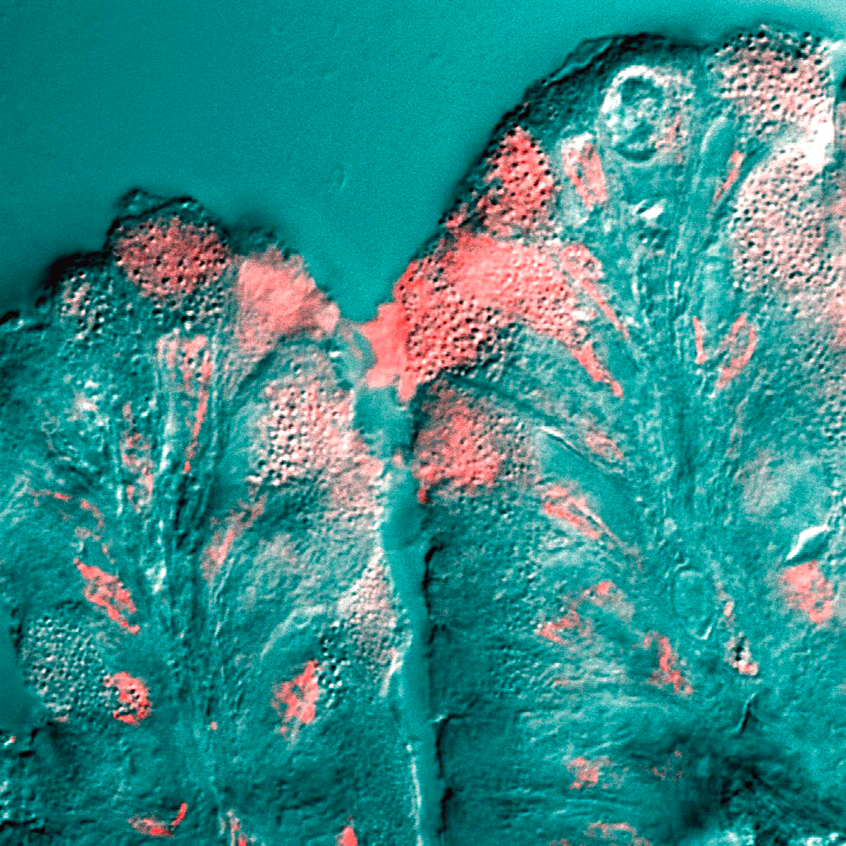
A micrograph is an image, captured photographically or digitally, taken through a microscope or similar device to show a magnify, magnified image of an object. This is opposed to a macrograph or photomacrograph, an image which is also taken on a microscope but is only slightly magnified, usually less than 10 times. Micrography is the practice or art of using microscopes to make photographs. A photographic micrograph is a photomicrograph, and one taken with an electron microscope is an electron micrograph. A micrograph contains extensive details of microstructure. A wealth of information can be obtained from a simple micrograph like behavior of the material under different conditions, the phases found in the system, failure analysis, grain size estimation, elemental analysis and so on. Micrographs are widely used in all fields of microscopy. Types Photomicrograph A light micrograph or photomicrograph is a micrograph prepared using an optical microscope, a process referred to ... [...More Info...] [...Related Items...] OR: [Wikipedia] [Google] [Baidu] |
Plasma Membrane
The cell membrane (also known as the plasma membrane or cytoplasmic membrane, and historically referred to as the plasmalemma) is a biological membrane that separates and protects the interior of a cell from the outside environment (the extracellular space). The cell membrane consists of a lipid bilayer, made up of two layers of phospholipids with cholesterols (a lipid component) interspersed between them, maintaining appropriate membrane fluidity at various temperatures. The membrane also contains membrane proteins, including integral proteins that span the membrane and serve as membrane transporters, and peripheral proteins that loosely attach to the outer (peripheral) side of the cell membrane, acting as enzymes to facilitate interaction with the cell's environment. Glycolipids embedded in the outer lipid layer serve a similar purpose. The cell membrane controls the movement of substances in and out of a cell, being selectively permeable to ions and organic molecu ... [...More Info...] [...Related Items...] OR: [Wikipedia] [Google] [Baidu] |
Conjunctiva
In the anatomy of the eye, the conjunctiva (: conjunctivae) is a thin mucous membrane that lines the inside of the eyelids and covers the sclera (the white of the eye). It is composed of non-keratinized, stratified squamous epithelium with goblet cells, stratified columnar epithelium and stratified cuboidal epithelium (depending on the zone). The conjunctiva is highly Angiogenesis, vascularised, with many microvessels easily accessible for imaging studies. Structure The conjunctiva is typically divided into three parts: Blood supply Blood to the bulbar conjunctiva is primarily derived from the ophthalmic artery. The blood supply to the palpebral conjunctiva (the eyelid) is derived from the external carotid artery. However, the circulations of the bulbar conjunctiva and palpebral conjunctiva are linked, so both bulbar conjunctival and palpebral conjunctival vessels are supplied by both the ophthalmic artery and the external carotid artery, to varying extents. Nerve supply Se ... [...More Info...] [...Related Items...] OR: [Wikipedia] [Google] [Baidu] |
Stomach
The stomach is a muscular, hollow organ in the upper gastrointestinal tract of Human, humans and many other animals, including several invertebrates. The Ancient Greek name for the stomach is ''gaster'' which is used as ''gastric'' in medical terms related to the stomach. The stomach has a dilated structure and functions as a vital organ in the digestive system. The stomach is involved in the gastric phase, gastric phase of digestion, following the cephalic phase in which the sight and smell of food and the act of chewing are stimuli. In the stomach a chemical breakdown of food takes place by means of secreted digestive enzymes and gastric acid. It also plays a role in regulating gut microbiota, influencing digestion and overall health. The stomach is located between the esophagus and the small intestine. The pyloric sphincter controls the passage of partially digested food (chyme) from the stomach into the duodenum, the first and shortest part of the small intestine, where p ... [...More Info...] [...Related Items...] OR: [Wikipedia] [Google] [Baidu] |
Respiratory Tract
The respiratory tract is the subdivision of the respiratory system involved with the process of conducting air to the alveoli for the purposes of gas exchange in mammals. The respiratory tract is lined with respiratory epithelium as respiratory mucosa. Air is breathed in through the nose to the nasal cavity, where a layer of nasal mucosa acts as a filter and traps pollutants and other harmful substances found in the air. Next, air moves into the pharynx, a passage that contains the intersection between the oesophagus and the larynx. The opening of the larynx has a special flap of cartilage, the epiglottis, that opens to allow air to pass through but closes to prevent food from moving into the airway. From the larynx, air moves into the trachea and down to the intersection known as the carina that branches to form the right and left primary (main) bronchi. Each of these bronchi branches into a secondary (lobar) bronchus that branches into tertiary (segmental) bronchi, t ... [...More Info...] [...Related Items...] OR: [Wikipedia] [Google] [Baidu] |
Endometrium
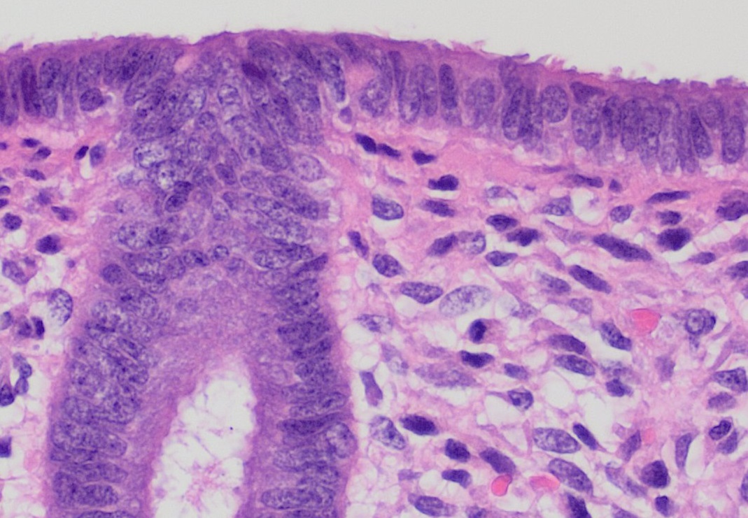
The endometrium is the inner epithelium, epithelial layer, along with its mucous membrane, of the mammalian uterus. It has a basal layer and a functional layer: the basal layer contains stem cells which regenerate the functional layer. The functional layer thickens and then is shed during menstruation in humans and some other mammals, including other apes, Old World monkeys, some species of bat, the elephant shrew and the Cairo spiny mouse. In most other mammals, the endometrium is reabsorbed in the estrous cycle. During pregnancy, the glands and blood vessels in the endometrium further increase in size and number. Vascular spaces fuse and become interconnected, forming the placenta, which supplies oxygen and nutrition to the embryo and fetus.Blue Histology - Female Reproductive System ... [...More Info...] [...Related Items...] OR: [Wikipedia] [Google] [Baidu] |
Ileum
The ileum () is the final section of the small intestine in most higher vertebrates, including mammals, reptiles, and birds. In fish, the divisions of the small intestine are not as clear and the terms posterior intestine or distal intestine may be used instead of ileum. Its main function is to absorb vitamin B12, vitamin B12, bile salts, and whatever products of digestion that were not absorbed by the jejunum. The ileum follows the duodenum and jejunum and is separated from the cecum by the ileocecal valve (ICV). In humans, the ileum is about 2–4 m long, and the pH is usually between 7 and 8 (neutral or slightly base (chemistry), basic). ''Ileum'' is derived from the Greek word εἰλεός (eileós), referring to a medical condition known as ileus. Structure The ileum is the third and final part of the small intestine. It follows the jejunum and ends at the ileocecal junction, where the wikt:terminal, terminal ileum communicates with the cecum of the large intestine thro ... [...More Info...] [...Related Items...] OR: [Wikipedia] [Google] [Baidu] |
Jejunum
The jejunum is the second part of the small intestine in humans and most higher vertebrates, including mammals, reptiles, and birds. Its lining is specialized for the absorption by enterocytes of small nutrient molecules which have been previously digested by enzymes in the duodenum. The jejunum lies between the duodenum and the ileum and is considered to start at the suspensory muscle of the duodenum, a location called the duodenojejunal flexure. The division between the jejunum and ileum is not anatomically distinct. In adult humans, the small intestine is usually long (post mortem), about two-fifths of which (about ) is the jejunum. Structure The interior surface of the jejunum—which is exposed to ingested food—is covered in finger–like projections of mucosa, called villi, which increase the surface area of tissue available to absorb nutrients from ingested foodstuffs. The epithelial cells which line these villi have microvilli. The transport of nutrien ... [...More Info...] [...Related Items...] OR: [Wikipedia] [Google] [Baidu] |


