|
Microvascular
Blood vessels are the tubular structures of a circulatory system that transport blood throughout many animals’ bodies. Blood vessels transport blood cells, nutrients, and oxygen to most of the tissues of a body. They also take waste and carbon dioxide away from the tissues. Some tissues such as cartilage, epithelium, and the lens and cornea of the eye are not supplied with blood vessels and are termed ''avascular''. There are five types of blood vessels: the arteries, which carry the blood away from the heart; the arterioles; the capillaries, where the exchange of water and chemicals between the blood and the tissues occurs; the venules; and the veins, which carry blood from the capillaries back towards the heart. The word ''vascular'', is derived from the Latin ''vas'', meaning ''vessel'', and is mostly used in relation to blood vessels. Etymology * artery – late Middle English; from Latin ''arteria'', from Greek ''artēria'', probably from ''airein'' ("raise"). * ve ... [...More Info...] [...Related Items...] OR: [Wikipedia] [Google] [Baidu] |
Capillaries
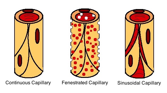
A capillary is a small blood vessel, from 5 to 10 micrometres in diameter, and is part of the microcirculation system. Capillaries are microvessels and the smallest blood vessels in the body. They are composed of only the tunica intima (the innermost layer of an artery or vein), consisting of a thin wall of simple squamous endothelial cells. They are the site of the exchange of many substances from the surrounding interstitial fluid, and they convey blood from the smallest branches of the arteries (arterioles) to those of the veins (venules). Other substances which cross capillaries include water, oxygen, carbon dioxide, urea, glucose, uric acid, lactic acid and creatinine. Lymph capillaries connect with larger lymph vessels to drain lymphatic fluid collected in microcirculation. Etymology ''Capillary'' comes from the Latin word , meaning "of or resembling hair", with use in English beginning in the mid-17th century. The meaning stems from the tiny, hairlike diameter of a ... [...More Info...] [...Related Items...] OR: [Wikipedia] [Google] [Baidu] |
Circulatory System
In vertebrates, the circulatory system is a system of organs that includes the heart, blood vessels, and blood which is circulated throughout the body. It includes the cardiovascular system, or vascular system, that consists of the heart and blood vessels (from Greek meaning ''heart'', and Latin meaning ''vessels''). The circulatory system has two divisions, a systemic circulation or circuit, and a pulmonary circulation or circuit. Some sources use the terms ''cardiovascular system'' and ''vascular system'' interchangeably with ''circulatory system''. The network of blood vessels are the great vessels of the heart including large elastic arteries, and large veins; other arteries, smaller arterioles, capillaries that join with venules (small veins), and other veins. The circulatory system is closed in vertebrates, which means that the blood never leaves the network of blood vessels. Many invertebrates such as arthropods have an open circulatory system with a he ... [...More Info...] [...Related Items...] OR: [Wikipedia] [Google] [Baidu] |
Tunica Intima
The tunica intima (Neo-Latin "inner coat"), or intima for short, is the innermost tunica (biology), tunica (layer) of an artery or vein. It is made up of one layer of endothelium, endothelial cells (and macrophages in areas of disturbed blood flow), and is supported by an internal elastic lamina. The endothelial cells are in direct contact with the blood flow. The three layers of a blood vessel are an inner layer (the tunica intima), a middle layer (the tunica media), and an outer layer (the tunica externa). In dissection, the inner coat (tunica intima) can be separated from the middle (tunica media) by a little maceration, or it may be stripped off in small pieces; but, because of its friability, it cannot be separated as a complete membrane. It is a fine, transparent, colorless structure which is highly elastic, and, after death, is commonly corrugated into longitudinal wrinkles. Structure The structure of the tunica intima depends on the blood vessel type. Elastic artery, E ... [...More Info...] [...Related Items...] OR: [Wikipedia] [Google] [Baidu] |
Circulatory Anastomosis
A circulatory anastomosis is a connection (an anastomosis) between two blood vessels, such as between arteries (arterio-arterial anastomosis), between veins (veno-venous anastomosis) or between an artery and a vein (arterio-venous anastomosis). Anastomoses between arteries and between veins result in a multitude of arteries and veins, respectively, serving the same volume of tissue. Such anastomoses occur normally in the body in the circulatory system, serving as back-up routes in a collateral circulation that allow blood to flow if one link is blocked or otherwise compromised, but may also occur pathologically. Physiologic Arterio-arterial anastomoses include actual (e.g., palmar and plantar arches) and potential varieties (e.g., coronary arteries and cortical branch of cerebral arteries). There are many examples of normal arterio-arterial anastomoses in the body. Clinically important examples include: * Circle of Willis (in the brain) * Coronary: anterior interventricul ... [...More Info...] [...Related Items...] OR: [Wikipedia] [Google] [Baidu] |
Basement Membrane
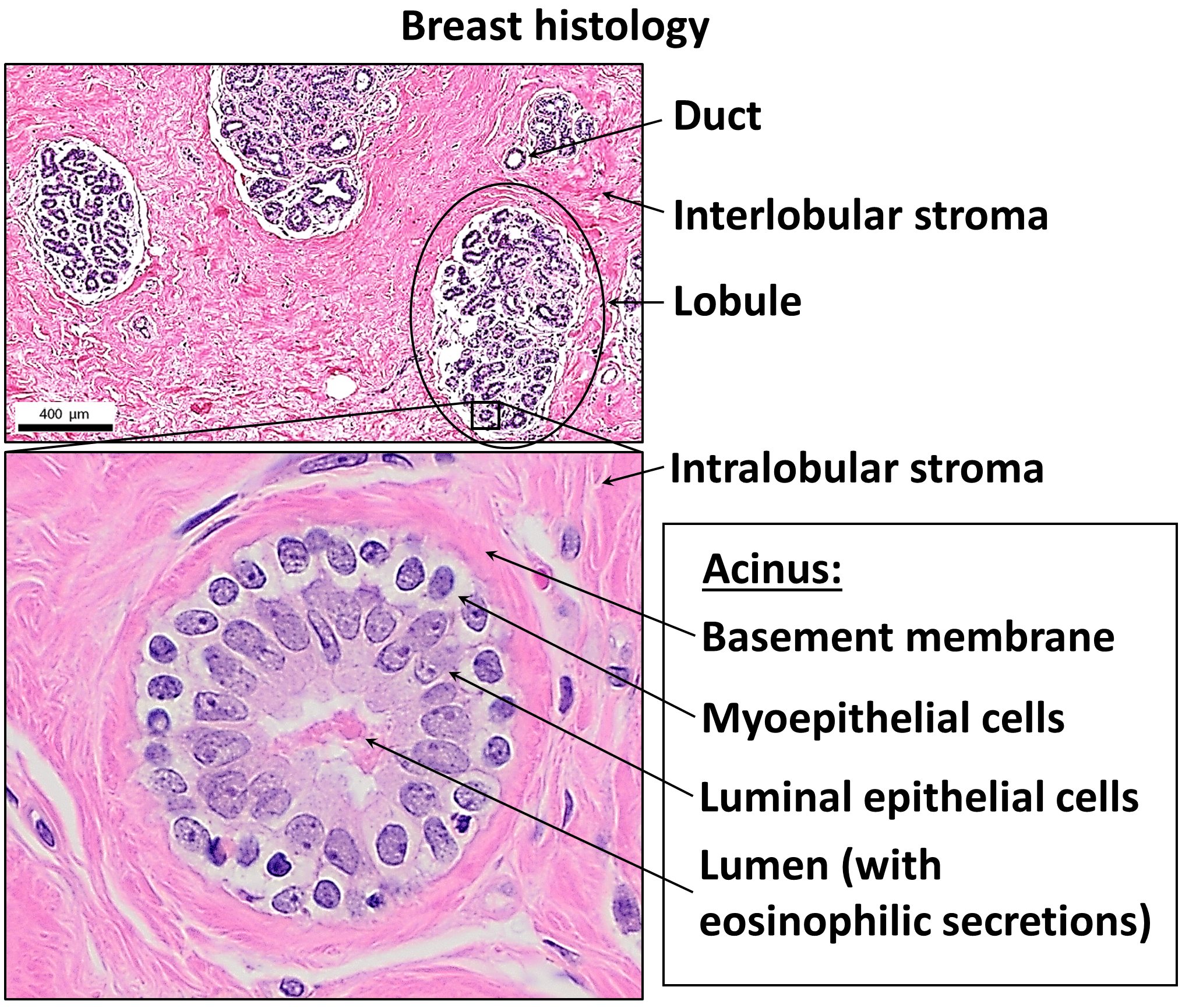
The basement membrane, also known as base membrane, is a thin, pliable sheet-like type of extracellular matrix that provides cell and tissue support and acts as a platform for complex signalling. The basement membrane sits between epithelial tissues including mesothelium and endothelium, and the underlying connective tissue. Structure As seen with the electron microscope, the basement membrane is composed of two layers, the basal lamina and the reticular lamina. The underlying connective tissue attaches to the basal lamina with collagen VII anchoring fibrils and fibrillin microfibrils. The basal lamina layer can further be subdivided into two layers based on their visual appearance in electron microscopy. The lighter-colored layer closer to the epithelium is called the lamina lucida, while the denser-colored layer closer to the connective tissue is called the lamina densa. The electron-dense lamina densa layer is about 30–70 nanometers thick and consists of an und ... [...More Info...] [...Related Items...] OR: [Wikipedia] [Google] [Baidu] |
Endothelial Cell
The endothelium (: endothelia) is a single layer of squamous endothelial cells that line the interior surface of blood vessels and lymphatic vessels. The endothelium forms an interface between circulating blood or lymph in the lumen and the rest of the vessel wall. Endothelial cells in direct contact with blood are called vascular endothelial cells whereas those in direct contact with lymph are known as lymphatic endothelial cells. Vascular endothelial cells line the entire circulatory system, from the heart to the smallest capillaries. These cells have unique functions that include fluid filtration, such as in the glomerulus of the kidney, blood vessel tone, hemostasis, neutrophil recruitment, and hormone trafficking. Endothelium of the interior surfaces of the heart chambers is called endocardium. An impaired function can lead to serious health issues throughout the body. Structure The endothelium is a thin layer of single flat ( squamous) cells that line the ... [...More Info...] [...Related Items...] OR: [Wikipedia] [Google] [Baidu] |
Vasa Vasorum
Vasa vasorum are networks of small blood vessels that supply the walls of large blood vessels, such as elastic arteries (e.g., the aorta) and large veins (e.g., the venae cavae). The name derives . Occasionally, two different singular forms are seen: vasa vasis () and vas vasis (). Structure Studies conducted with 3D micro-CT on pig and human arteries from different vascular beds have shown that there are three different types of vasa vasorum: * ''Vasa vasorum internae'', that originate directly from the main lumen of the artery and then branch into the vessel wall. * ''Vasa vasorum externae'', that originate from branches of the main artery and then dive back into the vessel wall of the main artery. * ''Venous vasa vasorae'', that originate within the vessel wall of the artery but then drain into the main lumen or branches of concomitant vein. Depending on the type of vasa vasorum, it penetrates the vessel wall starting at the intimal layer (vasa vasorum interna) or the ... [...More Info...] [...Related Items...] OR: [Wikipedia] [Google] [Baidu] |
Nerve
A nerve is an enclosed, cable-like bundle of nerve fibers (called axons). Nerves have historically been considered the basic units of the peripheral nervous system. A nerve provides a common pathway for the Electrochemistry, electrochemical nerve impulses called action potentials that are transmitted along each of the axons to peripheral organs or, in the case of sensory nerves, from the periphery back to the central nervous system. Each axon is an extension of an individual neuron, along with other supportive cells such as some Schwann cells that coat the axons in myelin. Each axon is surrounded by a layer of connective tissue called the endoneurium. The axons are bundled together into groups called Nerve fascicle, fascicles, and each fascicle is wrapped in a layer of connective tissue called the perineurium. The entire nerve is wrapped in a layer of connective tissue called the epineurium. Nerve cells (often called neurons) are further classified as either Sensory neuron, sens ... [...More Info...] [...Related Items...] OR: [Wikipedia] [Google] [Baidu] |
Tunica Adventitia
The tunica externa (Neo-Latin "outer coat"), also known as the tunica adventitia (Neo-Latin "additional coat"), is the outermost tunica (layer) of a blood vessel, surrounding the tunica media. It is mainly composed of collagen and, in arteries, is supported by external elastic lamina. The collagen serves to anchor the blood vessel to nearby organs, giving it stability. The three layers of the blood vessels are: an inner tunica intima, a middle tunica media, and an outer tunica externa. Structure The tunica externa is made from collagen and elastic fibers in a loose connective tissue. This is secreted by fibroblasts. This is normally the thickest tunic in veins and may be thicker than the tunica media in some larger arteries. The outer layers of the tunica externa are not distinct but rather blend with the surrounding connective tissue outside the vessel, helping to hold the vessel in relative position. Function The tunica externa provides basic structural support to blood v ... [...More Info...] [...Related Items...] OR: [Wikipedia] [Google] [Baidu] |
Vascular Smooth Muscle
Vascular smooth muscle is the type of smooth muscle that makes up most of the walls of blood vessels. Structure Vascular smooth muscle refers to the particular type of smooth muscle found within, and composing the majority of the wall of blood vessels. Nerve supply Vascular smooth muscle is innervated primarily by the sympathetic nervous system through adrenergic receptors (adrenoceptors). The three types present are: alpha-1, alpha-2 and beta-2 adrenergic receptors, . The main endogenous agonist of these cell receptors is norepinephrine (NE). The adrenergic receptors exert opposite physiologic effects in the vascular smooth muscle under activation: * alpha-1 receptors. Under NE binding alpha-1 receptors cause vasoconstriction (contraction of the vascular smooth muscle cells decreasing the diameter of the vessels). These receptors are activated in response to shock or low blood pressure as a defensive reaction trying to restore the normal blood pressure. Antagonists o ... [...More Info...] [...Related Items...] OR: [Wikipedia] [Google] [Baidu] |
Tunica Media
The tunica media (Neo-Latin "middle coat"), or media for short, is the middle tunica (layer) of an artery or vein. It lies between the internal elastic lamina of the tunica intima on the inside and the tunica externa on the outside. Artery The tunica media is made up of smooth muscle cells, elastic tissue and collagen. It lies between the tunica intima on the inside and the tunica externa on the outside. The middle coat (tunica media) is distinguished from the inner (tunica intima) by its color and by the transverse arrangement of its fibers. * In the ''smaller arteries'', it consists principally of smooth muscle fibers in fine bundles, arranged in lamellae and disposed circularly around the vessel. These lamellae vary in number according to the size of the vessel; the smallest arteries having only a single layer, and those slightly larger three or four layers - up to a maximum of six layers. It is to this coat that the thickness of the wall of the artery is mainly due. * I ... [...More Info...] [...Related Items...] OR: [Wikipedia] [Google] [Baidu] |


