|
Mesenchymal–epithelial Transition
A mesenchymal–epithelial transition (MET) is a reversible biological process that involves the transition from motile, multipolar or spindle-shaped mesenchymal cells to planar arrays of polarized cells called epithelia. MET is the reverse process of epithelial–mesenchymal transition (EMT) and it has been shown to occur in normal development, induced pluripotent stem cell reprogramming, cancer metastasis and wound healing. __TOC__ Introduction Unlike epithelial cells – which are stationary and characterized by an apico-basal polarity with binding by a basal lamina, tight junctions, gap junctions, adherent junctions and expression of cell-cell adhesion markers such as E-cadherin, mesenchymal cells do not make mature cell-cell contacts, can invade through the extracellular matrix, and express markers such as vimentin, fibronectin, N-cadherin, Twist, and Snail. MET plays also a critical role in metabolic switching and epigenetic modifications. In general, epithelium-asso ... [...More Info...] [...Related Items...] OR: [Wikipedia] [Google] [Baidu] |
Mesenchymal Cell
Mesenchymal stem cells (MSCs), also known as mesenchymal stromal cells or medicinal signaling cells, are multipotent stromal cells that can differentiate into a variety of cell types, including osteoblasts (bone cells), chondrocytes (cartilage cells), myocytes (muscle cells) and adipocytes (fat cells which give rise to marrow adipose tissue). The primary function of MSCs is to respond to injury and infection by secreting and recruiting a range of biological factors, as well as modulating inflammatory processes to facilitate tissue repair and regeneration. Extensive research interest has led to more than 80,000 peer-reviewed papers on MSCs. Structure Definition Mesenchymal stem cells (MSCs), a term first used (in 1991) by Arnold Caplan at Case Western Reserve University, are characterized morphologically by a small cell body with long, thin cell processes. While the terms ''mesenchymal stem cell'' (MSC) and ''marrow stromal cell'' have been used interchangeably for many ye ... [...More Info...] [...Related Items...] OR: [Wikipedia] [Google] [Baidu] |
Epigenetic Modifications
Embryonic stem cells are capable of self-renewing and differentiating to the desired fate depending on their position in the body. Stem cell homeostasis is maintained through epigenetic mechanisms that are highly dynamic in regulating the chromatin structure as well as specific gene transcription programs. Epigenetics has been used to refer to changes in gene expression, which are heritable through modifications not affecting the DNA sequence. * The mammalian epigenome undergoes global remodeling during early stem cell development that requires commitment of cells to be restricted to the desired lineage. There has been multiple evidence suggesting that the maintenance of the lineage commitment of stem cells is controlled by epigenetic mechanisms such as DNA methylation, histone modifications and regulation of ATP-dependent remolding of chromatin structure. Based on the ''histone code'' hypothesis, distinct covalent histone modifications can lead to functionally distinct chromati ... [...More Info...] [...Related Items...] OR: [Wikipedia] [Google] [Baidu] |
Nephron
The nephron is the minute or microscopic structural and functional unit of the kidney. It is composed of a renal corpuscle and a renal tubule. The renal corpuscle consists of a tuft of capillaries called a glomerulus and a cup-shaped structure called Bowman's capsule. The renal tubule extends from the capsule. The capsule and tubule are connected and are composed of epithelial cells with a lumen. A healthy adult has 1 to 1.5 million nephrons in each kidney. Blood is filtered as it passes through three layers: the endothelial cells of the capillary wall, its basement membrane, and between the podocyte foot processes of the lining of the capsule. The tubule has adjacent peritubular capillaries that run between the descending and ascending portions of the tubule. As the fluid from the capsule flows down into the tubule, it is processed by the epithelial cells lining the tubule: water is reabsorbed and substances are exchanged (some are added, others are removed); first wit ... [...More Info...] [...Related Items...] OR: [Wikipedia] [Google] [Baidu] |
Kidney Development
Kidney development, or nephrogenesis, describes the embryology, embryologic origins of the kidney, a major organ in the urinary system. This article covers a 3 part developmental process that is observed in most reptiles, birds and mammals, including humans. Nephrogenesis is often considered in the broader context of the development of the urinary and reproductive organs. Phases The development of the kidney proceeds through a series of successive phases, each marked by the development of a more advanced kidney: the archinephros, pronephros, mesonephros, and metanephros. The pronephros is the most immature form of kidney, while the metanephros is most developed. The metanephros persists as the definitive adult kidney. Archinephros The archinephros is considered as hypothetical or primitive kidney. Pronephros The pronephros develops in the cervical region of the embryo. During approximately day 22 of human gestation, the paired pronephri appears towards the cranial end of the int ... [...More Info...] [...Related Items...] OR: [Wikipedia] [Google] [Baidu] |
Mammalian Kidney
The mammalian kidneys are a pair of excretory organs of the urinary system of mammals, being functioning Kidney (vertebrates), kidneys in postnatal-to-adult individuals (i. e. Kidney (vertebrates)#Metanephros, metanephric kidneys). The kidneys in mammals are usually bean-shaped or externally lobulated. They are located behind the peritoneum (Retroperitoneal space, retroperitoneally) on the back (Dorsal (anatomy), dorsal) wall of the body. The typical mammalian kidney consists of a renal capsule, a peripheral Renal cortex, cortex, an internal Renal medulla, medulla, one or more Renal calyx, renal calyces, and a renal pelvis. Although the calyces or renal pelvis may be absent in some species. The medulla is made up of one or more renal pyramids, forming papillae with their innermost parts. Generally, urine produced by the cortex and medulla drains from the papillae into the calyces, and then into the renal pelvis, from which urine exits the kidney through the ureter. Nitrogen-contain ... [...More Info...] [...Related Items...] OR: [Wikipedia] [Google] [Baidu] |
Ontogenesis
Ontogeny (also ontogenesis) is the origination and development of an organism (both physical and psychological, e.g., moral development), usually from the time of fertilization of the egg to adult. The term can also be used to refer to the study of the entirety of an organism's lifespan. Ontogeny is the developmental history of an organism within its own lifetime, as distinct from phylogeny, which refers to the evolutionary history of a species. Another way to think of ontogeny is that it is the process of an organism going through all of the developmental stages over its lifetime. The developmental history includes all the developmental events that occur during the existence of an organism, beginning with the changes in the egg at the time of fertilization and events from the time of birth or hatching and afterward (i.e., growth, remolding of body shape, development of secondary sexual characteristics, etc.). While developmental (i.e., ontogenetic) processes can influence sub ... [...More Info...] [...Related Items...] OR: [Wikipedia] [Google] [Baidu] |
Foregut
The foregut in humans is the anterior part of the alimentary canal, from the distal esophagus to the first half of the duodenum, at the entrance of the bile duct. Beyond the stomach, the foregut is attached to the abdominal walls by mesentery. The foregut arises from the endoderm, developing from the folding primitive gut, and is developmentally distinct from the midgut and hindgut. Although the term “foregut” is typically used in reference to the anterior section of the primitive gut, components of the adult gut can also be described with this designation. Pain in the epigastric region, just below the intersection of the ribs, typically refers to structures in the adult foregut. Adult foregut Components * Esophagus * Respiratory tract (lower respiratory tract) * Stomach * Duodenum (up to ampulla of vater) * Liver * Gallbladder * Pancreas * Spleen – The spleen arises from the mesodermal dorsal mesentery (the foregut arises from the endoderm not mesoderm). But the sp ... [...More Info...] [...Related Items...] OR: [Wikipedia] [Google] [Baidu] |
Heart Development
Heart development, also known as cardiogenesis, refers to the prenatal development of the heart. This begins with the formation of two endocardial tubes which merge to form the tubular heart, also called the primitive heart tube. The heart is the first functional organ in vertebrate embryos. The tubular heart quickly differentiates into the truncus arteriosus, bulbus cordis, primitive ventricle, primitive atrium, and the sinus venosus. The truncus arteriosus splits into the ascending aorta and the pulmonary trunk. The bulbus cordis forms part of the ventricles. The sinus venosus connects to the fetal circulation. The heart tube elongates on the right side, looping and becoming the first visual sign of left-right asymmetry of the body. Septa form within the atria and ventricles to separate the left and right sides of the heart. Early development The heart derives from embryonic mesodermal germ layer cells that differentiate after gastrulation into mesothelium ... [...More Info...] [...Related Items...] OR: [Wikipedia] [Google] [Baidu] |
Carcinogenesis
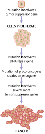
Carcinogenesis, also called oncogenesis or tumorigenesis, is the formation of a cancer, whereby normal cell (biology), cells are malignant transformation, transformed into cancer cells. The process is characterized by changes at the cellular, Genetics, genetic, and Epigenetics, epigenetic levels and abnormal cell division. Cell division is a physiological process that occurs in almost all Tissue (biology), tissues and under a variety of circumstances. Normally, the balance between proliferation and programmed cell death, in the form of apoptosis, is maintained to ensure the integrity of tissues and Organ (anatomy), organs. According to the prevailing accepted theory of carcinogenesis, the somatic mutation theory, mutations in DNA and Epigenetics, epimutations that lead to cancer disrupt these orderly processes by interfering with the programming regulating the processes, upsetting the normal balance between proliferation and cell death. This results in uncontrolled cell division ... [...More Info...] [...Related Items...] OR: [Wikipedia] [Google] [Baidu] |
Nephrogenesis
Kidney development, or nephrogenesis, describes the embryologic origins of the kidney, a major organ in the urinary system. This article covers a 3 part developmental process that is observed in most reptiles, birds and mammals, including humans. Nephrogenesis is often considered in the broader context of the development of the urinary and reproductive organs. Phases The development of the kidney proceeds through a series of successive phases, each marked by the development of a more advanced kidney: the archinephros, pronephros, mesonephros, and metanephros. The pronephros is the most immature form of kidney, while the metanephros is most developed. The metanephros persists as the definitive adult kidney. Archinephros The archinephros is considered as hypothetical or primitive kidney. Pronephros The pronephros develops in the cervical region of the embryo. During approximately day 22 of human gestation, the paired pronephri appears towards the cranial end of the intermediate me ... [...More Info...] [...Related Items...] OR: [Wikipedia] [Google] [Baidu] |
Somitogenesis
Somitogenesis is the process by which somites form. Somites are bilaterally paired blocks of paraxial mesoderm that form along the anterior-posterior axis of the developing embryo in vertebrates. The somites give rise to skeletal muscle, cartilage, tendons, endothelium, and dermis. Overview During somitogenesis, somites form from the pre-somitic mesoderm, a region of mesoderm at the posterior of the developing embryo. This tissue undergoes convergent extension as the primitive streak regresses, or as the embryo gastrulates. The notochord extends from the base of the head to the tail; with it extend thick bands of paraxial mesoderm. As the primitive streak continues to regress, somites form from the pre-somitic mesoderm by 'budding off' periodically from the anterior end of the pre-somitic mesoderm. The underlying developmental signals controlling this periodic formation are thought to conform to a clock-wavefront model. These immature somites then are compacted into an oute ... [...More Info...] [...Related Items...] OR: [Wikipedia] [Google] [Baidu] |
Embryonic Development
In developmental biology, animal embryonic development, also known as animal embryogenesis, is the developmental stage of an animal embryo. Embryonic development starts with the fertilization of an egg cell (ovum) by a sperm, sperm cell (spermatozoon). Once fertilized, the ovum becomes a single diploid cell known as a zygote. The zygote undergoes mitosis, mitotic cell division, divisions with no significant growth (a process known as cleavage (embryo), cleavage) and cellular differentiation, leading to development of a multicellular embryo after passing through an organizational checkpoint during mid-embryogenesis. In mammals, the term refers chiefly to the early stages of prenatal development, whereas the terms fetus and fetal development describe later stages. The main stages of animal embryonic development are as follows: * The zygote undergoes a series of cell divisions (called cleavage) to form a structure called a morula. * The morula develops into a structure called a bla ... [...More Info...] [...Related Items...] OR: [Wikipedia] [Google] [Baidu] |


