|
Maxillary First Premolar
The maxillary first premolar is one of two premolars that exist in the maxilla. Premolars are only found in the adult dentition and typically erupt at the age of 10–11, replacing the first molars in primary dentition. The maxillary first premolar is located behind the canine and in front of the second premolar. Its function is to bite and chew food. Notation For Palmer notation, the right maxillary premolar is known as “4⏌” and the left maxillary premolar is known as “⎿4”. For FDI notation, the right maxillary premolar is known as “14” and the left maxillary premolar is known as “24”. Tooth morphology The maxillary first premolar has two well-defined cusps, a buccal cusp and a lingual cusp, with the buccal cusp often 1mm taller than the lingual cusp, giving the tooth an angular crown. This crown is broader buccolingually than mesiodistally, resulting in a characteristic hexagonal or six-sided shape when viewed from the occlusal aspect. It is typically ... [...More Info...] [...Related Items...] OR: [Wikipedia] [Google] [Baidu] |
Premolar
The premolars, also called premolar Tooth (human), teeth, or bicuspids, are transitional teeth located between the Canine tooth, canine and Molar (tooth), molar teeth. In humans, there are two premolars per dental terminology#Quadrant, quadrant in the permanent teeth, permanent set of teeth, making eight premolars total in the mouth. They have at least two Cusp (dentistry), cusps. Premolars can be considered transitional teeth during chewing, or mastication. They have properties of both the canines, that lie anterior and molars that lie Posterior (anatomy), posterior, and so food can be transferred from the canines to the premolars and finally to the molars for grinding, instead of directly from the canines to the molars. Human anatomy The premolars in humans are the maxillary first premolar, maxillary second premolar, mandibular first premolar, and the mandibular second premolar. Premolar teeth by definition are permanent teeth Anatomical terms of location#Proximal and distal, ... [...More Info...] [...Related Items...] OR: [Wikipedia] [Google] [Baidu] |
Incisor
Incisors (from Latin ''incidere'', "to cut") are the front teeth present in most mammals. They are located in the premaxilla above and on the mandible below. Humans have a total of eight (two on each side, top and bottom). Opossums have 18, whereas armadillos, anteaters and other animals in the order Edentata have none. Structure Adult humans normally have eight incisors, two of each type. The types of incisors are: * maxillary central incisor (upper jaw, closest to the center of the lips) * maxillary lateral incisor (upper jaw, beside the maxillary central incisor) * mandibular central incisor (lower jaw, closest to the center of the lips) * mandibular lateral incisor (lower jaw, beside the mandibular central incisor) Children with a full set of deciduous teeth (primary teeth) also have eight incisors, named the same way as in permanent teeth. Young children may have from zero to eight incisors depending on the stage of their tooth eruption and tooth development. Typic ... [...More Info...] [...Related Items...] OR: [Wikipedia] [Google] [Baidu] |
Clinical Attachment Loss
Clinical attachment loss (CAL) is the predominant clinical manifestation and determinant of periodontal disease. Anatomy of the attachment Teeth are attached to the surrounding and supporting alveolar bone by periodontal ligament (PDL) fibers; these fibers run from the bone into the cementum that naturally exists on the entire root surface of teeth. They are also attached to the gingival (gum) tissue that covers the alveolar bone by an attachment apparatus; because this attachment exists superficial to the crest, or height, of the alveolar bone, it is termed the ''supracrestal attachment apparatus''. The supracrestal attachment apparatus is composed of two layers: the coronal junctional epithelium In dental anatomy, the junctional epithelium (JE) is that epithelium which lies at, and in health also defines, the base of the gingival sulcus (i.e. where the gums attach to a tooth). The probing depth of the gingival sulcus is measured by a cal ... and the more apical gingival c ... [...More Info...] [...Related Items...] OR: [Wikipedia] [Google] [Baidu] |
Periodontal Disease
Periodontal disease, also known as gum disease, is a set of inflammatory conditions affecting the tissues surrounding the teeth. In its early stage, called gingivitis, the gums become swollen and red and may bleed. It is considered the main cause of tooth loss for adults worldwide. In its more serious form, called periodontitis, the gums can pull away from the tooth, bone can be lost, and the teeth may loosen or fall out. Halitosis (bad breath) may also occur. Periodontal disease typically arises from the development of plaque biofilm, which harbors harmful bacteria such as ''Porphyromonas gingivalis'' and ''Treponema denticola''. These bacteria infect the gum tissue surrounding the teeth, leading to inflammation and, if left untreated, progressive damage to the teeth and gum tissue. Recent meta-analysis have shown that the composition of the oral microbiota and its response to periodontal disease differ between men and women. These differences are particularly notable in t ... [...More Info...] [...Related Items...] OR: [Wikipedia] [Google] [Baidu] |
Calculus (dental)
In dentistry, calculus or tartar is a form of hardened dental plaque. It is caused by precipitation of minerals from saliva and Gingival sulcus, gingival crevicular fluid (GCF) in plaque on the teeth. This process of precipitation kills the bacterial cells within dental plaque, but the rough and hardened surface that is formed provides an ideal surface for further plaque formation. This leads to calculus buildup, which compromises the health of the gingiva (gums). Calculus can form both along the gumline, where it is referred to as supragingival (), and within the narrow Sulcus (morphology), sulcus that exists between the teeth and the gingiva, where it is referred to as subgingival (). Calculus formation is associated with a number of clinical manifestations, including halitosis, bad breath, receding gums and Chronic (medicine), chronically inflamed gingiva. Brushing and flossing can remove plaque from which calculus forms; however, once formed, calculus is too hard (firmly att ... [...More Info...] [...Related Items...] OR: [Wikipedia] [Google] [Baidu] |
Dental Plaque
Dental plaque is a biofilm of microorganisms (mostly bacteria, but also fungi) that grows on surfaces within the mouth. It is a sticky colorless deposit at first, but when it forms Calculus (dental), tartar, it is often brown or pale yellow. It is commonly found between the teeth, on the front of teeth, behind teeth, on chewing surfaces, along the gums, gumline (supragingival), or below the gumline cervical margins (subgingival). Dental plaque is also known as microbial plaque, oral biofilm, dental biofilm, dental plaque biofilm or bacterial plaque biofilm. Bacterial plaque is one of the major causes for dental decay and gum disease. It has been observed that differences in the composition of dental plaque microbiota exist between men and women, particularly in the presence of periodontal disease, periodontitis. Progression and build-up of dental plaque can give rise to tooth decay – the localised destruction of the tissues of the tooth by acid produced from the bacterial degrad ... [...More Info...] [...Related Items...] OR: [Wikipedia] [Google] [Baidu] |
Cementoenamel Junction
In dental anatomy, the cementoenamel junction (CEJ) is the location where the enamel, which covers the anatomical crown of a tooth, and the cementum, which covers the anatomical root of a tooth, meet. Informally it is known as the neck of the tooth. The border created by these two dental tissues has much significance as it is usually the location where the gingiva (gums) attaches to a healthy tooth by fibers called the gingival fibers. Active recession of the gingiva reveals the cementoenamel junction in the mouth and is usually a sign of an unhealthy condition. The loss of attachment is considered a more reliable indicator of periodontal disease. The CEJ is the site of major tooth resorption. A significant proportion of tooth loss is caused by tooth resorption, which occurs in 5 to 10 percent of the population. The clinical location of CEJ which is a static landmark, serves as a crucial anatomical site for the measurement of probing pocket depth (PPD) and clinical attachment l ... [...More Info...] [...Related Items...] OR: [Wikipedia] [Google] [Baidu] |
Molar (tooth)
The molars or molar teeth are large, flat teeth at the back of the mouth. They are more developed in mammals. They are used primarily to grind food during chewing. The name ''molar'' derives from Latin, ''molaris dens'', meaning "millstone tooth", from ''mola'', millstone and ''dens'', tooth. Molars show a great deal of diversity in size and shape across the mammal groups. The third molar of humans is sometimes vestigial. Human anatomy In humans, the molar teeth have either four or five cusps. Adult humans have 12 molars, in four groups of three at the back of the mouth. The third, rearmost molar in each group is called a wisdom tooth. It is the last tooth to appear, breaking through the front of the gum at about the age of 20, although this varies among individuals and populations, and in many cases the tooth is missing. The human mouth contains upper (maxillary) and lower (mandibular) molars. They are: maxillary first molar, maxillary second molar, maxillary third mol ... [...More Info...] [...Related Items...] OR: [Wikipedia] [Google] [Baidu] |
Tooth Enamel
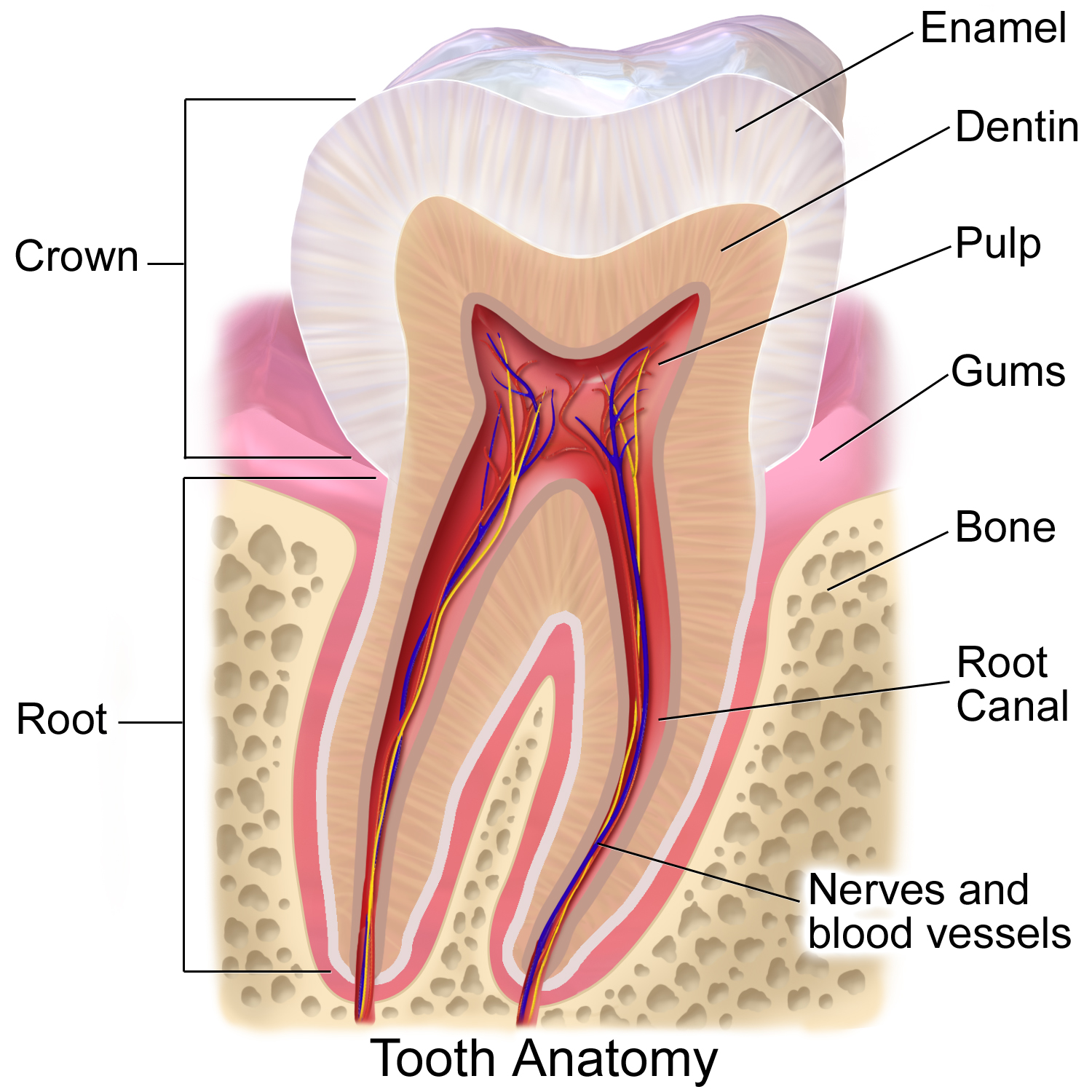
Tooth enamel is one of the four major Tissue (biology), tissues that make up the tooth in humans and many animals, including some species of fish. It makes up the normally visible part of the tooth, covering the Crown (tooth), crown. The other major tissues are dentin, cementum, and Pulp (tooth), dental pulp. It is a very hard, white to off-white, highly mineralised substance that acts as a barrier to protect the tooth but can become susceptible to degradation, especially by acids from food and drink. In rare circumstances enamel fails to form, leaving the underlying dentin exposed on the surface. Features Enamel is the hardest substance in the human body and contains the highest percentage of minerals (at 96%),Ross ''et al.'', p. 485 with water and organic material composing the rest.Ten Cate's Oral Histology, Nancy, Elsevier, pp. 70–94 The primary mineral is hydroxyapatite, which is a crystalline calcium phosphate. Enamel is formed on the tooth while the tooth develops wit ... [...More Info...] [...Related Items...] OR: [Wikipedia] [Google] [Baidu] |
Calcification
Calcification is the accumulation of calcium salts in a body tissue. It normally occurs in the formation of bone, but calcium can be deposited abnormally in soft tissue,Miller, J. D. Cardiovascular calcification: Orbicular origins. ''Nature Materials'' 12, 476-478 (2013). causing it to harden. Calcifications may be classified on whether there is mineral balance or not, and the location of the calcification. Calcification may also refer to the processes of normal mineral deposition in biological systems, such as the formation of stromatolites or mollusc shells (see Biomineralization). Signs and symptoms Calcification can manifest itself in many ways in the body depending on the location. In the pulpal structure of a tooth, calcification often presents asymptomatically, and is diagnosed as an incidental finding during radiographic interpretation. Individual teeth with calcified pulp will typically respond negatively to vitality testing; teeth with calcified pulp often lack ... [...More Info...] [...Related Items...] OR: [Wikipedia] [Google] [Baidu] |
Pulp (tooth)
The pulp is the connective tissue, nerves, blood vessels, and odontoblasts that comprise the innermost layer of a tooth. The pulp's activity and signalling processes regulate its behaviour. Anatomy The pulp is the neurovascular bundle central to each tooth, permanent tooth, permanent or primary tooth, primary. It is composed of a central pulp chamber, pulp horns, and radicular canals. The large mass of the pulp is contained within the pulp chamber, which is contained in and mimics the overall shape of the crown of the tooth.Fehrenbach, MJ. and Popowics, T. (2026), ''Illustrated Dental Embryology, Histology, and Anatomy'', Elsevier, page 185-86. Because of the continuous deposition of the dentine, the pulp chamber becomes smaller with the age. This is not uniform throughout the coronal pulp but progresses faster on the floor than on the roof or sidewalls. Radicular pulp canals extend down from the cervical region of the crown to the Root apex (dental), root apex. They are not ... [...More Info...] [...Related Items...] OR: [Wikipedia] [Google] [Baidu] |
Maxilla
In vertebrates, the maxilla (: maxillae ) is the upper fixed (not fixed in Neopterygii) bone of the jaw formed from the fusion of two maxillary bones. In humans, the upper jaw includes the hard palate in the front of the mouth. The two maxillary bones are fused at the intermaxillary suture, forming the anterior nasal spine. This is similar to the mandible (lower jaw), which is also a fusion of two mandibular bones at the mandibular symphysis. The mandible is the movable part of the jaw. Anatomy Structure The maxilla is a paired bone - the two maxillae unite with each other at the intermaxillary suture. The maxilla consists of: * The body of the maxilla: pyramid-shaped; has an orbital, a nasal, an infratemporal, and a facial surface; contains the maxillary sinus. * Four processes: ** the zygomatic process ** the frontal process ** the alveolar process ** the palatine process It has three surfaces: * the anterior, posterior, medial Features of the maxilla include: * t ... [...More Info...] [...Related Items...] OR: [Wikipedia] [Google] [Baidu] |


