|
Lupus Band Test
Lupus band test is done upon skin biopsy, with direct immunofluorescence staining, in which, if positive, IgG and complement depositions are found at the dermoepidermal junction.Marks, James G; Miller, Jeffery (2006). ''Lookingbill and Marks' Principles of Dermatology'' (4th ed.). Elsevier Inc. . This test can be helpful in distinguishing systemic lupus erythematosus (SLE) from cutaneous lupus, because in SLE the lupus band test will be positive in both involved and uninvolved skin, whereas with cutaneous lupus only the involved skin will be positive. The minimum criteria for positivity are:Ther Clin Risk Manag. 2011; 7: 27–32. The lupus band test in systemic lupus erythematosus patients. Adam Reich, Katarzyna Marcinow, and Rafal Bialynicki-Birula * In sun-exposed skin: presence of a band of deposits of IgM along the epidermal basement membrane in 50% of the biopsy, intermediate (2+) intensity or more. * In sun-protected skin : presence of interrupted (i.e. less than 50%) de ... [...More Info...] [...Related Items...] OR: [Wikipedia] [Google] [Baidu] |
Histology
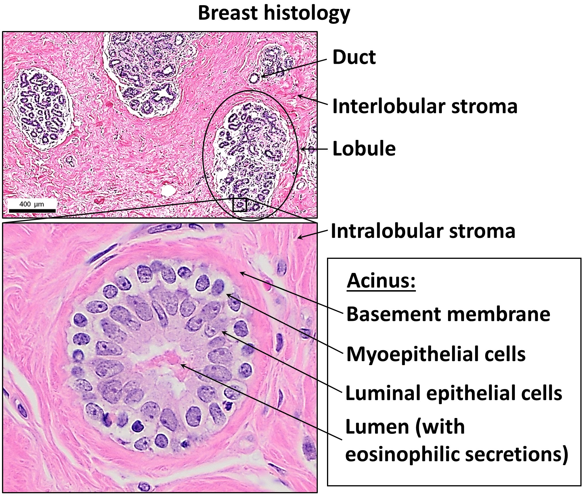
Histology, also known as microscopic anatomy or microanatomy, is the branch of biology that studies the microscopic anatomy of biological tissue (biology), tissues. Histology is the microscopic counterpart to gross anatomy, which looks at larger structures visible without a microscope. Although one may divide microscopic anatomy into ''organology'', the study of organs, ''histology'', the study of tissues, and ''cytology'', the study of cell (biology), cells, modern usage places all of these topics under the field of histology. In medicine, histopathology is the branch of histology that includes the microscopic identification and study of diseased tissue. In the field of paleontology, the term paleohistology refers to the histology of fossil organisms. Biological tissues Animal tissue classification There are four basic types of animal tissues: muscle tissue, nervous tissue, connective tissue, and epithelial tissue. All animal tissues are considered to be subtypes of these ... [...More Info...] [...Related Items...] OR: [Wikipedia] [Google] [Baidu] |
Direct Immunofluorescence
A direct fluorescent antibody (DFA or dFA), also known as "direct immunofluorescence", is an antibody that has been tagged in a direct fluorescent antibody test. Its name derives from the fact that it directly tests the presence of an antigen with the tagged antibody, unlike western blotting, which uses an indirect method of detection, where the primary antibody binds the target antigen, with a secondary antibody directed against the primary, and a tag attached to the secondary antibody. Commercial DFA testing kits are available, which contain fluorescently labelled antibodies, designed to specifically target unique antigens present in the bacteria or virus, but not present in mammals (Eukaryotes). This technique can be used to quickly determine if a subject has a specific viral or bacterial infection. In the case of respiratory viruses, many of which have similar broad symptoms, detection can be carried out using nasal wash samples from the subject with the suspected infection ... [...More Info...] [...Related Items...] OR: [Wikipedia] [Google] [Baidu] |
IGG Antibodies
Immunoglobulin G (IgG) is a type of antibody. Representing approximately 75% of serum antibodies in humans, IgG is the most common type of antibody found in blood circulation. IgG molecules are created and released by plasma B cells. Each IgG antibody has two paratopes. Function Antibodies are major components of humoral immunity. IgG is the main type of antibody found in blood and extracellular fluid, allowing it to control infection of body tissues. By binding many kinds of pathogens such as viruses, bacteria, and fungi, IgG protects the body from infection. It does this through several mechanisms: * IgG-mediated binding of pathogens causes their immobilization and binding together via agglutination; IgG coating of pathogen surfaces (known as opsonization) allows their recognition and ingestion by phagocytic immune cells leading to the elimination of the pathogen itself; * IgG activates the classical pathway of the complement system, a cascade of immune protein production ... [...More Info...] [...Related Items...] OR: [Wikipedia] [Google] [Baidu] |
Systemic Lupus Erythematosus
Lupus, formally called systemic lupus erythematosus (SLE), is an autoimmune disease in which the body's immune system mistakenly attacks healthy tissue in many parts of the body. Symptoms vary among people and may be mild to severe. Common symptoms include painful and swollen joints, fever, chest pain, hair loss, mouth ulcers, swollen lymph nodes, feeling tired, and a red rash which is most commonly on the face. Often there are periods of illness, called flares, and periods of remission during which there are few symptoms. Children up to 18 years old develop a more severe form of SLE termed childhood-onset systemic lupus erythematosus. The cause of SLE is not clear. It is thought to involve a combination of genetics and environmental factors. Among identical twins, if one is affected there is a 24% chance the other one will also develop the disease. Female sex hormones, sunlight, smoking, vitamin D deficiency, and certain infections are also believed to increa ... [...More Info...] [...Related Items...] OR: [Wikipedia] [Google] [Baidu] |
Basement Membrane
The basement membrane, also known as base membrane, is a thin, pliable sheet-like type of extracellular matrix that provides cell and tissue support and acts as a platform for complex signalling. The basement membrane sits between epithelial tissues including mesothelium and endothelium, and the underlying connective tissue. Structure As seen with the electron microscope, the basement membrane is composed of two layers, the basal lamina and the reticular lamina. The underlying connective tissue attaches to the basal lamina with collagen VII anchoring fibrils and fibrillin microfibrils. The basal lamina layer can further be subdivided into two layers based on their visual appearance in electron microscopy. The lighter-colored layer closer to the epithelium is called the lamina lucida, while the denser-colored layer closer to the connective tissue is called the lamina densa. The electron-dense lamina densa layer is about 30–70 nanometers thick and consists of an und ... [...More Info...] [...Related Items...] OR: [Wikipedia] [Google] [Baidu] |
Cell Nucleus
The cell nucleus (; : nuclei) is a membrane-bound organelle found in eukaryote, eukaryotic cell (biology), cells. Eukaryotic cells usually have a single nucleus, but a few cell types, such as mammalian red blood cells, have #Anucleated_cells, no nuclei, and a few others including osteoclasts have Multinucleate, many. The main structures making up the nucleus are the nuclear envelope, a double membrane that encloses the entire organelle and isolates its contents from the cellular cytoplasm; and the nuclear matrix, a network within the nucleus that adds mechanical support. The cell nucleus contains nearly all of the cell's genome. Nuclear DNA is often organized into multiple chromosomes – long strands of DNA dotted with various proteins, such as histones, that protect and organize the DNA. The genes within these chromosomes are Nuclear organization, structured in such a way to promote cell function. The nucleus maintains the integrity of genes and controls the activities of the ... [...More Info...] [...Related Items...] OR: [Wikipedia] [Google] [Baidu] |
Epidermis (skin)
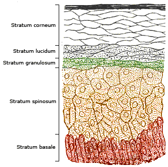
The epidermis is the outermost of the three layers that comprise the skin, the inner layers being the dermis and hypodermis. The epidermal layer provides a barrier to infection from environmental pathogens and regulates the amount of water released from the body into the atmosphere through transepidermal water loss. The epidermis is composed of multiple layers of flattened cells that overlie a base layer ( stratum basale) composed of columnar cells arranged perpendicularly. The layers of cells develop from stem cells in the basal layer. The thickness of the epidermis varies from 31.2μm for the penis to 596.6μm for the sole of the foot with most being roughly 90μm. Thickness does not vary between the sexes but becomes thinner with age. The human epidermis is an example of epithelium, particularly a stratified squamous epithelium. The word epidermis is derived through Latin , itself and . Something related to or part of the epidermis is termed epidermal. Structure ... [...More Info...] [...Related Items...] OR: [Wikipedia] [Google] [Baidu] |
Skin Biopsy
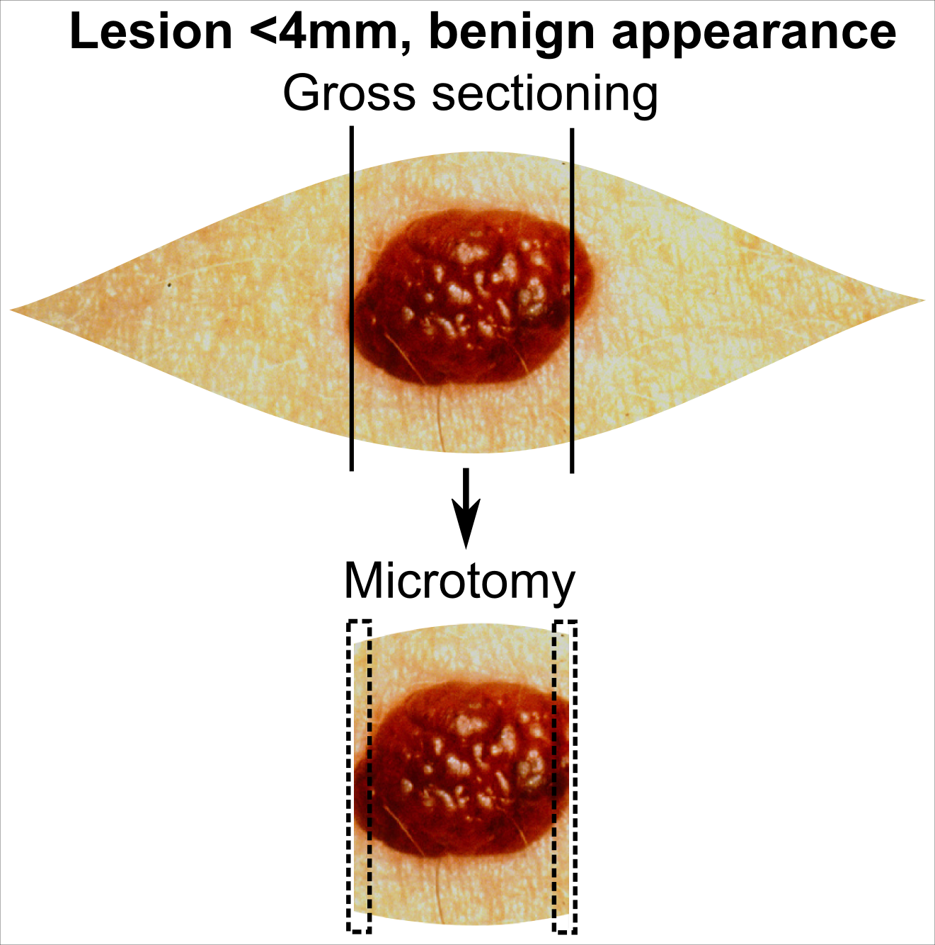
Skin biopsy is a biopsy technique in which a skin lesion is removed to be sent to a pathologist to render a microscopic diagnosis. It is usually done under local anesthetic in a physician's office, and results are often available in 4 to 10 days. It is commonly performed by dermatologists. Skin biopsies are also done by family physicians, internists, surgeons, and other specialties. However, performed incorrectly, and without appropriate clinical information, a pathologist's interpretation of a skin biopsy can be severely limited, and therefore doctors and patients may forgo traditional biopsy techniques and instead choose Mohs surgery. There are four main types of skin biopsies: shave biopsy, punch biopsy, excisional biopsy, and incisional biopsy. The choice of the different skin biopsies is dependent on the suspected diagnosis of the skin lesion. Like most biopsies, patient consent and anesthesia (usually lidocaine injected into the skin) are prerequisites. Types Shave ... [...More Info...] [...Related Items...] OR: [Wikipedia] [Google] [Baidu] |
Immunoglobulin G
Immunoglobulin G (IgG) is a type of antibody. Representing approximately 75% of serum antibodies in humans, IgG is the most common type of antibody found in blood circulation. IgG molecules are created and released by plasma B cells. Each IgG antibody has two paratopes. Function Antibodies are major components of humoral immunity. IgG is the main type of antibody found in blood and extracellular fluid, allowing it to control infection of body tissues. By binding many kinds of pathogens such as viruses, bacteria, and fungi, IgG protects the body from infection. It does this through several mechanisms: * IgG-mediated binding of pathogens causes their immobilization and binding together via agglutination; IgG coating of pathogen surfaces (known as opsonization) allows their recognition and ingestion by phagocytic immune cells leading to the elimination of the pathogen itself; * IgG activates the classical pathway of the complement system, a cascade of immune protein product ... [...More Info...] [...Related Items...] OR: [Wikipedia] [Google] [Baidu] |
Complement System (immunology)
The complement system, also known as complement cascade, is a part of the humoral, innate immune system and enhances (complements) the ability of antibodies and phagocytic cells to clear microbes and damaged cells from an organism, promote inflammation, and attack the pathogen's cell membrane. Despite being part of the innate immune system, the complement system can be recruited and brought into action by antibodies generated by the adaptive immune system. The complement system consists of a number of small, inactive, liver synthesized protein precursors circulating in the blood. When stimulated by one of several triggers, proteases in the system cleave specific proteins to release cytokines and initiate an amplifying cascade of further cleavages. The end result of this ''complement activation'' or ''complement fixation'' cascade is stimulation of phagocytes to clear foreign and damaged material, inflammation to attract additional phagocytes, and activation of the cell-killing ... [...More Info...] [...Related Items...] OR: [Wikipedia] [Google] [Baidu] |
Dermoepidermal Junction
The dermoepidermal junction or dermal-epidermal junction (DEJ) is the interface between the epidermal and the dermal layers of the skin. The basal cells of the epidermis connect to the basement membrane by the anchoring filaments of hemidesmosomes; the cells of the papillary layer of the dermis are attached to the basement membrane by anchoring fibrils, which consist of type VII collagen. Clinical significance * Interface dermatitis includes conditions which primarily involve the dermoepidermal junction. * Stevens–Johnson syndrome and toxic epidermal necrolysis Toxic epidermal necrolysis (TEN), also known as Lyell's syndrome, is a type of severe skin reaction. Together with Stevens–Johnson syndrome (SJS) it forms a spectrum of disease, with TEN being more severe. Early symptoms include fever and f ... are diseases where there is a breakdown of the dermoepidermal junction. References Skin anatomy {{dermatology-stub ... [...More Info...] [...Related Items...] OR: [Wikipedia] [Google] [Baidu] |
Complement Component 4
Complement component 4 (C4), in humans, is a protein involved in the intricate complement system, originating from the human leukocyte antigen (HLA) system. It serves a number of critical functions in immunity, tolerance, and autoimmunity with the other numerous components. Furthermore, it is a crucial factor in connecting the recognition pathways of the overall system instigated by antibody-antigen (Ab-Ag) complexes to the other effector proteins of the innate immune response. For example, the severity of a dysfunctional complement system can lead to fatal diseases and infections. Complex variations of it can also lead to schizophrenia. The C4 protein was thought to derive from a simple two-locus allelic model, which however has been replaced by a much more sophisticated multimodular RCCX gene complex model which contain long and short forms of the C4A or C4B genes usually in tandem RCCX cassettes with copy number variation, that somewhat parallels variation in the levels of the ... [...More Info...] [...Related Items...] OR: [Wikipedia] [Google] [Baidu] |


