|
Leukocoria
Leukocoria (also white pupillary reflex) is an abnormal white reflection from the retina of the eye. Leukocoria resembles eyeshine, but leukocoria can also occur in animals that lack eyeshine because their retina lacks a ''tapetum lucidum''. Leukocoria is a medical sign for a number of conditions, including Coats disease, congenital cataract, corneal scarring, melanoma of the ciliary body, Norrie disease, ocular toxocariasis, persistence of the tunica vasculosa lentis (PFV/PHPV), retinoblastoma, and retrolental fibroplasia. Because of the potentially life-threatening nature of retinoblastoma, a cancer, that condition is usually considered in the evaluation of leukocoria. In some rare cases (1%) the leukocoria is caused by Coats' disease (leaking retinal vessels). Diagnosis On photographs taken using a flash, instead of the familiar red-eye effect, leukocoria can cause a bright white reflection in an affected eye. Leukocoria may appear also in low indirect light, similar to e ... [...More Info...] [...Related Items...] OR: [Wikipedia] [Google] [Baidu] |
Retinoblastoma
Retinoblastoma (Rb) is a rare form of cancer that rapidly develops from the immature cells of a retina, the light-detecting tissue of the eye. It is the most common primary malignant intraocular cancer in children, and it is almost exclusively found in young children. Though most children survive this cancer, they may lose their vision in the affected eye(s) or need to have the eye removed. Almost half of children with retinoblastoma have a hereditary genetic defect associated with retinoblastoma. In other cases, it is caused by a congenital mutation in the chromosome 13 gene 13q14 ( retinoblastoma protein). Signs and symptoms Retinoblastoma is universally known as the most intrusive intraocular cancer among children. The chance of survival and preservation of the eye depends fully on the severity. Retinoblastoma is extremely rare as there are only about 200 to 300 cases every year in the United States. Looking at retinoblastoma globally, only 1 in about 15,000 children have ... [...More Info...] [...Related Items...] OR: [Wikipedia] [Google] [Baidu] |
Norrie Disease
Norrie disease is a rare disease and genetic disorder that primarily affects the eyes and almost always leads to blindness. It is caused by mutations in the ''Norrin cystine knot growth factor (NDP)'' gene, which is located on the X chromosome. In addition to the congenital ocular symptoms, the majority of patients experience a progressive hearing loss starting mostly in their 2nd decade of life, and some may have learning difficulties among other additional characteristics. Patients with Norrie disease may develop cataracts, leukocoria (where the pupils appear white when light is shone on them), along with other developmental issues in the eye, such as shrinking of the globe and the wasting away of the iris. Around 30 to 50% of them will also have developmental delay or learning difficulties, psychotic-like features, incoordination of movements or behavioral abnormalities. Most patients are born with normal hearing; however, the onset of hearing loss is very common in early ado ... [...More Info...] [...Related Items...] OR: [Wikipedia] [Google] [Baidu] |
Coats' Disease
Coats' disease is a rare congenital, nonhereditary eye disorder, causing full or partial blindness, characterized by abnormal development of blood vessels behind the retina. Coats' disease can also fall under glaucoma. It can have a similar presentation to that of retinoblastoma. Signs and symptoms The most common sign at presentation is leukocoria (abnormal white reflection of the retina). Symptoms typically begin as blurred vision, usually pronounced when one eye is closed (due to the unilateral nature of the disease). Often the unaffected eye will compensate for the loss of vision in the other eye; however, this results in some loss of depth perception and parallax. Deterioration of sight may begin in either the central or peripheral vision. Deterioration is likely to begin in the upper part of the vision field as this corresponds with the bottom of the eye where blood usually pools. Flashes of light, known as photopsia, and floaters are common symptoms. Persistent co ... [...More Info...] [...Related Items...] OR: [Wikipedia] [Google] [Baidu] |
Tapetum Lucidum
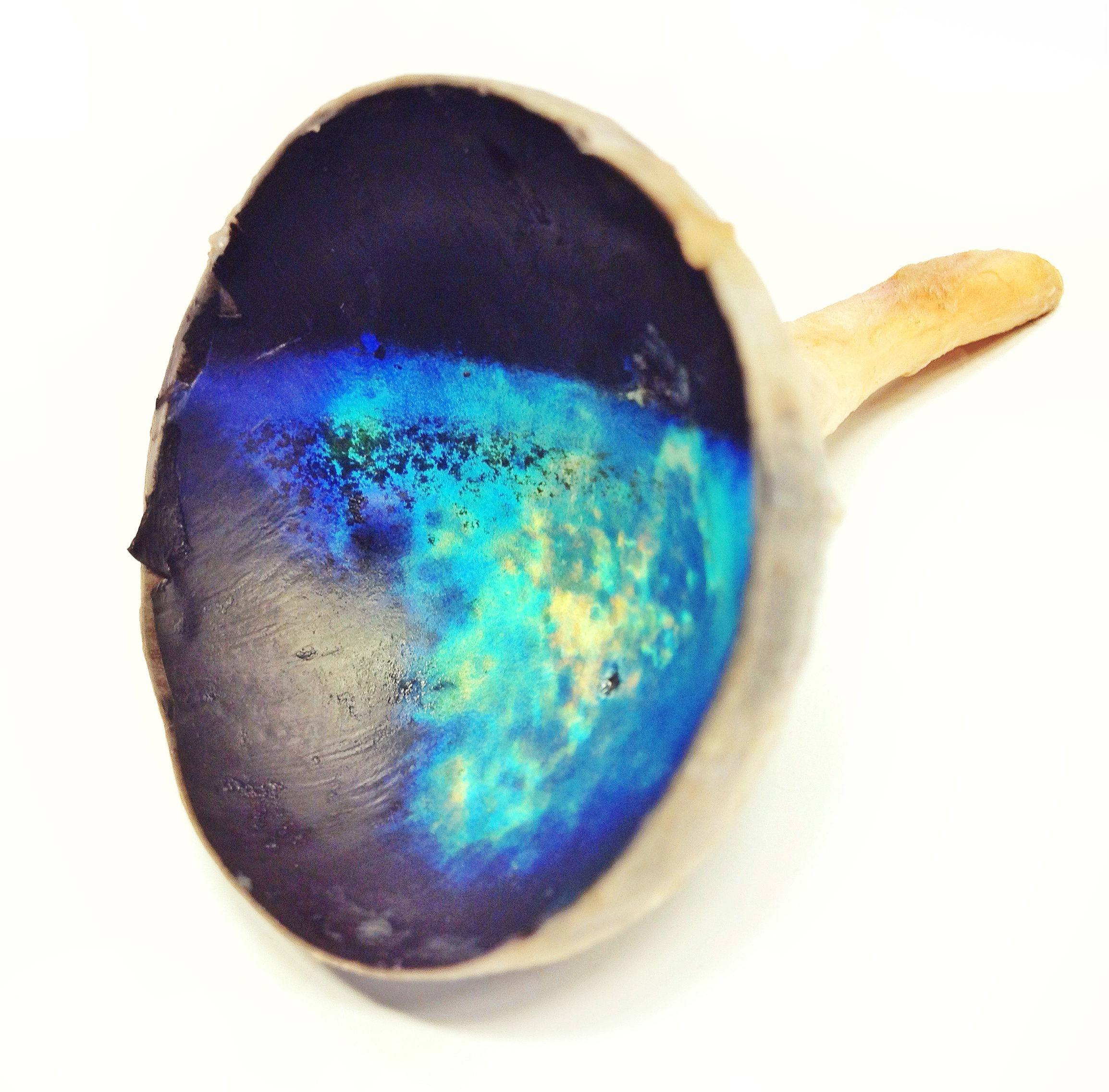
The ''tapetum lucidum'' ( ; ; ) is a layer of tissue in the eye of many vertebrates and some other animals. Lying immediately behind the retina, it is a retroreflector. It reflects visible light back through the retina, increasing the light available to the photoreceptors (although slightly blurring the image). The tapetum lucidum contributes to the superior night vision of some animals. Many of these animals are nocturnal, especially carnivores, while others are deep sea animals. Similar adaptations occur in some species of spiders. Haplorhine primates, including humans, are diurnal and lack a ''tapetum lucidum''. Function and mechanism Presence of a ''tapetum lucidum'' enables animals to see in dimmer light than would otherwise be possible. The ''tapetum lucidum'', which is iridescent, reflects light roughly on the interference principles of thin-film optics, as seen in other iridescent tissues. However, the ''tapetum lucidum'' cells are leucophores, not iridophores ... [...More Info...] [...Related Items...] OR: [Wikipedia] [Google] [Baidu] |
Coats Disease
Coats' disease is a rare congenital, nonhereditary eye disorder, causing full or partial blindness, characterized by abnormal development of blood vessels behind the retina. Coats' disease can also fall under glaucoma. It can have a similar presentation to that of retinoblastoma. Signs and symptoms The most common sign at presentation is leukocoria (abnormal white reflection of the retina). Symptoms typically begin as blurred vision, usually pronounced when one eye is closed (due to the unilateral nature of the disease). Often the unaffected eye will compensate for the loss of vision in the other eye; however, this results in some loss of depth perception and parallax. Deterioration of sight may begin in either the central or peripheral vision. Deterioration is likely to begin in the upper part of the vision field as this corresponds with the bottom of the eye where blood usually pools. Flashes of light, known as photopsia, and floaters are common symptoms. Persistent color ... [...More Info...] [...Related Items...] OR: [Wikipedia] [Google] [Baidu] |
Red Reflex
The red reflex refers to the reddish-orange reflection of light from the back of the eye, or fundus, observed when using an ophthalmoscope or retinoscope. The reflex relies on the transparency of optical media (tear film, cornea, aqueous humor, crystalline lens, vitreous humor) and reflects off the fundus back through media into the aperture of the ophthalmoscope. The red reflex is considered abnormal if there is any asymmetry between the eyes, dark spots, or white reflex ( Leukocoria). Generally, it is a physical exam done on neonates and children by healthcare providers but occasionally occurs in flash photography seen when the pupil does not have enough time to constrict and reflects the fundus known as the red-eye effect. This is a recommended screening by the American Academy of Pediatrics and American Academy of Family Physicians for neonates and children at every office visit. The objective is to detect ocular pathology that needs early intervention and ophthalmology ... [...More Info...] [...Related Items...] OR: [Wikipedia] [Google] [Baidu] |
Red-eye Effect
The red-eye effect in photography is the common appearance of red pupils in color photographs of the eyes of humans and several other animals. It occurs when using a photographic flash that is very close to the camera lens (as with most compact cameras) in ambient low light. Causes In flash photography the light of the flash occurs too fast for the pupil to close, so much of the very bright light from the flash passes into the eye through the pupil, reflects off the fundus at the back of the eyeball and out through the pupil. The camera records this reflected light. The main cause of the red color is the ample amount of blood in the choroid which nourishes the back of the eye and is behind the retina. The blood in the retinal circulation is far less than in the choroid, and plays virtually no role. The eye contains several photostable pigments that all absorb in the short wavelength region, and hence contribute somewhat to the red eye effect. The lens cuts off deep blue and v ... [...More Info...] [...Related Items...] OR: [Wikipedia] [Google] [Baidu] |
Ophthalmology
Ophthalmology ( ) is a surgical subspecialty within medicine that deals with the diagnosis and treatment of eye disorders. An ophthalmologist is a physician who undergoes subspecialty training in medical and surgical eye care. Following a medical degree, a doctor specialising in ophthalmology must pursue additional postgraduate residency training specific to that field. This may include a one-year integrated internship that involves more general medical training in other fields such as internal medicine or general surgery. Following residency, additional specialty training (or fellowship) may be sought in a particular aspect of eye pathology. Ophthalmologists prescribe medications to treat eye diseases, implement laser therapy, and perform surgery when needed. Ophthalmologists provide both primary and specialty eye care - medical and surgical. Most ophthalmologists participate in academic research on eye diseases at some point in their training and many include research as p ... [...More Info...] [...Related Items...] OR: [Wikipedia] [Google] [Baidu] |
The Collection (film)
''The Collection'' is a 2012 American horror film, serving as the sequel to the 2009 film, ''The Collector''. Written by Patrick Melton and Marcus Dunstan, and directed by Dunstan, the film stars Randall Archer, Emma Fitzpatrick, Christopher McDonald, Lee Tergesen, and Josh Stewart, who reprises his role as Arkin from the first film. The story follows a young woman who gets captured by The Collector, while Arkin escapes but is recruited shortly after by a group of mercenaries whose mission is to save her at the Collector's base. ''The Collection'' was released on November 30, 2012, by LD Entertainment. The film received generally negative reviews by critics, who deemed it a slight improvement over its predecessor. A third film, ''The Collected'', was in production in 2019 but was suspended shortly after and shelved indefinitely. Plot A few weeks after the events of the first film, teenager Elena Peters and her friends, Missy and Josh, go to a party. Elena witnesses her boyfri ... [...More Info...] [...Related Items...] OR: [Wikipedia] [Google] [Baidu] |
The Collector (2009 Film)
''The Collector'' is a 2009 American horror film written by Patrick Melton and Marcus Dunstan, and directed by Dunstan. It stars Josh Stewart, Michael Reilly Burke, Andrea Roth, Juan Fernandez, Karley Scott Collins, Madeline Zima, and Robert Wisdom. The film follows a man who, in order to pay a debt, decides to rob a house, only to find out somebody with far more sinister intentions has already broken in. The original script, titled ''The Midnight Man'', was at one point shopped as a spin-off prequel to the ''Saw'' franchise, as an origin story for the villain John Kramer/Jigsaw, but the producers opposed the idea and dismissed it, leading to the script getting reworked to an original story. ''The Collector'' was released on July 31, 2009, by LD Entertainment. It received generally negative reviews from critics. A sequel, ''The Collection'', was released in 2012. Plot Married couple Larry and Gena Wharton return home to find the power is out. They discover a large trunk ups ... [...More Info...] [...Related Items...] OR: [Wikipedia] [Google] [Baidu] |
Fundus (eye)
The fundus of the eye is the interior surface of the eye opposite the lens and includes the retina, optic disc, macula, fovea, and posterior pole.Cassin, B. and Solomon, S. ''Dictionary of Eye Terminology''. Gainesville, Florida: Triad Publishing Company, 1990. The fundus can be examined by ophthalmoscopy and/or fundus photography. Variation The color of the fundus varies both between and within species. In one study of primates the retina is blue, green, yellow, orange, and red; only the human fundus (from a lightly pigmented blond person) is red. The major differences noted among the "higher" primate species were size and regularity of the border of macular area, size and shape of the optic disc, apparent 'texturing' of retina, and pigmentation of retina. Clinical significance Medical signs that can be detected from observation of eye fundus (generally by funduscopy) include hemorrhages, exudates, cotton wool spots, blood vessel abnormalities (tortuosity, pulsation an ... [...More Info...] [...Related Items...] OR: [Wikipedia] [Google] [Baidu] |


