|
Lactofuchsin Mount
A Lactofuchsin mount (also spelled Lacto-fuchsin or Lacto-Fuchsin) is a technique used for mounting fungi with hyphae on a microscope slide for examination. The main advantage of a lactofuchsin mount is that if performed correctly, it preserves the structure and arrangement of any hyphae that are present. Advantages To examine the hyphae of fungi under a microscope, a wet mount is essential. While this is possible to do with a water based mount, a better result can be obtained with lactofuchsin mounting fluid, which both sticks to the cell walls and colours the cell walls red in the process. Lactofuchsin, a 1% solution of basic fuchsine in lactic acid, dries much slower than water, so the slide may be preserved for a longer period, particularly if the edges of the finished slide are sealed, for example with clear nail polish. In addition, the refractive index of the fluid is significantly different to that of the cell walls, which provides a stronger visual contrast of the cell w ... [...More Info...] [...Related Items...] OR: [Wikipedia] [Google] [Baidu] |
Microscope Slide
A microscope slide is a thin flat piece of glass, typically 75 by 26 mm (3 by 1 inches) and about 1 mm thick, used to hold objects for examination under a microscope. Typically the object is mounted (secured) on the slide, and then both are inserted together in the microscope for viewing. This arrangement allows several slide-mounted objects to be quickly inserted and removed from the microscope, labeled, transported, and stored in appropriate slide cases or folders etc. Microscope slides are often used together with a cover slip or cover glass, a smaller and thinner sheet of glass that is placed over the specimen. Slides are held in place on the microscope's stage by slide clips, slide clamps or a cross-table which is used to achieve precise, remote movement of the slide upon the microscope's stage (such as in an automated/computer operated system, or where touching the slide with fingers is inappropriate either due to the risk of contamination or lack of precision ... [...More Info...] [...Related Items...] OR: [Wikipedia] [Google] [Baidu] |
Fungi
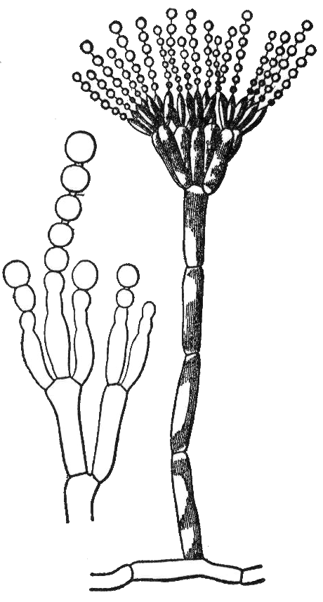
A fungus (plural, : fungi or funguses) is any member of the group of Eukaryote, eukaryotic organisms that includes microorganisms such as yeasts and Mold (fungus), molds, as well as the more familiar mushrooms. These organisms are classified as a Kingdom (biology), kingdom, separately from the other eukaryotic kingdoms, which by one traditional classification include Plantae, Animalia, Protozoa, and Chromista. A characteristic that places fungi in a different kingdom from plants, bacteria, and some protists is chitin in their cell walls. Fungi, like animals, are heterotrophs; they acquire their food by absorbing dissolved molecules, typically by secreting digestive enzymes into their environment. Fungi do not photosynthesize. Growth is their means of motility, mobility, except for spores (a few of which are flagellated), which may travel through the air or water. Fungi are the principal decomposers in ecological systems. These and other differences place fungi in a single gro ... [...More Info...] [...Related Items...] OR: [Wikipedia] [Google] [Baidu] |
Hyphae
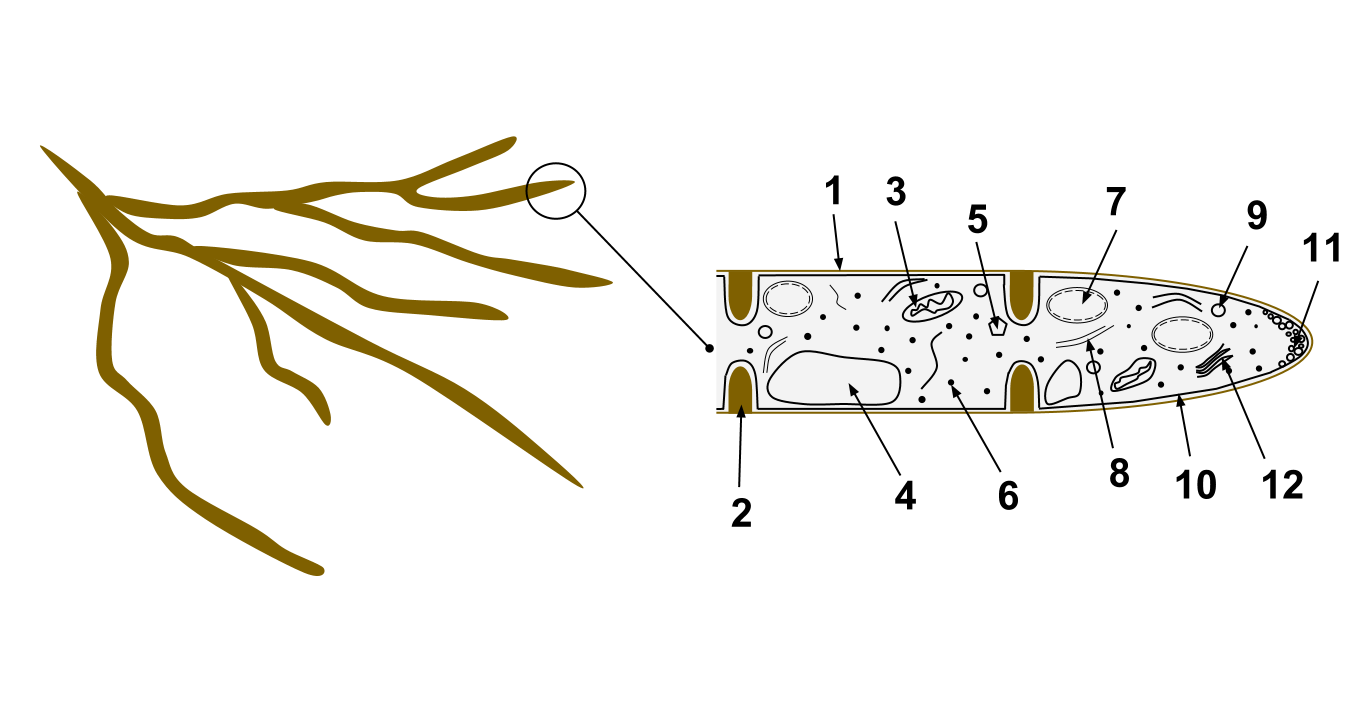
A hypha (; ) is a long, branching, filamentous structure of a fungus, oomycete, or actinobacterium. In most fungi, hyphae are the main mode of vegetative growth, and are collectively called a mycelium. Structure A hypha consists of one or more cells surrounded by a tubular cell wall. In most fungi, hyphae are divided into cells by internal cross-walls called "septa" (singular septum). Septa are usually perforated by pores large enough for ribosomes, mitochondria, and sometimes nuclei to flow between cells. The major structural polymer in fungal cell walls is typically chitin, in contrast to plants and oomycetes that have cellulosic cell walls. Some fungi have aseptate hyphae, meaning their hyphae are not partitioned by septa. Hyphae have an average diameter of 4–6 µm. Growth Hyphae grow at their tips. During tip growth, cell walls are extended by the external assembly and polymerization of cell wall components, and the internal production of new cell membrane. Th ... [...More Info...] [...Related Items...] OR: [Wikipedia] [Google] [Baidu] |
Lactofuchsin Mount
A Lactofuchsin mount (also spelled Lacto-fuchsin or Lacto-Fuchsin) is a technique used for mounting fungi with hyphae on a microscope slide for examination. The main advantage of a lactofuchsin mount is that if performed correctly, it preserves the structure and arrangement of any hyphae that are present. Advantages To examine the hyphae of fungi under a microscope, a wet mount is essential. While this is possible to do with a water based mount, a better result can be obtained with lactofuchsin mounting fluid, which both sticks to the cell walls and colours the cell walls red in the process. Lactofuchsin, a 1% solution of basic fuchsine in lactic acid, dries much slower than water, so the slide may be preserved for a longer period, particularly if the edges of the finished slide are sealed, for example with clear nail polish. In addition, the refractive index of the fluid is significantly different to that of the cell walls, which provides a stronger visual contrast of the cell w ... [...More Info...] [...Related Items...] OR: [Wikipedia] [Google] [Baidu] |
Microscope Slide
A microscope slide is a thin flat piece of glass, typically 75 by 26 mm (3 by 1 inches) and about 1 mm thick, used to hold objects for examination under a microscope. Typically the object is mounted (secured) on the slide, and then both are inserted together in the microscope for viewing. This arrangement allows several slide-mounted objects to be quickly inserted and removed from the microscope, labeled, transported, and stored in appropriate slide cases or folders etc. Microscope slides are often used together with a cover slip or cover glass, a smaller and thinner sheet of glass that is placed over the specimen. Slides are held in place on the microscope's stage by slide clips, slide clamps or a cross-table which is used to achieve precise, remote movement of the slide upon the microscope's stage (such as in an automated/computer operated system, or where touching the slide with fingers is inappropriate either due to the risk of contamination or lack of precision ... [...More Info...] [...Related Items...] OR: [Wikipedia] [Google] [Baidu] |
Cell Wall
A cell wall is a structural layer surrounding some types of cells, just outside the cell membrane. It can be tough, flexible, and sometimes rigid. It provides the cell with both structural support and protection, and also acts as a filtering mechanism. Cell walls are absent in many eukaryotes, including animals, but are present in some other ones like fungi, algae and plants, and in most prokaryotes (except mollicute bacteria). A major function is to act as pressure vessels, preventing over-expansion of the cell when water enters. The composition of cell walls varies between taxonomic group and species and may depend on cell type and developmental stage. The primary cell wall of land plants is composed of the polysaccharides cellulose, hemicelluloses and pectin. Often, other polymers such as lignin, suberin or cutin are anchored to or embedded in plant cell walls. Algae possess cell walls made of glycoproteins and polysaccharides such as carrageenan and agar that are ... [...More Info...] [...Related Items...] OR: [Wikipedia] [Google] [Baidu] |
Basic Fuchsine
Fuchsine (sometimes spelled fuchsin) or rosaniline hydrochloride is a magenta dye with chemical formula C20H19N3·HCl."Basic chemical data" ''Discovery Series'' online database, Developmental Therapeutics Program, U.S. National Institutes of Health. Retrieved on 2007-10-08. There are other similar chemical formulations of products sold as fuchsine, and several dozen other synonyms of this molecule. It becomes magenta when dissolved in ; as a , it forms dark |
Lactic Acid
Lactic acid is an organic acid. It has a molecular formula . It is white in the solid state and it is miscible with water. When in the dissolved state, it forms a colorless solution. Production includes both artificial synthesis as well as natural sources. Lactic acid is an alpha-hydroxy acid (AHA) due to the presence of a hydroxyl group adjacent to the carboxyl group. It is used as a synthetic intermediate in many organic synthesis industries and in various biochemical industries. The conjugate base of lactic acid is called lactate (or the lactate anion). The name of the derived acyl group is lactoyl. In solution, it can ionize by loss of a proton to produce the lactate ion . Compared to acetic acid, its p''K'' is 1 unit less, meaning lactic acid is ten times more acidic than acetic acid. This higher acidity is the consequence of the intramolecular hydrogen bonding between the α-hydroxyl and the carboxylate group. Lactic acid is chiral, consisting of two enantiomers. One ... [...More Info...] [...Related Items...] OR: [Wikipedia] [Google] [Baidu] |
Refractive Index
In optics, the refractive index (or refraction index) of an optical medium is a dimensionless number that gives the indication of the light bending ability of that medium. The refractive index determines how much the path of light is bent, or refracted, when entering a material. This is described by Snell's law of refraction, , where ''θ''1 and ''θ''2 are the angle of incidence and angle of refraction, respectively, of a ray crossing the interface between two media with refractive indices ''n''1 and ''n''2. The refractive indices also determine the amount of light that is reflected when reaching the interface, as well as the critical angle for total internal reflection, their intensity (Fresnel's equations) and Brewster's angle. The refractive index can be seen as the factor by which the speed and the wavelength of the radiation are reduced with respect to their vacuum values: the speed of light in a medium is , and similarly the wavelength in that medium is , where ''� ... [...More Info...] [...Related Items...] OR: [Wikipedia] [Google] [Baidu] |
Poison
Poison is a chemical substance that has a detrimental effect to life. The term is used in a wide range of scientific fields and industries, where it is often specifically defined. It may also be applied colloquially or figuratively, with a broad sense. Whether something is considered a poison may change depending on the amount, the circumstances, and what living things are present. Poisoning could be accidental or deliberate, and if the cause can be identified there may be ways to neutralise the effects or minimise the symptoms. In biology, a poison is a chemical substance causing death, injury or harm to organisms or their parts. In medicine, poisons are a kind of toxin that are delivered passively, not actively. In industry the term may be negative, something to be removed to make a thing safe, or positive, an agent to limit unwanted pests. In ecological terms, poisons introduced into the environment can later cause unwanted effects elsewhere, or in other parts of the fo ... [...More Info...] [...Related Items...] OR: [Wikipedia] [Google] [Baidu] |
Microscopy
Microscopy is the technical field of using microscopes to view objects and areas of objects that cannot be seen with the naked eye (objects that are not within the resolution range of the normal eye). There are three well-known branches of microscopy: optical, electron, and scanning probe microscopy, along with the emerging field of X-ray microscopy. Optical microscopy and electron microscopy involve the diffraction, reflection, or refraction of electromagnetic radiation/electron beams interacting with the specimen, and the collection of the scattered radiation or another signal in order to create an image. This process may be carried out by wide-field irradiation of the sample (for example standard light microscopy and transmission electron microscopy) or by scanning a fine beam over the sample (for example confocal laser scanning microscopy and scanning electron microscopy). Scanning probe microscopy involves the interaction of a scanning probe with the surface of the ... [...More Info...] [...Related Items...] OR: [Wikipedia] [Google] [Baidu] |


