|
Keratohyalin
Keratohyalin is a protein structure found in cytoplasmic granules of the keratinocytes in the stratum granulosum of the epidermis. Keratohyalin granules (KHG) mainly consist of keratin, profilaggrin, loricrin and trichohyalin proteins which contribute to cornification or keratinization, the process of the formation of epidermal cornified cell envelope. During the keratinocyte differentiation, these granules maturate and expand in size, which leads to the conversion of keratin tonofilaments into a homogenous keratin matrix, an important step in cornification. Keratohyalin granules can be divided in three classes: globular KHG (found in quickly dividing epithelia, such as the oral mucose), stellate KHG (found in the slowly dividing normal epidermis) and KHG of Hassall's corpuscles or type VI epithelioreticular cells of the thymus gland. The exact purpose of the keratinization of Hassall's corpuscles remains unknown. During skin differentiation process, keratohyaline granules disch ... [...More Info...] [...Related Items...] OR: [Wikipedia] [Google] [Baidu] |
Stratum Granulosum
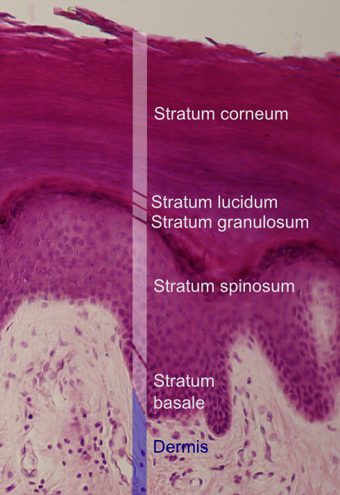
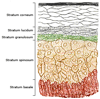
The stratum granulosum (or granular layer) is a thin layer of cells in the epidermis lying above the stratum spinosum and below the stratum corneum ( stratum lucidum on the soles and palms).James, William; Berger, Timothy; Elston, Dirk (2005) ''Andrews' Diseases of the Skin: Clinical Dermatology'' (10th ed.). Saunders. Page 2. . Keratinocytes migrating from the underlying stratum spinosum become known as granular cells in this layer. These cells contain keratohyalin granules, which are filled with histidine- and cysteine-rich proteins that appear to bind the keratin filaments together. Therefore, the main function of keratohyalin granules is to bind intermediate keratin filaments together.Marks, James G; Miller, Jeffery (2006). ''Lookingbill and Marks' Principles of Dermatology'' (4th ed.). Elsevier Inc. Page 7. . At the transition between this layer and the stratum corneum, cells secrete lamellar bodies (containing lipids and proteins) into the extracellular space. This results ... [...More Info...] [...Related Items...] OR: [Wikipedia] [Google] [Baidu] |
Filaggrin
Filaggrin (filament aggregating protein) is a filament-associated protein that binds to keratin fibers in epithelial cells. Ten to twelve filaggrin units are post-translationally hydrolyzed from a large profilaggrin precursor protein during terminal differentiation of epidermal cells. In humans, profilaggrin is encoded by the ''FLG'' gene, which is part of the S100 fused-type protein (SFTP) family within the epidermal differentiation complex on chromosome 1q21. In cetaceans and sirenians, the ''FLG'' family has lost its function, with the curious exception of manatees in the latter clade: manatees still retain some functional ''FLG'' genes. Profilaggrin Filaggrin monomers are tandemly clustered into a large, 350kDa protein precursor known as profilaggrin. In the epidermis, these structures are present in the keratohyalin granules in cells of the stratum granulosum. Profilaggrin undergoes proteolytic processing to yield individual filaggrin monomers at the transition between the ... [...More Info...] [...Related Items...] OR: [Wikipedia] [Google] [Baidu] |
Hassall's Corpuscles
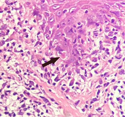
Hassall's corpuscles (also known as thymic bodies) are structures found in the medulla of the human thymus, formed from eosinophilic type VI thymic epithelial cells arranged concentrically. These concentric corpuscles are composed of a central mass, consisting of one or more granular cells, and of a capsule formed of epithelioid cells. They vary in size with diameters from 20 to more than 100μm, and tend to grow larger with age. They can be spherical or ovoid and their epithelial cells contain keratohyalin and bundles of cytoplasmic fibres. Later studies indicate that Hassall's corpuscles differentiate from medullary thymic epithelial cells after they lose autoimmune regulator (AIRE) expression. This makes them an example of Thymic mimetic cells. They are named for Arthur Hill Hassall, who discovered them in 1846. The function of Hassall's corpuscles is currently unclear, and the absence of this structure in the thymus of most murine species (except for the New Zealand White ... [...More Info...] [...Related Items...] OR: [Wikipedia] [Google] [Baidu] |
Keratinocyte
Keratinocytes are the primary type of cell found in the epidermis, the outermost layer of the skin. In humans, they constitute 90% of epidermal skin cells. Basal cells in the basal layer (''stratum basale'') of the skin are sometimes referred to as basal keratinocytes. Keratinocytes form a barrier against environmental damage by heat, UV radiation, water loss, pathogenic bacteria, fungi, parasites, and viruses. A number of structural proteins, enzymes, lipids, and antimicrobial peptides contribute to maintain the important barrier function of the skin. Keratinocytes differentiate from epidermal stem cells in the lower part of the epidermis and migrate towards the surface, finally becoming corneocytes and eventually being shed, which happens every 40 to 56 days in humans. Function The primary function of keratinocytes is the formation of a barrier against environmental damage by heat, UV radiation, dehydration, pathogenic bacteria, fungi, parasites, and viruses. Pathoge ... [...More Info...] [...Related Items...] OR: [Wikipedia] [Google] [Baidu] |
Epidermis (skin)
The epidermis is the outermost of the three layers that comprise the skin, the inner layers being the dermis and hypodermis. The epidermal layer provides a barrier to infection from environmental pathogens and regulates the amount of water released from the body into the atmosphere through transepidermal water loss. The epidermis is composed of multiple layers of flattened cells that overlie a base layer ( stratum basale) composed of columnar cells arranged perpendicularly. The layers of cells develop from stem cells in the basal layer. The thickness of the epidermis varies from 31.2μm for the penis to 596.6μm for the sole of the foot with most being roughly 90μm. Thickness does not vary between the sexes but becomes thinner with age. The human epidermis is an example of epithelium, particularly a stratified squamous epithelium. The word epidermis is derived through Latin , itself and . Something related to or part of the epidermis is termed epidermal. Structure ... [...More Info...] [...Related Items...] OR: [Wikipedia] [Google] [Baidu] |
Keratin
Keratin () is one of a family of structural fibrous proteins also known as ''scleroproteins''. It is the key structural material making up Scale (anatomy), scales, hair, Nail (anatomy), nails, feathers, horn (anatomy), horns, claws, Hoof, hooves, and the outer layer of skin in vertebrates. Keratin also protects epithelial cells from damage or stress. Keratin is extremely insoluble in water and organic solvents. Keratin monomers assemble into bundles to form intermediate filaments, which are tough and form strong mineralization (biology), unmineralized epidermal appendages found in reptiles, birds, amphibians, and mammals. Excessive keratinization participate in fortification of certain tissues such as in horns of cattle and rhinos, and armadillos' osteoderm. The only other biology, biological matter known to approximate the toughness of keratinized tissue is chitin. Keratin comes in two types: the primitive, softer forms found in all vertebrates and the harder, derived forms fou ... [...More Info...] [...Related Items...] OR: [Wikipedia] [Google] [Baidu] |
Loricrin
Loricrin is a protein that in humans is encoded by the ''LOR'' gene. Function Loricrin is a major protein component of the stratum corneum, cornified cell envelope found in terminally differentiated epidermis (skin), epidermal cells. Loricrin is expressed in the granular layer of all keratinized epithelial cells of mammals tested including oral, esophageal and stomach mucosa of rodents, tracheal squamous metaplasia of vitamin A deficient hamster and estrogen induced squamous vaginal epithelium of rats. Clinical significance Mutations in the LOR gene are associated with Vohwinkel's syndrome and Camisa disease, both inherited skin diseases. See also * List of cutaneous conditions caused by mutations in keratins References Further reading * * * * * * * * * * * * * * * * * {{gene-1-stub Structural proteins Cytoskeleton Skin ... [...More Info...] [...Related Items...] OR: [Wikipedia] [Google] [Baidu] |
Trichohyalin
Trichohyalin is a protein that in mammals is encoded by the ''TCHH'' gene. Discovery In 1903 the name ''trichohyalin'' was assigned to the granules of the inner root sheath (IRS) of hair follicles discovered by Hans Vörner. In 1986 the name was reassigned to a protein isolated from sheep wool follicles. Gene location The human TCHH is located on the long (q) arm of chromosome 1 at region 2 band 1 sub-band 3 (1q21.3), from base pair 152,105,403 to base pair 152,116,368map. This region in chromosome 1q21 is known as the epidermal differentiation complex, since it harbors over fifty other genes involved in keratinocyte differentiation. Gene coding sequence contains 5829 nucleotides. Gene orthologs were identified in most mammals including mice, chickens, rats, pigs, sheep, horses and other species. Protein localisation Trichohyalin is highly expressed in the inner root sheath cells of the hair follicle and medulla. It was also detected in the granular layer and stratum corn ... [...More Info...] [...Related Items...] OR: [Wikipedia] [Google] [Baidu] |
Oral Mucosa
The oral mucosa is the mucous membrane lining the inside of the mouth. It comprises stratified squamous epithelium, termed "oral epithelium", and an underlying connective tissue termed '' lamina propria''. The oral cavity has sometimes been described as a mirror that reflects the health of the individual. Changes indicative of disease are seen as alterations in the oral mucosa lining the mouth, which can reveal systemic conditions, such as diabetes or vitamin deficiency, or the local effects of chronic tobacco or alcohol use. The oral mucosa tends to heal faster and with less scar formation compared to the skin. The underlying mechanism remains unknown, but research suggests that extracellular vesicles might be involved. Classification Oral mucosa can be divided into three main categories based on function and histology: * ''Lining mucosa'', nonkeratinized stratified squamous epithelium, found almost everywhere else in the oral cavity, including the: ** ''Alveolar mucosa'', ... [...More Info...] [...Related Items...] OR: [Wikipedia] [Google] [Baidu] |
Epidermis
The epidermis is the outermost of the three layers that comprise the skin, the inner layers being the dermis and Subcutaneous tissue, hypodermis. The epidermal layer provides a barrier to infection from environmental pathogens and regulates the amount of water released from the body into the atmosphere through transepidermal water loss. The epidermis is composed of stratified squamous epithelium, multiple layers of flattened cells that overlie a base layer (stratum basale) composed of Epithelium#Cell types, columnar cells arranged perpendicularly. The layers of cells develop from stem cells in the basal layer. The thickness of the epidermis varies from 31.2μm for the penis to 596.6μm for the Sole (foot), sole of the foot with most being roughly 90μm. Thickness does not vary between the sexes but becomes thinner with age. The human epidermis is an example of epithelium, particularly a stratified squamous epithelium. The word epidermis is derived through Latin , itself and . ... [...More Info...] [...Related Items...] OR: [Wikipedia] [Google] [Baidu] |
Thymus
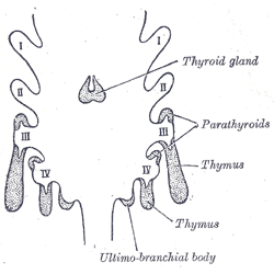
The thymus (: thymuses or thymi) is a specialized primary lymphoid organ of the immune system. Within the thymus, T cells mature. T cells are critical to the adaptive immune system, where the body adapts to specific foreign invaders. The thymus is located in the upper front part of the chest, in the anterior superior mediastinum, behind the sternum, and in front of the heart. It is made up of two lobes, each consisting of a central medulla and an outer cortex, surrounded by a capsule. The thymus is made up of immature T cells called thymocytes, as well as lining cells called epithelial cells which help the thymocytes develop. T cells that successfully develop react appropriately with Major histocompatibility complex, MHC immune receptors of the body (called ''positive selection'') and not against proteins of the body (called ''negative selection''). The thymus is the largest and most active during the neonatal and pre-adolescent periods. By the early teens, the Thymic involuti ... [...More Info...] [...Related Items...] OR: [Wikipedia] [Google] [Baidu] |
Stratum Corneum
The stratum corneum (Latin language, Latin for 'horny layer') is the outermost layer of the epidermis (skin), epidermis. Consisting of dead tissue, it protects underlying tissue from infection, dehydration, chemicals and mechanical stress. It is composed of 15–20 layers of flattened cells with no nuclei and cell organelles. Among its properties are mechanical shear, impact resistance, water flux and hydration regulation, microbial proliferation and invasion regulation, initiation of inflammation through cytokine activation and dendritic cell activity, and selective permeability to exclude toxins, irritants, and allergens. The cytoplasm of its cells shows filamentous keratin. These corneocytes are embedded in a lipid matrix composed of ceramides, cholesterol, and fatty acids. Desquamation is the process of cell shedding from the surface of the stratum corneum, balancing proliferating keratinocytes that form in the stratum basale. These cells migrate through the epidermis tow ... [...More Info...] [...Related Items...] OR: [Wikipedia] [Google] [Baidu] |
