|
Jugular
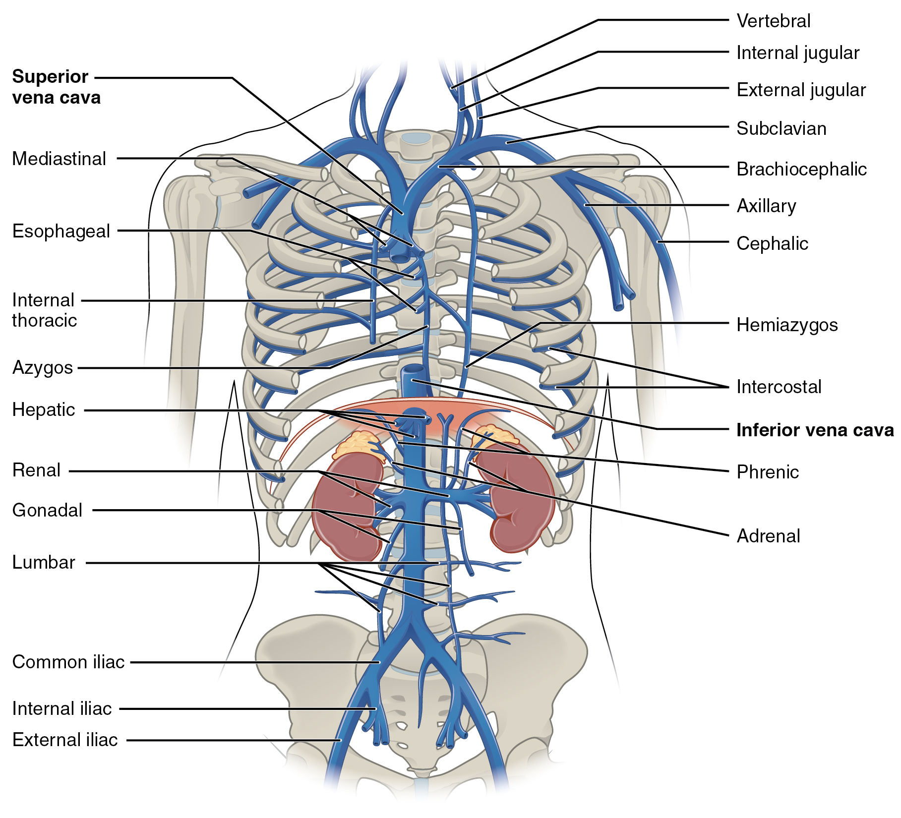
The jugular veins () are veins that take blood from the head back to the heart via the superior vena cava. The internal jugular vein descends next to the internal carotid artery and continues posteriorly to the sternocleidomastoid muscle. Structure and function There are two sets of jugular veins: external and internal. The left and right external jugular veins drain into the subclavian veins. The internal jugular veins join with the subclavian veins more medially to form the brachiocephalic veins. Finally, the left and right brachiocephalic veins join to form the superior vena cava, which delivers deoxygenated blood to the right atrium of the heart. The jugular vein has tributaries consisting of petrosal sinus, facial, lingual, pharylingual, the thyroid, and sometimes the occipital vein. Internal The internal jugular vein is formed by the anastomosis of blood from the sigmoid sinus of the dura mater and the ... [...More Info...] [...Related Items...] OR: [Wikipedia] [Google] [Baidu] |
Internal Jugular Vein
The internal jugular vein is a paired jugular vein that collects blood from the brain and the superficial parts of the face and neck. This vein runs in the carotid sheath with the common carotid artery and vagus nerve. It begins in the posterior compartment of the jugular foramen, at the base of the skull. It is somewhat dilated at its origin, which is called the ''superior bulb''. This vein also has a common trunk into which drains the anterior branch of the retromandibular vein, the facial vein, and the lingual vein. It runs down the side of the neck in a vertical direction, being at one end lateral to the internal carotid artery, and then lateral to the common carotid artery, and at the root of the neck, it unites with the subclavian vein to form the brachiocephalic vein (innominate vein); a little above its termination is a second dilation, the ''inferior bulb''. Above, it lies upon the rectus capitis lateralis, behind the internal carotid artery and the nerves pa ... [...More Info...] [...Related Items...] OR: [Wikipedia] [Google] [Baidu] |
Anterior Jugular Vein
The anterior jugular vein is a vein in the neck. Structure The anterior jugular vein lies lateral to the cricothyroid membrane. It begins near the hyoid bone by the confluence of several superficial veins from the submandibular region. Its tributaries are some laryngeal veins, and occasionally a small thyroid vein. It descends between the median line and the anterior border of the sternocleidomastoid muscle, and, at the lower part of the neck, passes beneath that muscle to open into the termination of the external jugular vein, or, in some instances, into the subclavian vein. Just above the sternum the two anterior jugular veins communicate by a transverse trunk, the venous jugular arch, which receive tributaries from the inferior thyroid veins; each also communicates with the internal jugular. There are no valves in this vein. The pretracheal lymph nodes follow the anterior jugular vein on each side of the midline. Variation The anterior jugular vein varies consider ... [...More Info...] [...Related Items...] OR: [Wikipedia] [Google] [Baidu] |
External Jugular Vein
The external jugular vein is a paired jugular vein which receives the greater part of the blood from the exterior of the cranium and the deep parts of the face, being formed by the junction of the posterior division of the retromandibular vein with the posterior auricular vein. Structure The external jugular vein commences in the substance of the parotid gland, on a level with the angle of the mandible, and runs perpendicularly down the neck, in the direction of a line drawn from the angle of the mandible to the middle of the clavicle superficial to the sternocleidomastoid muscle. In its course, it crosses the sternocleidomastoid muscle obliquely, and in the subclavian triangle perforates the deep fascia, and ends in the subclavian vein lateral to or in front of the scalenus anterior, piercing the roof of the posterior triangle. It is separated from the sternocleidomastoid muscle by the investing layer of the deep cervical fascia, and is covered by the platysma, the superfici ... [...More Info...] [...Related Items...] OR: [Wikipedia] [Google] [Baidu] |
Common Carotid Artery
In anatomy, the left and right common carotid arteries (carotids) () are artery, arteries that supply the head and neck with oxygenated blood; they divide in the neck to form the external carotid artery, external and internal carotid artery, internal carotid arteries. Structure The common carotid arteries are present on the left and right sides of the body. These arteries originate from different arteries but follow symmetrical courses. The right common carotid originates in the neck from the brachiocephalic trunk; the left from the aortic arch in the thorax. These split into the external and internal carotid arteries at the upper border of the thyroid cartilage, at around the level of the fourth cervical vertebra. The left common carotid artery can be thought of as having two parts: a thoracic (chest) part and a cervical (neck) part. The right common carotid originates in or close to the neck and contains only a small thoracic portion. There are studies in the bioengineering l ... [...More Info...] [...Related Items...] OR: [Wikipedia] [Google] [Baidu] |
Vagus Nerve
The vagus nerve, also known as the tenth cranial nerve (CN X), plays a crucial role in the autonomic nervous system, which is responsible for regulating involuntary functions within the human body. This nerve carries both sensory and motor fibers and serves as a major pathway that connects the brain to various organs, including the heart, lungs, and digestive tract. As a key part of the parasympathetic nervous system, the vagus nerve helps regulate essential involuntary functions like heart rate, breathing, and digestion. By controlling these processes, the vagus nerve contributes to the body's "rest and digest" response, helping to calm the body after stress, lower heart rate, improve digestion, and maintain homeostasis. The vagus nerve consists of two branches: the right and left vagus nerves. In the neck, the right vagus nerve contains approximately 105,000 fibers, while the left vagus nerve has about 87,000 fibers, according to one source. However, other sources report sl ... [...More Info...] [...Related Items...] OR: [Wikipedia] [Google] [Baidu] |
Common Carotid Artery
In anatomy, the left and right common carotid arteries (carotids) () are artery, arteries that supply the head and neck with oxygenated blood; they divide in the neck to form the external carotid artery, external and internal carotid artery, internal carotid arteries. Structure The common carotid arteries are present on the left and right sides of the body. These arteries originate from different arteries but follow symmetrical courses. The right common carotid originates in the neck from the brachiocephalic trunk; the left from the aortic arch in the thorax. These split into the external and internal carotid arteries at the upper border of the thyroid cartilage, at around the level of the fourth cervical vertebra. The left common carotid artery can be thought of as having two parts: a thoracic (chest) part and a cervical (neck) part. The right common carotid originates in or close to the neck and contains only a small thoracic portion. There are studies in the bioengineering l ... [...More Info...] [...Related Items...] OR: [Wikipedia] [Google] [Baidu] |
Subclavian Veins
The subclavian vein is a paired large vein, one on either side of the body, that is responsible for draining blood from the upper extremities, allowing this blood to return to the heart. The left subclavian vein plays a key role in the absorption of lipids, by allowing products that have been carried by lymph in the thoracic duct to enter the bloodstream. The diameter of the subclavian veins is approximately 1–2 cm, depending on the individual. Structure Each subclavian vein is a continuation of the axillary vein and runs from the outer border of the first rib to the medial border of anterior scalene muscle. From here it joins with the internal jugular vein to form the brachiocephalic vein (also known as "innominate vein"). The angle of union is termed the venous angle. The subclavian vein follows the subclavian artery and is separated from the subclavian artery by the insertion of anterior scalene. Thus, the subclavian vein lies anterior to the anterior scalene while th ... [...More Info...] [...Related Items...] OR: [Wikipedia] [Google] [Baidu] |
Brachiocephalic Vein
The left and right brachiocephalic veins (previously called innominate veins) are major veins in the Thorax, upper chest, formed by the union of the ipsilateral internal jugular vein and subclavian vein (the so-called venous angle) behind the sternoclavicular joint. The left brachiocephalic vein is more than twice the length of the right brachiocephalic vein. These veins merge to form the superior vena cava, a great vessel, posterior to the junction of the first costal cartilage with the Manubrium, manubrium of the sternum. The brachiocephalic veins are the major veins returning blood to the superior vena cava. Left and right veins Left brachiocephalic vein The left brachiocephalic vein is about 6cm, more than twice the length of the right brachiocephalic vein. and is formed by the confluence of the left subclavian vein, subclavian and left internal jugular veins. In addition the left vein receives drainage from the following tributaries: * The left vertebral vein, internal thor ... [...More Info...] [...Related Items...] OR: [Wikipedia] [Google] [Baidu] |
Carotid Sheath
The carotid sheath is a condensation of the deep cervical fascia enveloping multiple vital neurovascular structures of the neck, including the common and internal carotid arteries, the internal jugular vein, the vagus nerve (CN X), and ansa cervicalis. The carotid sheath helps protects the structures contained therein. Anatomy One carotid sheath is situated on each side of the neck, extending between the base of the skull superiorly and the thorax inferiorly. Superiorly, the carotid sheath encircles the margins of the carotid canal and jugular foramen. Inferiorly, it terminates at the arch of the aorta; it is continuous inferiorly with the axillary sheath at the venous angle. Its inferior end occurs at the level of the first rib and sternum inferiorly (varying between the levels of C7 and T4). Structure The carotid sheath is a fibrous connective tissue formation surrounding several important structures of the neck. It is thicker around the arteries than around the ... [...More Info...] [...Related Items...] OR: [Wikipedia] [Google] [Baidu] |


