|
Joint Replacement
Joint replacement is a procedure of orthopedic surgery known also as arthroplasty, in which an arthritic or dysfunctional joint surface is replaced with an orthopedic prosthesis. Joint replacement is considered as a treatment when severe joint pain or dysfunction is not alleviated by less-invasive therapies. Joint replacement surgery is often indicated from various Arthropathy, joint diseases, including osteoarthritis and rheumatoid arthritis. Joint replacement has become more common, mostly with knee replacement, knee and hip replacements. About 773,000 Americans had a hip or knee replaced in 2009.Joint Replacement Surgery and You. (April, 2009) In ''Arthritis, Musculoskeletal and Skin Disease online''. Retrieved from http://www.niams.nih.gov/#. Uses Shoulder For shoulder replacement, there are a few major approaches to access the shoulder joint. The first is the deltopectoral approach, which saves the deltoid, but requires the supraspinatus to be cut. The second is the transde ... [...More Info...] [...Related Items...] OR: [Wikipedia] [Google] [Baidu] |
Hip Joint Replacement
Hip replacement is a surgical procedure in which the hip joint is replaced by a prosthetic implant, that is, a hip prosthesis. Hip replacement surgery can be performed as a total replacement or a hemi/semi(half) replacement. Such joint replacement orthopaedic surgery is generally conducted to relieve arthritis pain or in some hip fractures. A total hip replacement (total hip arthroplasty) consists of replacing both the acetabulum and the femoral head while hemiarthroplasty generally only replaces the femoral head. Hip replacement is one of the most common orthopaedic operations, though patient satisfaction varies widely between different techniques and implants. Approximately 58% of total hip replacements are estimated to last 25 years. The average cost of a total hip replacement in 2012 was $40,364 in the United States (€37,307.44 in euros), and about $7,700 to $12,000 in most European countries. NOTE: In euros, that is from €7,116.92 to €11,091.30 euros. Medical uses Tot ... [...More Info...] [...Related Items...] OR: [Wikipedia] [Google] [Baidu] |
Vastus Medialis
The vastus medialis (vastus internus or teardrop muscle) is an extensor muscle located medially in the thigh that extends the knee. The vastus medialis is part of the quadriceps muscle group. Structure The vastus medialis is a muscle present in the anterior compartment of thigh, and is one of the four muscles that make up the quadriceps muscle. The others are the vastus lateralis, vastus intermedius and rectus femoris. It is the most medial of the "vastus" group of muscles. The vastus medialis arises medially along the entire length of the femur, and attaches with the other muscles of the quadriceps in the quadriceps tendon. The vastus medialis muscle originates from a continuous line of attachment on the femur, which begins on the front and middle side (anteromedially) on the intertrochanteric line of the femur. It continues down and back (posteroinferiorly) along the pectineal line and then descends along the inner (medial) lip of the linea aspera and onto the medi ... [...More Info...] [...Related Items...] OR: [Wikipedia] [Google] [Baidu] |
Ankle Replacement
Ankle replacement, or ankle arthroplasty, is a surgical procedure to replace the damaged articular surfaces of the human ankle joint with prosthetic components. This procedure is becoming the treatment of choice for patients requiring arthroplasty, replacing the conventional use of arthrodesis, i.e. fusion of the bones. The restoration of range of motion is the key feature in favor of ankle replacement with respect to arthrodesis. However, clinical evidence of the superiority of the former has only been demonstrated for particular isolated implant designs. History Since the early 1970s, the disadvantages of ankle arthrodesis and the excellent results attained by arthroplasty at other human joints have encouraged numerous prosthesis designs also for the ankle. In the following decade, the disappointing results of long-term follow-up clinical studies of the pioneering designs has left ankle arthrodesis as the surgical treatment of choice for these patients. More modern designs hav ... [...More Info...] [...Related Items...] OR: [Wikipedia] [Google] [Baidu] |
Metal Foam
Regular foamed aluminium In materials science, a metal foam is a material or structure consisting of a solid metal (frequently aluminium) with gas-filled pores comprising a large portion of the volume. The pores can be sealed (closed-cell foam) or interconnected (open-cell foam). The defining characteristic of metal foams is a high porosity: typically only 5–25% of the volume is the base metal. The strength of the material is due to the square–cube law. Metal foams typically retain some physical properties of their base material. Foam made from non-flammable metal remains non-flammable and can generally be recycled as the base material. Its coefficient of thermal expansion is similar while thermal conductivity is likely reduced. Definitions Open-cell Open-celled metal foam, also called metal sponge, can be used in heat exchangers (compact electronics cooling, cryogen tanks, PCM heat exchangers), energy absorption, flow diffusion, scrubbers, flame arrestors, an ... [...More Info...] [...Related Items...] OR: [Wikipedia] [Google] [Baidu] |
Osseointegration
Osseointegration (from Latin " bony" and "to make whole") is the direct structural and functional connection between living bone and the surface of a load-bearing artificial implant ("load-bearing" as defined by Albrektsson et al. in 1981). A more recent definition (by Schroeder et al.) defines osseointegration as "functional ankylosis (bone adherence)", where new bone is laid down directly on the implant surface and the implant exhibits mechanical stability (i.e., resistance to destabilization by mechanical agitation or shear forces). Osseointegration has enhanced the science of medical bone and joint replacement techniques as well as dental implants and improving prosthetics for amputees. Definitions Osseointegration is also defined as: "the formation of a direct interface between an implant and bone, without intervening soft tissue". An osseointegrated implant is a type of implant defined as "an endosteal implant containing pores into which osteoblasts and supportin ... [...More Info...] [...Related Items...] OR: [Wikipedia] [Google] [Baidu] |
Polymethylmethacrylate
Poly(methyl methacrylate) (PMMA) is a synthetic polymer derived from methyl methacrylate. It is a transparent thermoplastic, used as an engineering plastic. PMMA is also known as acrylic, acrylic glass, as well as by the trade names and brands Crylux, Walcast, Hesalite, Plexiglas, Acrylite, Lucite, PerClax, and Perspex, among several others ( see below). This plastic is often used in sheet form as a lightweight or shatter-resistant alternative to glass. It can also be used as a casting resin, in inks and coatings, and for many other purposes. It is often technically classified as a type of glass, in that it is a non-crystalline vitreous substance—hence its occasional historic designation as ''acrylic glass''. History The first acrylic acid was created in 1843. Methacrylic acid, derived from acrylic acid, was formulated in 1865. The reaction between methacrylic acid and methanol results in the ester methyl methacrylate. It was developed in 1928 in several different lab ... [...More Info...] [...Related Items...] OR: [Wikipedia] [Google] [Baidu] |
Fibular Collateral Ligament
The lateral collateral ligament (LCL, long external lateral ligament or fibular collateral ligament) is an extrinsic ligament of the knee located on the lateral side of the knee. Its superior attachment is at the lateral epicondyle of the femur (superoposterior to the popliteal groove); its inferior attachment is at the lateral aspect of the head of fibula (anterior to the apex). The LCL is not fused with the joint capsule. Inferiorly, the LCL splits the tendon of insertion of the biceps femoris muscle. Structure The LCL measures some 5 cm in length. It is rounded, and is more narrow and less broad compared to the medial collateral ligament. It extends obliquely inferoposteriorly from its superior attachment to its inferior attachment. In contrast to the medial collateral ligament, it is not fused with either the capsular ligament nor the lateral meniscus. Because of this, the LCL is more flexible than its medial counterpart, and is therefore less susceptible to injury. ... [...More Info...] [...Related Items...] OR: [Wikipedia] [Google] [Baidu] |
Medial Collateral Ligament
The medial collateral ligament (MCL), also called the superficial medial collateral ligament (sMCL) or tibial collateral ligament (TCL), is one of the major ligaments of the knee. It is on the medial (inner) side of the knee joint and occurs in humans and other primates. Its primary function is to resist valgus (inward bending) forces on the knee. Structure It is a broad, flat, membranous band, situated slightly posterior on the medial side of the knee joint. It is attached proximally to the medial epicondyle of the femur, immediately below the adductor tubercle; below to the medial condyle of the tibia and medial surface of its body. It resists forces that would push the knee medially, which would otherwise produce valgus deformity. It provides up to 78% of the restraining force that resists valgus (inward pressing) loads on the knee. The fibers of the posterior part of the ligament are short and incline backward as they descend; they are inserted into the tibia above t ... [...More Info...] [...Related Items...] OR: [Wikipedia] [Google] [Baidu] |
Posterior Cruciate Ligament
The posterior cruciate ligament (PCL) is a ligament in each knee of humans and various other animals. It works as a counterpart to the anterior cruciate ligament (ACL). It connects the posterior intercondylar area of the tibia to the Medial condyle of femur, medial condyle of the femur. This configuration allows the PCL to resist forces pushing the tibia posteriorly relative to the femur. The PCL and ACL are intracapsular ligaments because they lie deep within the knee joint. They are both isolated from the fluid-filled synovial cavity, with the synovial membrane wrapped around them. The PCL gets its name by attaching to the posterior portion of the tibia. The PCL, Anterior cruciate ligament, ACL, medial collateral ligament, MCL, and fibular collateral ligament, LCL are the four main ligaments of the knee in primates. Structure The PCL is located within the knee joint where it stabilizes the articulating bones, particularly the femur and the tibia, during movement. It originates ... [...More Info...] [...Related Items...] OR: [Wikipedia] [Google] [Baidu] |
Anterior Cruciate Ligament
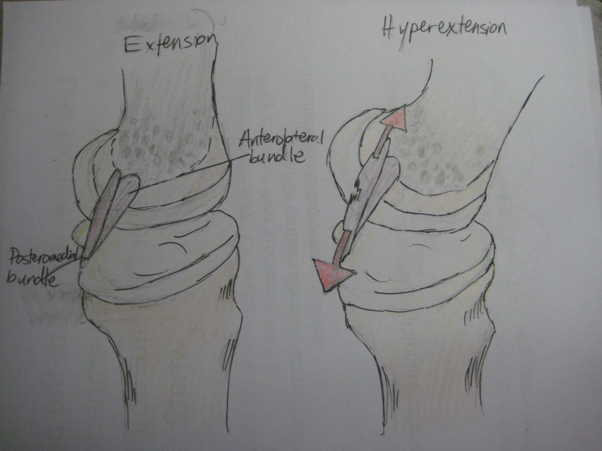
The anterior cruciate ligament (ACL) is one of a pair of cruciate ligaments (the other being the posterior cruciate ligament) in the human knee. The two ligaments are called "cruciform" ligaments, as they are arranged in a crossed formation. In the quadruped stifle joint (analogous to the knee), based on its anatomical position, it is also referred to as the cranial cruciate ligament. The term cruciate is Latin for cross. This name is fitting because the ACL crosses the posterior cruciate ligament to form an "X". It is composed of strong, fibrous material and assists in controlling excessive motion by limiting mobility of the joint. The anterior cruciate ligament is one of the four main ligaments of the knee, providing 85% of the restraining force to anterior tibial displacement at 30 and 90° of knee flexion. The ACL is the most frequently injured ligament in the knee. Structure The ACL originates from deep within the notch of the distal femur. Its proximal fibers fan out alo ... [...More Info...] [...Related Items...] OR: [Wikipedia] [Google] [Baidu] |
Cartilage
Cartilage is a resilient and smooth type of connective tissue. Semi-transparent and non-porous, it is usually covered by a tough and fibrous membrane called perichondrium. In tetrapods, it covers and protects the ends of long bones at the joints as articular cartilage, and is a structural component of many body parts including the rib cage, the neck and the bronchial tubes, and the intervertebral discs. In other taxa, such as chondrichthyans and cyclostomes, it constitutes a much greater proportion of the skeleton. It is not as hard and rigid as bone, but it is much stiffer and much less flexible than muscle. The matrix of cartilage is made up of glycosaminoglycans, proteoglycans, collagen fibers and, sometimes, elastin. It usually grows quicker than bone. Because of its rigidity, cartilage often serves the purpose of holding tubes open in the body. Examples include the rings of the trachea, such as the cricoid cartilage and carina. Cartilage is composed of specialized c ... [...More Info...] [...Related Items...] OR: [Wikipedia] [Google] [Baidu] |
Tibia
The tibia (; : tibiae or tibias), also known as the shinbone or shankbone, is the larger, stronger, and anterior (frontal) of the two Leg bones, bones in the leg below the knee in vertebrates (the other being the fibula, behind and to the outside of the tibia); it connects the knee with the ankle bones, ankle. The tibia is found on the anatomical terms of location#Medial, medial side of the leg next to the fibula and closer to the median plane. The tibia is connected to the fibula by the interosseous membrane of leg, forming a type of fibrous joint called a syndesmosis with very little movement. The tibia is named for the flute ''aulos, tibia''. It is the second largest bone in the human body, after the femur. The leg bones are the strongest long bones as they support the rest of the body. Structure In human anatomy, the tibia is the second largest bone next to the femur. As in other vertebrates the tibia is one of two bones in the lower leg, the other being the fibula, and is a ... [...More Info...] [...Related Items...] OR: [Wikipedia] [Google] [Baidu] |


