|
Ileum
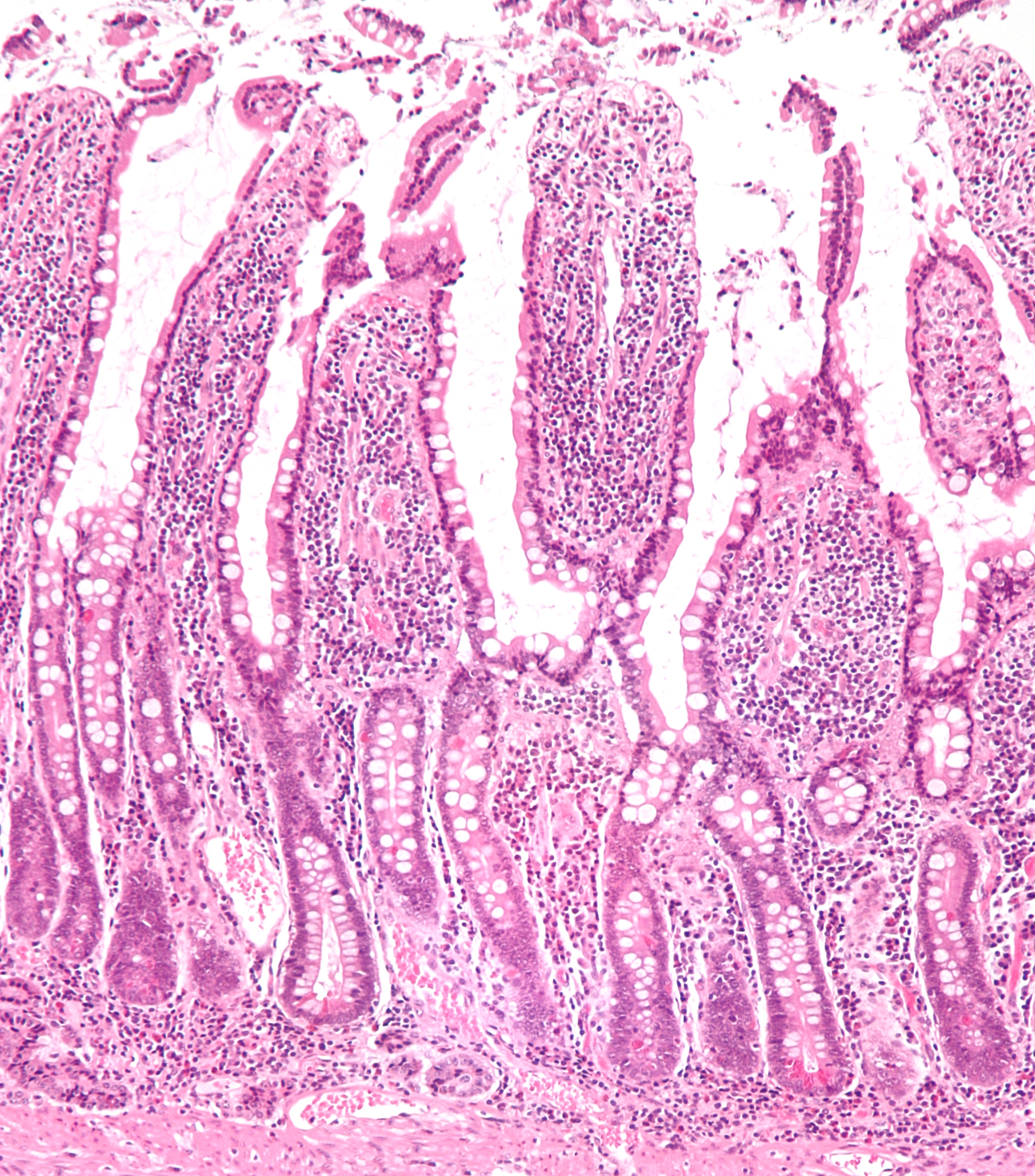
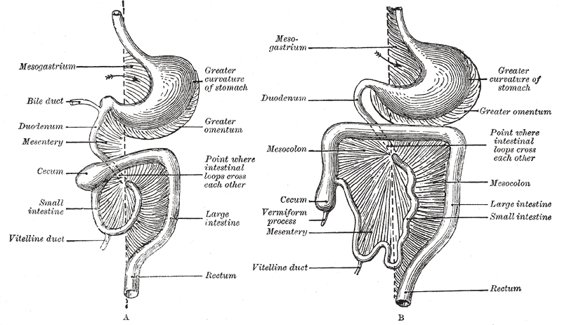
The ileum () is the final section of the small intestine in most higher vertebrates, including mammals, reptiles, and birds. In fish, the divisions of the small intestine are not as clear and the terms posterior intestine or distal intestine may be used instead of ileum. Its main function is to absorb vitamin B12, bile salts, and whatever products of digestion that were not absorbed by the jejunum. The ileum follows the duodenum and jejunum and is separated from the cecum by the ileocecal valve (ICV). In humans, the ileum is about 2–4 m long, and the pH is usually between 7 and 8 (neutral or slightly basic). ''Ileum ''is derived from the Greek word ''eilein'', meaning "to twist up tightly". Structure The ileum is the third and final part of the small intestine. It follows the jejunum and ends at the ileocecal junction, where the terminal ileum communicates with the cecum of the large intestine through the ileocecal valve. The ileum, along with the jejunum, is suspended ... [...More Info...] [...Related Items...] OR: [Wikipedia] [Google] [Baidu] |
Small Intestine
The small intestine or small bowel is an organ in the gastrointestinal tract where most of the absorption of nutrients from food takes place. It lies between the stomach and large intestine, and receives bile and pancreatic juice through the pancreatic duct to aid in digestion. The small intestine is about long and folds many times to fit in the abdomen. Although it is longer than the large intestine, it is called the small intestine because it is narrower in diameter. The small intestine has three distinct regions – the duodenum, jejunum, and ileum. The duodenum, the shortest, is where preparation for absorption through small finger-like protrusions called villi begins. The jejunum is specialized for the absorption through its lining by enterocytes: small nutrient particles which have been previously digested by enzymes in the duodenum. The main function of the ileum is to absorb vitamin B12, bile salts, and whatever products of digestion that were not absorbed by the ... [...More Info...] [...Related Items...] OR: [Wikipedia] [Google] [Baidu] |
Jejunum
The jejunum is the second part of the small intestine in humans and most higher vertebrates, including mammals, reptiles, and birds. Its lining is specialised for the absorption by enterocytes of small nutrient molecules which have been previously digested by enzymes in the duodenum. The jejunum lies between the duodenum and the ileum and is considered to start at the suspensory muscle of the duodenum, a location called the duodenojejunal flexure. The division between the jejunum and ileum is not anatomically distinct. In adult humans, the small intestine is usually long (post mortem), about two-fifths of which (about ) is the jejunum. Structure The interior surface of the jejunum—which is exposed to ingested food—is covered in finger–like projections of mucosa, called villi, which increase the surface area of tissue available to absorb nutrients from ingested foodstuffs. The epithelial cells which line these villi have microvilli. The transport of nutrients across epi ... [...More Info...] [...Related Items...] OR: [Wikipedia] [Google] [Baidu] |
Cecum
The cecum or caecum is a pouch within the peritoneum that is considered to be the beginning of the large intestine. It is typically located on the right side of the body (the same side of the body as the appendix (anatomy), appendix, to which it is joined). The word cecum (, plural ceca ) stems from the Latin ''wikt:caecus, caecus'' meaning blindness, blind. It receives chyme from the ileum, and connects to the ascending colon of the large intestine. It is separated from the ileum by the ileocecal valve (ICV) or Bauhin's valve. It is also separated from the Large intestine#Structure, colon by the cecocolic junction. While the cecum is usually intraperitoneal, the ascending colon is Retroperitoneal space, retroperitoneal. In herbivores, the cecum stores food material where bacteria are able to break down the cellulose. In humans, the cecum is involved in absorption of salts and electrolytes and lubricates the solid waste that passes into the large intestine. Structure Develo ... [...More Info...] [...Related Items...] OR: [Wikipedia] [Google] [Baidu] |
Peyer's Patches
Peyer's patches (or aggregated lymphoid nodules) are organized lymphoid follicles, named after the 17th-century Swiss anatomist Johann Conrad Peyer. * Reprinted as: * Peyer referred to Peyer's patches as ''plexus'' or ''agmina glandularum'' (clusters of glands). From (Peyer, 1681), p. 7: ''"Tenui a perfectiorum animalium Intestina accuratius perlustranti, crebra hinc inde, variis intervallis, corpusculorum glandulosorum Agmina sive Plexus se produnt, diversae Magnitudinis atque Figurae."'' (I knew from careful study of more advanced animals, the intestines bear — often here and there, at various intervals — clusters of glandular small bodies or "plexuses" of diverse size and shape.) From p. 15: ''"(has Plexus seu agmina Glandularum voco)"'' (I call them "plexuses" or clusters of glands) He described their appearance. From p. 8: ''"Horum vero Plexuum facies modo in orbem concinnata; modo in Ovi aut Olivae oblongam, aliamve angulosam ac magis anomalam disposita figuram cerni ... [...More Info...] [...Related Items...] OR: [Wikipedia] [Google] [Baidu] |
Ileocecal Valve
The ileocecal valve (ileal papilla, ileocaecal valve, Tulp's valve, Tulpius valve, Bauhin's valve, ileocecal eminence, valve of Varolius or colic valve) is a sphincter muscle valve that separates the small intestine and the large intestine. Its critical function is to limit the reflux of colonic contents into the ileum. Approximately two liters of fluid enters the colon daily through the ileocecal valve. Microanatomy The histology of the ileocecal valve shows an abrupt change from a villous mucosa pattern of the ileum to a more colonic mucosa. A thickening of the muscularis mucosa, which is the smooth muscle tissue found beneath the mucosal layer of the digestive tract. A thickening of the muscularis externa is also noted. There is also a variable amount of lymphatic tissue found at the valve. The ileocecal valve has a papillose structure. Clinical significance Colonoscopy During colonoscopy, the ileocecal valve is used, along with the appendiceal orifice, in the identifi ... [...More Info...] [...Related Items...] OR: [Wikipedia] [Google] [Baidu] |
Mesentery
The mesentery is an organ that attaches the intestines to the posterior abdominal wall in humans and is formed by the double fold of peritoneum. It helps in storing fat and allowing blood vessels, lymphatics, and nerves to supply the intestines, among other functions. The mesocolon was thought to be a fragmented structure, with all named parts—the ascending, transverse, descending, and sigmoid mesocolons, the mesoappendix, and the mesorectum—separately terminating their insertion into the posterior abdominal wall. However, in 2012, new microscopic and electron microscopic examinations showed the mesocolon to be a single structure derived from the duodenojejunal flexure and extending to the distal mesorectal layer. Thus, the mesentery is an internal organ. Structure The mesentery of the small intestine arises from the root of the mesentery (or mesenteric root) and is the part connected with the structures in front of the vertebral column. The root is narrow, about 15 ... [...More Info...] [...Related Items...] OR: [Wikipedia] [Google] [Baidu] |
Human Gastrointestinal Tract
The gastrointestinal tract (GI tract, digestive tract, alimentary canal) is the tract or passageway of the digestive system that leads from the mouth to the anus. The GI tract contains all the major organs of the digestive system, in humans and other animals, including the esophagus, stomach, and intestines. Food taken in through the mouth is digested to extract nutrients and absorb energy, and the waste expelled at the anus as feces. ''Gastrointestinal'' is an adjective meaning of or pertaining to the stomach and intestines. Most animals have a "through-gut" or complete digestive tract. Exceptions are more primitive ones: sponges have small pores ( ostia) throughout their body for digestion and a larger dorsal pore (osculum) for excretion, comb jellies have both a ventral mouth and dorsal anal pores, while cnidarians and acoels have a single pore for both digestion and excretion. The human gastrointestinal tract consists of the esophagus, stomach, and intestines, and is ... [...More Info...] [...Related Items...] OR: [Wikipedia] [Google] [Baidu] |
Peritoneum
The peritoneum is the serous membrane forming the lining of the abdominal cavity or coelom in amniotes and some invertebrates, such as annelids. It covers most of the intra-abdominal (or coelomic) organs, and is composed of a layer of mesothelium supported by a thin layer of connective tissue. This peritoneal lining of the cavity supports many of the abdominal organs and serves as a conduit for their blood vessels, lymphatic vessels, and nerves. The abdominal cavity (the space bounded by the vertebrae, abdominal muscles, diaphragm, and pelvic floor) is different from the intraperitoneal space (located within the abdominal cavity but wrapped in peritoneum). The structures within the intraperitoneal space are called "intraperitoneal" (e.g., the stomach and intestines), the structures in the abdominal cavity that are located behind the intraperitoneal space are called "retroperitoneal" (e.g., the kidneys), and those structures below the intraperitoneal space are called "subp ... [...More Info...] [...Related Items...] OR: [Wikipedia] [Google] [Baidu] |
Circular Folds
The circular folds (also known as valves of Kerckring, valves of Kerchkring, plicae circulares, ''plicae circulae, and'' ''valvulae conniventes'') are large valvular flaps projecting into the lumen of the small intestine. Structure The entire small intestine has circular folds of mucous membrane. The majority extend transversely around the cylinder of the small intestine, for about one-half or two-thirds of its circumference. Some form complete circles. Others have a spiral direction. The latter usually extend a little more than once around the bowel, but occasionally two or three times. The larger folds are about 1 cm. in depth at their broadest part; but the greater number are smaller. The larger and smaller folds alternate with each other. These can increase the surface area of the small intestine threefold. These larger folds are called ''plica circularis''. Distribution They are not found at the commencement of the duodenum, but begin to appear about 2.5 or 5 cm b ... [...More Info...] [...Related Items...] OR: [Wikipedia] [Google] [Baidu] |
Superior Mesenteric Artery
In human anatomy, the superior mesenteric artery (SMA) is an artery which arises from the anterior surface of the abdominal aorta, just inferior to the origin of the celiac trunk, and supplies blood to the intestine from the lower part of the duodenum through two-thirds of the transverse colon, as well as the pancreas. Structure It arises anterior to lower border of vertebra L1 in an adult. It is usually 1 cm lower than the celiac trunk. It initially travels in an anterior/inferior direction, passing behind/under the neck of the pancreas and the splenic vein. Located under this portion of the superior mesenteric artery, between it and the aorta, are the following: * left renal vein - travels between the left kidney and the inferior vena cava (can be compressed between the SMA and the abdominal aorta at this location, leading to nutcracker syndrome). * the third part of the duodenum, a segment of the small intestines (can be compressed by the SMA at this location, lea ... [...More Info...] [...Related Items...] OR: [Wikipedia] [Google] [Baidu] |
Duodenum
The duodenum is the first section of the small intestine in most higher vertebrates, including mammals, reptiles, and birds. In fish, the divisions of the small intestine are not as clear, and the terms anterior intestine or proximal intestine may be used instead of duodenum. In mammals the duodenum may be the principal site for iron absorption. The duodenum precedes the jejunum and ileum and is the shortest part of the small intestine. In humans, the duodenum is a hollow jointed tube about 25–38 cm (10–15 inches) long connecting the stomach to the middle part of the small intestine. It begins with the duodenal bulb and ends at the suspensory muscle of duodenum. Duodenum can be divided into four parts: the first (superior), the second (descending), the third (horizontal) and the fourth (ascending) parts. Structure The duodenum is a C-shaped structure lying adjacent to the stomach. It is divided anatomically into four sections. The first part of the duodenum lies ... [...More Info...] [...Related Items...] OR: [Wikipedia] [Google] [Baidu] |


