|
Intestinal Villus
Intestinal villi (: villus) are small, finger-like projections that extend into the lumen of the small intestine. Each villus is approximately 0.5–1.6 mm in length (in humans), and has many microvilli projecting from the enterocytes of its epithelium which collectively form the striated or brush border. Each of these microvilli are about 1 μm in length, around 1000 times shorter than a single villus. The intestinal villi are much smaller than any of the circular folds in the intestine. Villi increase the internal surface area of the intestinal walls making available a greater surface area for absorption. An increased absorptive area is useful because digested nutrients (including monosaccharide and amino acids) pass into the semipermeable villi through diffusion, which is effective only at short distances. In other words, increased surface area (in contact with the fluid in the lumen) decreases the average distance travelled by nutrient molecules, so effectiveness of ... [...More Info...] [...Related Items...] OR: [Wikipedia] [Google] [Baidu] |
Micrograph
A micrograph is an image, captured photographically or digitally, taken through a microscope or similar device to show a magnify, magnified image of an object. This is opposed to a macrograph or photomacrograph, an image which is also taken on a microscope but is only slightly magnified, usually less than 10 times. Micrography is the practice or art of using microscopes to make photographs. A photographic micrograph is a photomicrograph, and one taken with an electron microscope is an electron micrograph. A micrograph contains extensive details of microstructure. A wealth of information can be obtained from a simple micrograph like behavior of the material under different conditions, the phases found in the system, failure analysis, grain size estimation, elemental analysis and so on. Micrographs are widely used in all fields of microscopy. Types Photomicrograph A light micrograph or photomicrograph is a micrograph prepared using an optical microscope, a process referred to ... [...More Info...] [...Related Items...] OR: [Wikipedia] [Google] [Baidu] |
Goblet Cells
Goblet cells are simple columnar epithelial cells that secrete gel-forming mucins, like mucin 2 in the lower gastrointestinal tract, and mucin 5AC in the respiratory tract. The goblet cells mainly use the merocrine method of secretion, secreting vesicles into a duct, but may use apocrine methods, budding off their secretions, when under stress. The term ''goblet'' refers to the cell's goblet-like shape. The apical portion is shaped like a cup, as it is distended by abundant mucus laden granules; its basal portion lacks these granules and is shaped like a stem. The goblet cell is highly polarized with the nucleus and other organelles concentrated at the base of the cell and secretory granules containing mucin, at the apical surface. The apical plasma membrane projects short microvilli to give an increased surface area for secretion. Goblet cells are typically found in the respiratory, reproductive and lower gastrointestinal tract and are surrounded by other columnar cells. Bia ... [...More Info...] [...Related Items...] OR: [Wikipedia] [Google] [Baidu] |
Chylomicron
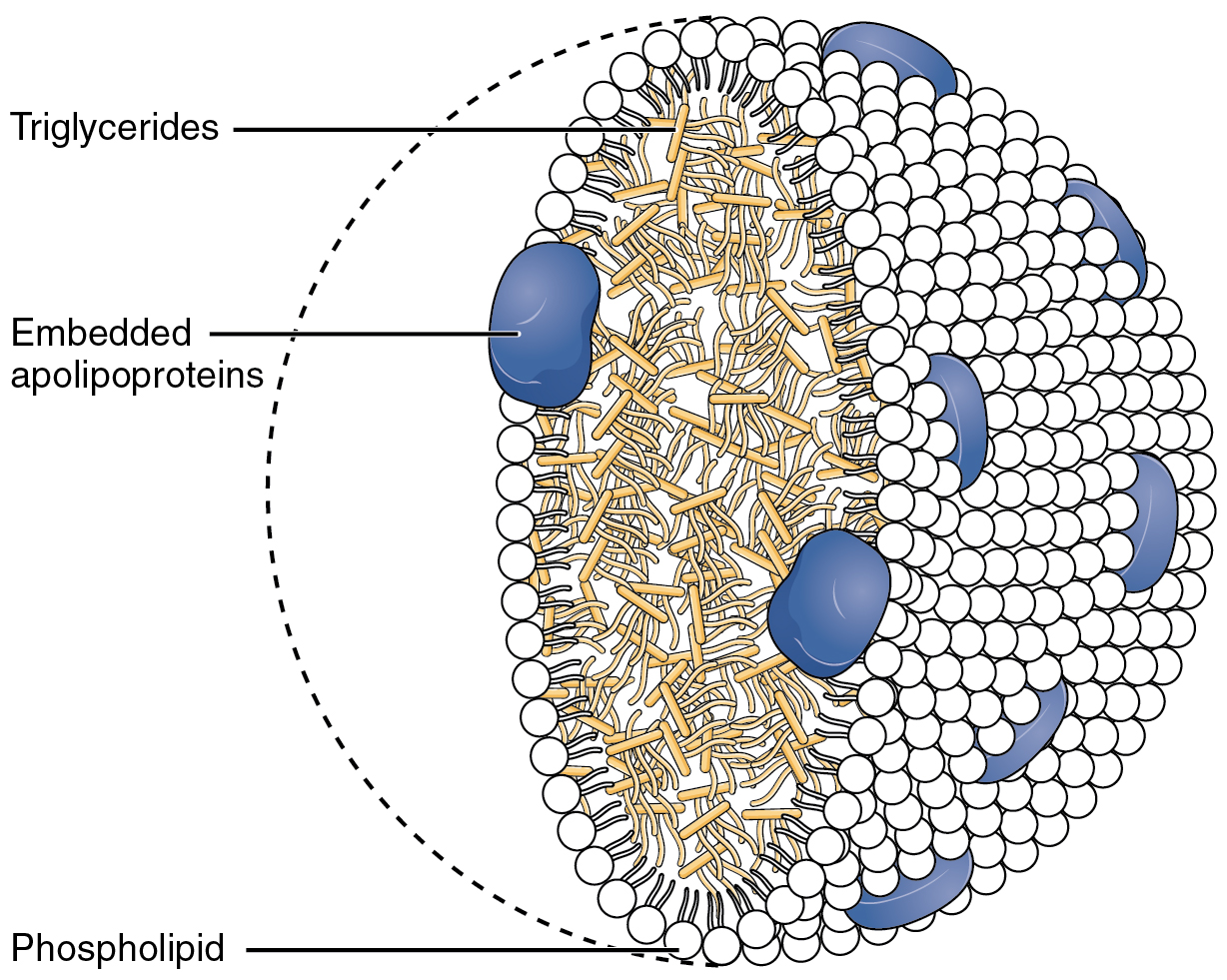
Chylomicrons (from the Greek χυλός, chylos, meaning ''juice'' (of plants or animals), and micron, meaning ''small''), also known as ultra low-density lipoproteins (ULDL), are lipoprotein particles that consist of triglycerides (85–92%), phospholipids (6–12%), cholesterol (1–3%), and proteins (1–2%). They transport dietary lipids, such as fats and cholesterol, from the intestines to other locations in the body, within the water-based solution of the bloodstream. ULDLs are one of the five major groups lipoproteins are divided into based on their density. A protein specific to chylomicrons is ApoB48. There is an inverse relationship in the density and size of lipoprotein particles: fats have a lower density than water or smaller protein molecules, and the larger particles have a higher ratio of internal fat molecules with respect to the outer emulsifying protein molecules in the shell. ULDLs, if in the region of 1,000 nm or more, are the only lipoprotein particl ... [...More Info...] [...Related Items...] OR: [Wikipedia] [Google] [Baidu] |
Lymph Capillaries
Lymph capillaries or lymphatic capillaries are tiny, thin-walled microvessels located in the spaces between cells (except in the central nervous system and non-vascular tissues) which serve to drain and process extracellular fluid. Upon entering the lumen of a lymphatic capillary, the collected fluid is known as lymph. Each lymphatic capillary carries lymph into a lymphatic vessel, which in turn connects to a lymph node, a small bean-shaped gland that filters and monitors the lymphatic fluid for infections. Lymph is ultimately returned to the venous circulation. Lymphatic capillaries are slightly larger in diameter than blood capillaries, and have closed ends (unlike the loop structure of blood capillaries). Lymph capillaries are strategically placed among the blood-related capillaries in order to have efficient and effective uptake from the interstitial fluid during capillary exchange. This intentional formation allows for a more rapid and continuous collection. Their uniq ... [...More Info...] [...Related Items...] OR: [Wikipedia] [Google] [Baidu] |
Lacteal
A lacteal is a Lymph capillary, lymphatic capillary that absorbs dietary fats in the Intestinal villus, villi of the small intestine. Triglycerides are emulsified by bile and hydrolyzed by the enzyme lipase, resulting in a mixture of fatty acids, di- and monoglycerides. These then pass from the intestinal lumen into the enterocyte, where they are re-esterified to form triglyceride. The triglyceride is then combined with phospholipids, cholesterol ester, and apolipoprotein B48 to form chylomicrons. These chylomicrons then pass into the lacteals, forming a milky substance known as chyle. The lacteals merge to form larger lymphatic vessels that transport the chyle to the thoracic duct where it is emptied into the bloodstream at the subclavian vein. At this point, the fats are in the bloodstream in the form of chylomicrons. Once in the blood, chylomicrons are subject to delipidation by lipoprotein lipase. Eventually, enough lipid has been lost and additional apolipoproteins gained, th ... [...More Info...] [...Related Items...] OR: [Wikipedia] [Google] [Baidu] |
Digestion
Digestion is the breakdown of large insoluble food compounds into small water-soluble components so that they can be absorbed into the blood plasma. In certain organisms, these smaller substances are absorbed through the small intestine into the blood stream. Digestion is a form of catabolism that is often divided into two processes based on how food is broken down: mechanical and chemical digestion. The term mechanical digestion refers to the physical breakdown of large pieces of food into smaller pieces which can subsequently be accessed by digestive enzymes. Mechanical digestion takes place in the mouth through Chewing, mastication and in the small intestine through segmentation contractions. In chemical digestion, enzymes break down food into the small compounds that the body can use. In the human digestive system, food enters the mouth and mechanical digestion of the food starts by the action of mastication (chewing), a form of mechanical digestion, and the wetting contact ... [...More Info...] [...Related Items...] OR: [Wikipedia] [Google] [Baidu] |
Enzyme
An enzyme () is a protein that acts as a biological catalyst by accelerating chemical reactions. The molecules upon which enzymes may act are called substrate (chemistry), substrates, and the enzyme converts the substrates into different molecules known as product (chemistry), products. Almost all metabolism, metabolic processes in the cell (biology), cell need enzyme catalysis in order to occur at rates fast enough to sustain life. Metabolic pathways depend upon enzymes to catalyze individual steps. The study of enzymes is called ''enzymology'' and the field of pseudoenzyme, pseudoenzyme analysis recognizes that during evolution, some enzymes have lost the ability to carry out biological catalysis, which is often reflected in their amino acid sequences and unusual 'pseudocatalytic' properties. Enzymes are known to catalyze more than 5,000 biochemical reaction types. Other biocatalysts include Ribozyme, catalytic RNA molecules, also called ribozymes. They are sometimes descr ... [...More Info...] [...Related Items...] OR: [Wikipedia] [Google] [Baidu] |
Nutrient
A nutrient is a substance used by an organism to survive, grow and reproduce. The requirement for dietary nutrient intake applies to animals, plants, fungi and protists. Nutrients can be incorporated into cells for metabolic purposes or excreted by cells to create non-cellular structures such as hair, scales, feathers, or exoskeletons. Some nutrients can be metabolically converted into smaller molecules in the process of releasing energy such as for carbohydrates, lipids, proteins and fermentation products ( ethanol or vinegar) leading to end-products of water and carbon dioxide. All organisms require water. Essential nutrients for animals are the energy sources, some of the amino acids that are combined to create proteins, a subset of fatty acids, vitamins and certain minerals. Plants require more diverse minerals absorbed through roots, plus carbon dioxide and oxygen absorbed through leaves. Fungi live on dead or living organic matter and meet nutrient needs from th ... [...More Info...] [...Related Items...] OR: [Wikipedia] [Google] [Baidu] |
Goblet Cells
Goblet cells are simple columnar epithelial cells that secrete gel-forming mucins, like mucin 2 in the lower gastrointestinal tract, and mucin 5AC in the respiratory tract. The goblet cells mainly use the merocrine method of secretion, secreting vesicles into a duct, but may use apocrine methods, budding off their secretions, when under stress. The term ''goblet'' refers to the cell's goblet-like shape. The apical portion is shaped like a cup, as it is distended by abundant mucus laden granules; its basal portion lacks these granules and is shaped like a stem. The goblet cell is highly polarized with the nucleus and other organelles concentrated at the base of the cell and secretory granules containing mucin, at the apical surface. The apical plasma membrane projects short microvilli to give an increased surface area for secretion. Goblet cells are typically found in the respiratory, reproductive and lower gastrointestinal tract and are surrounded by other columnar cells. Bia ... [...More Info...] [...Related Items...] OR: [Wikipedia] [Google] [Baidu] |
Enterocytes
Enterocytes, or intestinal absorptive cells, are simple columnar epithelial cells which line the inner surface of the small and large intestines. A glycocalyx surface coat contains digestive enzymes. Microvilli on the apical surface increase its surface area. This facilitates transport of numerous small molecules into the enterocyte from the intestinal lumen. These include broken down proteins, fats, and sugars, as well as water, electrolytes, vitamins, and bile salts. Enterocytes also have an endocrine role, secreting hormones such as leptin. Function The major functions of enterocytes include: *Ion uptake, including sodium, calcium, magnesium, iron, zinc, and copper. This typically occurs through active transport. *Water uptake. This follows the osmotic gradient established by Na+/K+ ATPase on the basolateral surface. This can occur transcellularly or paracellularly. *Sugar uptake. Polysaccharidases and disaccharidases in the glycocalyx break down large sugar molecules, ... [...More Info...] [...Related Items...] OR: [Wikipedia] [Google] [Baidu] |
X-ray Microtomography
In radiography, X-ray microtomography uses X-rays to create cross-sections of a physical object that can be used to recreate a virtual model (3D model) without destroying the original object. It is similar to tomography and X-ray computed tomography. The prefix ''micro-'' (symbol: μ) is used to indicate that the pixel sizes of the cross-sections are in the micrometre range. These pixel sizes have also resulted in creation of its synonyms high-resolution X-ray tomography, micro-computed tomography (micro-CT or μCT), and similar terms. Sometimes the terms high-resolution computed tomography (HRCT) and micro-CT are differentiated, but in other cases the term high-resolution micro-CT is used. Virtually all tomography today is computed tomography. Micro-CT has applications both in medical imaging and in industrial computed tomography. In general, there are two types of scanner setups. In one setup, the X-ray source and detector are typically stationary during the scan while the s ... [...More Info...] [...Related Items...] OR: [Wikipedia] [Google] [Baidu] |
Terminal Ileum
The ileum () is the final section of the small intestine in most higher vertebrates, including mammals, reptiles, and birds. In fish, the divisions of the small intestine are not as clear and the terms posterior intestine or distal intestine may be used instead of ileum. Its main function is to absorb vitamin B12, vitamin B12, bile salts, and whatever products of digestion that were not absorbed by the jejunum. The ileum follows the duodenum and jejunum and is separated from the cecum by the ileocecal valve (ICV). In humans, the ileum is about 2–4 m long, and the pH is usually between 7 and 8 (neutral or slightly base (chemistry), basic). ''Ileum'' is derived from the Greek word εἰλεός (eileós), referring to a medical condition known as ileus. Structure The ileum is the third and final part of the small intestine. It follows the jejunum and ends at the ileocecal junction, where the wikt:terminal, terminal ileum communicates with the cecum of the large intestine thr ... [...More Info...] [...Related Items...] OR: [Wikipedia] [Google] [Baidu] |


