|
Interventricular Septum
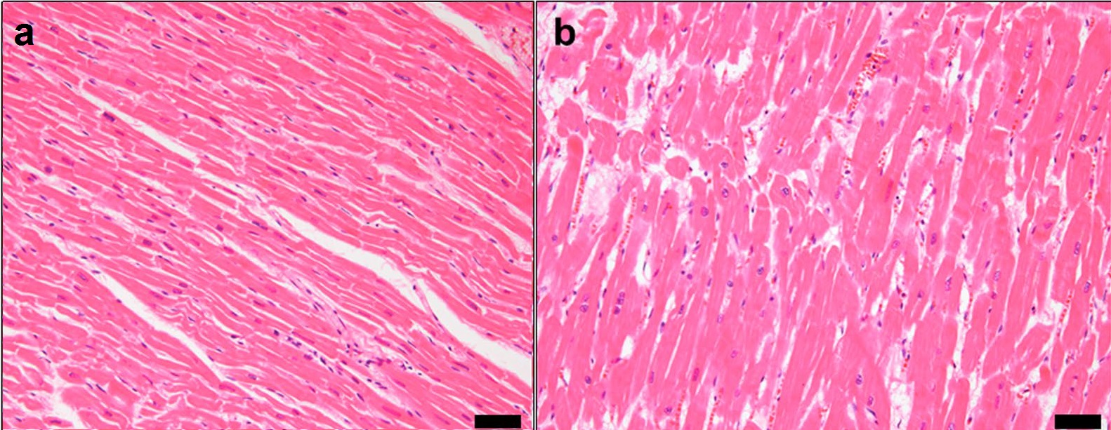
The interventricular septum (IVS, or ventricular septum, or during development septum inferius) is the stout wall separating the ventricle (heart), ventricles, the lower chambers of the heart, from one another. The interventricular septum is directed obliquely backward to the right and curved with the convexity toward the right ventricle; its margins correspond with the anterior interventricular sulcus, anterior and Posterior interventricular sulcus, posterior interventricular sulci. The lower part of the septum, which is the major part, is thick and muscular, and its much smaller upper part is thin and membraneous. During each cardiac cycle the interventricular septum contracts by shortening longitudinally and becoming thicker. Structure The interventricular septum is the stout wall separating the Ventricle (heart), ventricles, the lower chambers of the heart, from one another. The ventricular septum is directed obliquely backward to the right and curved with the convexity t ... [...More Info...] [...Related Items...] OR: [Wikipedia] [Google] [Baidu] |
Heart
The heart is a muscular Organ (biology), organ found in humans and other animals. This organ pumps blood through the blood vessels. The heart and blood vessels together make the circulatory system. The pumped blood carries oxygen and nutrients to the tissue, while carrying metabolic waste such as carbon dioxide to the lungs. In humans, the heart is approximately the size of a closed fist and is located between the lungs, in the middle compartment of the thorax, chest, called the mediastinum. In humans, the heart is divided into four chambers: upper left and right Atrium (heart), atria and lower left and right Ventricle (heart), ventricles. Commonly, the right atrium and ventricle are referred together as the right heart and their left counterparts as the left heart. In a healthy heart, blood flows one way through the heart due to heart valves, which prevent cardiac regurgitation, backflow. The heart is enclosed in a protective sac, the pericardium, which also contains a sma ... [...More Info...] [...Related Items...] OR: [Wikipedia] [Google] [Baidu] |
Right Atrium
The atrium (; : atria) is one of the two upper chambers in the heart that receives blood from the circulatory system. The blood in the atria is pumped into the heart ventricles through the atrioventricular mitral and tricuspid heart valves. There are two atria in the human heart – the left atrium receives blood from the pulmonary circulation, and the right atrium receives blood from the venae cavae of the systemic circulation. During the cardiac cycle, the atria receive blood while relaxed in diastole, then contract in systole to move blood to the ventricles. Each atrium is roughly cube-shaped except for an ear-shaped projection called an atrial appendage, previously known as an auricle. All animals with a closed circulatory system have at least one atrium. The atrium was formerly called the 'auricle'. That term is still used to describe this chamber in some other animals, such as the ''Mollusca''. Auricles in this modern terminology are distinguished by having thicker ... [...More Info...] [...Related Items...] OR: [Wikipedia] [Google] [Baidu] |
Ventricular Hypertrophy
Ventricular hypertrophy (VH) is thickening of the walls of a ventricle (lower chamber) of the heart. Although left ventricular hypertrophy (LVH) is more common, right ventricular hypertrophy (RVH), as well as concurrent hypertrophy of both ventricles can also occur. Ventricular hypertrophy can result from a variety of conditions, both adaptive and maladaptive. For example, it occurs in what is regarded as a physiologic, adaptive process in pregnancy in response to increased blood volume; but can also occur as a consequence of ventricular remodeling following a heart attack. Importantly, pathologic and physiologic remodeling engage different cellular pathways in the heart and result in different gross cardiac phenotypes. Presentation In individuals with eccentric hypertrophy there may be little or no indication that hypertrophy has occurred as it is generally a healthy response to increased demands on the heart. Conversely, concentric hypertrophy can make itself known in a vari ... [...More Info...] [...Related Items...] OR: [Wikipedia] [Google] [Baidu] |
Acyanotic Heart Defect
An acyanotic heart defect, is a class of congenital heart defects. In these, blood is shunted (flows) from the left side of the heart to the right side of the heart, most often due to a structural defect (hole) in the interventricular septum. People often retain normal levels of oxyhemoglobin saturation in systemic circulation. This term is outdated, because a person with an acyanotic heart defect may show cyanosis (turn blue due to insufficient oxygen in the blood). Signs and symptoms Presentation is the following: * Shortness of breath * Congested cough * Diaphoresis * Fatigue * Frequent respiratory infections * Machine-like heart murmur * Tachycardia * Tachypnea * Respiratory distress * Mild cyanosis (in right sided heart failure) * Poor growth and development (from increased energy spent on breathing) Complications This condition can cause congestive heart failure. Diagnosis Types Left to right shunting heart defects include: * Ventricular septal defect (VSD) (30% o ... [...More Info...] [...Related Items...] OR: [Wikipedia] [Google] [Baidu] |
Cardiac Shunt
In cardiology, a cardiac shunt is a pattern of blood flow in the heart that deviates from the normal circuit of the circulatory system. It may be described as right-left, left-right or bidirectional, or as systemic-to-pulmonary or pulmonary-to-systemic. The direction may be controlled by left and/or right heart pressure, a biological or artificial heart valve or both. The presence of a shunt may also affect left and/or right heart pressure either beneficially or detrimentally. Terminology The left and right sides of the heart are named from a dorsal view, i.e., looking at the heart from the back or from the perspective of the person whose heart it is. There are four chambers in a heart: an atrium (upper) and a ventricle (lower) on both the left and right sides. In mammals and birds, blood from the body goes to the right side of the heart first. Blood enters the upper right atrium, is pumped down to the right ventricle and from there to the lungs via the pulmonary artery. Blood ... [...More Info...] [...Related Items...] OR: [Wikipedia] [Google] [Baidu] |
Tetralogy Of Fallot
Tetralogy of Fallot (TOF), formerly known as Steno-Fallot tetralogy, is a congenital heart defect characterized by four specific cardiac defects. Classically, the four defects are: * Pulmonary stenosis, which is narrowing of the exit from the right ventricle; * A ventricular septal defect, which is a hole allowing blood to flow between the two ventricles; * Right ventricular hypertrophy, which is thickening of the right ventricular muscle; and * an overriding aorta, which is where the aorta expands to allow blood from both ventricles to enter. At birth, children may be asymptomatic or present with many severe symptoms. Later in infancy, there are typically episodes of bluish colour to the skin due to a lack of sufficient oxygenation, known as cyanosis. When affected babies cry or have a bowel movement, they may undergo a "tet spell" where they turn cyanotic, have difficulty breathing, become limp, and occasionally lose consciousness. Other symptoms may include a heart mur ... [...More Info...] [...Related Items...] OR: [Wikipedia] [Google] [Baidu] |
Congenital Heart Defect
A congenital heart defect (CHD), also known as a congenital heart anomaly, congenital cardiovascular malformation, and congenital heart disease, is a defect in the structure of the heart or great vessels that is present at birth. A congenital heart defect is classed as a cardiovascular disease. Signs and symptoms depend on the specific type of defect. Symptoms can vary from none to life-threatening. When present, symptoms are variable and may include rapid breathing, bluish skin (cyanosis), poor weight gain, and feeling tired. CHD does not cause chest pain. Most congenital heart defects are not associated with other diseases. A complication of CHD is heart failure. Congenital heart defects are the most common birth defect. In 2015, they were present in 48.9 million people globally. They affect between 4 and 75 per 1,000 live births, depending upon how they are diagnosed. In about 6 to 19 per 1,000 they cause a moderate to severe degree of problems. Congenital heart defects are t ... [...More Info...] [...Related Items...] OR: [Wikipedia] [Google] [Baidu] |
Ventricular Septal Defect
A ventricular septal defect (VSD) is a defect in the ventricular septum, the wall dividing the left and right ventricles of the heart. It's a common heart problem present at birth ( congenital heart defect). The extent of the opening may vary from pin size to complete absence of the ventricular septum, creating one common ventricle. The ventricular septum consists of an inferior muscular and superior membranous portion and is extensively innervated with conducting cardiomyocytes. The membranous portion, which is close to the atrioventricular node, is most commonly affected in adults and older children in the United States. It is also the type that will most commonly require surgical intervention, comprising over 80% of cases. Membranous ventricular septal defects are more common than muscular ventricular septal defects, and are the most common congenital cardiac anomaly. Signs and symptoms Ventricular septal defect is usually symptomless at birth. It usually manifests a fe ... [...More Info...] [...Related Items...] OR: [Wikipedia] [Google] [Baidu] |
Left Coronary Artery
The left coronary artery (LCA, also known as the left main coronary artery, or left main stem coronary artery) is a coronary artery that arises from the aorta above the left cusp of the aortic valve, and supplies blood to the left side of the heart muscle. The left coronary artery typically runs for 10–25 mm, then bifurcates into the left anterior descending artery, and the left circumflex artery. The part that is between the aorta and the bifurcation only is known as the left main artery (LM), while the term "LCA" might refer to just the left main, or to the left main and all its eventual branches. Structure Variation Sometimes, an additional artery arises at the bifurcation of the left main artery, forming a trifurcation; this extra artery is called the ''ramus'' or ''intermediate artery''. A "first septal branch" is sometimes described. Additional images File:Coronary arteries 1.jpg, Left coronary artery File:Cardiac vessels.png, Cardiac vessels File:Gray50 ... [...More Info...] [...Related Items...] OR: [Wikipedia] [Google] [Baidu] |
Left Anterior Descending Artery
The left anterior descending artery (LAD, or anterior descending branch), also called anterior interventricular artery (IVA, or anterior interventricular branch of left coronary artery) is a branch of the left coronary artery. It supplies the anterior portion of the left ventricle. It provides about half of the arterial supply to the left ventricle and is thus considered the most important vessel supplying the left ventricle. Blockage of this artery is often called the ''widow-maker infarction'' due to a high risk of death. Structure Course It first passes at posterior to the pulmonary artery, then passes anteriorward between that pulmonary artery and the left atrium to reach the anterior interventricular sulcus, along which it descends to the notch of cardiac apex. In 78% of cases, it reaches the apex of the heart. Although rare, multiple anomalous courses of the LAD have been described. These include the origin of the artery from the right aortic sinus. Branches The LAD g ... [...More Info...] [...Related Items...] OR: [Wikipedia] [Google] [Baidu] |
Right Coronary Artery
In the coronary circulation, blood supply of the heart, the right coronary artery (RCA) is an artery originating above the right cusp of the aortic valve, at the Aortic sinus, right aortic sinus in the heart. It travels down the right coronary sulcus, towards the crux cordis, crux of the heart. It gives off many branches, including the sinoatrial nodal artery, Right marginal branch of right coronary artery, right marginal artery, posterior interventricular artery, conus artery, and atrioventricular nodal branch. It contributes the right side of the heart, and parts of the interventricular septum. Structure The right coronary artery originates above the Aortic sinus, right aortic sinus above the aortic valve. It passes along the right coronary sulcus, coronary sulcus (right atrioventricular groove) towards the Crux cordis, crux of the heart. Segments * Proximal: starting at RCA origin, spanning half the distance to the Right border of heart, acute margin * Middle: from proxim ... [...More Info...] [...Related Items...] OR: [Wikipedia] [Google] [Baidu] |
Posterior Interventricular Artery
In the coronary circulation, the posterior descending artery (PDA), also called the posterior interventricular artery (PIV, PIA, or PIVA), is an artery running in the posterior interventricular sulcus to the apex of the heart where it meets with the left anterior descending artery also known as the anterior interventricular artery. The PDA supplies the posterior third of the interventricular septum. The remaining anterior two-thirds is supplied by the left anterior descending artery, which is a branch of left coronary artery. It is typically a branch of the right coronary artery (70%, known as right dominance). Alternately, the PDA can be a branch of the circumflex coronary artery (10%, known as left dominance) which itself is a branch of the left coronary artery. It can also be supplied by an anastomosis An anastomosis (, : anastomoses) is a connection or opening between two things (especially cavities or passages) that are normally diverging or branching, such as between blo ... [...More Info...] [...Related Items...] OR: [Wikipedia] [Google] [Baidu] |


